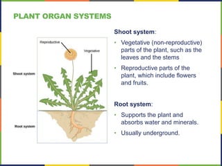
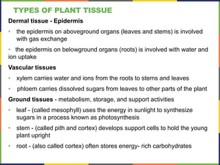
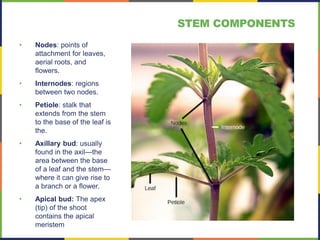
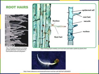
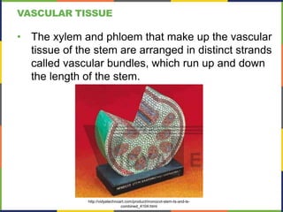
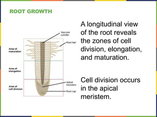
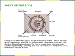
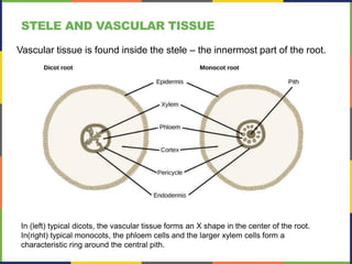
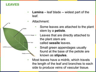
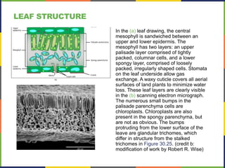
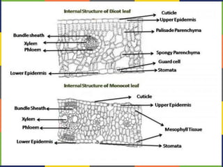
PLANT FORM AND PHYSIOLOGY
This work is licensed under
a Creative Commons
Attribution-NonCommercial-
ShareAlike 4.0 International
License.
Shoot system:
• Vegetative (non-reproductive)
parts of the plant, such as the
leaves and the stems
• Reproductive parts of the
plant, which include flowers
and fruits.
Root system:
• Supports the plant and
absorbs water and minerals.
• Usually underground.
PLANT ORGAN SYSTEMS
TYPES OF PLANT TISSUE
Dermal tissue - Epidermis
• the epidermis on aboveground organs (leaves and stems) is involved
with gas exchange
• the epidermis on belowground organs (roots) is involved with water and
ion uptake
Vascular tissues
• xylem carries water and ions from the roots to stems and leaves
• phloem carries dissolved sugars from leaves to other parts of the plant
Ground tissues - metabolism, storage, and support activities
• leaf - (called mesophyll) uses the energy in sunlight to synthesize
sugars in a process known as photosynthesis
• stem - (called pith and cortex) develops support cells to hold the young
plant upright
• root - (also called cortex) often stores energy- rich carbohydrates
PLANT CELLS
• Parenchyma - undifferentiated cell with potential to
become other cell types. Contains organelles, thin primary
cell wall.
• Collenchyma - similar to parenchyma cell, but with
thickened cell walls. Provides support.
• Sclerenchyma - contain secondary cell walls in addition to
primary cell wall. Provides more support and strength to
cells, as well as xylem and phloem.
• fibers - long slender cells
• sclereids - often branched, spherical shaped.
https://brainly.in/question/3350134
• Nodes: points of
attachment for leaves,
aerial roots, and
flowers.
• Internodes: regions
between two nodes.
• Petiole: stalk that
extends from the stem
to the base of the leaf is
the.
• Axillary bud: usually
found in the axil—the
area between the base
of a leaf and the stem—
where it can give rise to
a branch or a flower.
• Apical bud: The apex
(tip) of the shoot
contains the apical
meristem
STEM COMPONENTS
The stem of common St John's Wort (Hypericum perforatum) is shown in cross section in this
light micrograph. The central pith (greenish-blue, in the center) and peripheral cortex (narrow
zone 3–5 cells thick just inside the epidermis) are composed of parenchyma cells. Vascular
tissue composed of xylem (red) and phloem tissue (green, between the xylem and cortex)
surrounds the pith. (credit: Rolf-Dieter Mueller)
STEM ANATOMY
This light micrograph shows a cross section of a squash (Curcurbita maxima) stem. Each teardrop-shaped vascular
bundle consists of large xylem vessels toward the inside and smaller phloem cells toward the outside. Xylem cells,
which transport water and nutrients from the roots to the rest of the plant, are dead at functional maturity. Phloem
cells, which transport sugars and other organic compounds from photosynthetic tissue to the rest of the plant, are
living. The vascular bundles are encased in ground tissue and surrounded by dermal tissue. (credit: modification of
work by “(biophotos)”/Flickr; scale-bar data from Matt Russell)
STEM COMPONENTS
DERMAL TISSUE
• Guard cells - found mainly in leaf epidermis
• Regulate carbon dioxide uptake and loss of
water from plant.
• Guard cell complex is the stoma (stomata is
plural).
• Trichomes - hair-like outgrowths of epidermis.
• Root hairs - hair-like extensions of root epidermis.
Facilitate the uptake of water and nutrients.
Openings called stomata (singular: stoma) allow a plant to take up carbon dioxide and release oxygen and
water vapor. The (a) colorized scanning-electron micrograph shows a closed stoma of a dicot. Each stoma
is flanked by two guard cells that regulate its (b) opening and closing. The (c) guard cells sit within the layer
of epidermal cells (credit a: modification of work by Louisa Howard, Rippel Electron Microscope Facility,
Dartmouth College; credit b: modification of work by June Kwak, University of Maryland; scale-bar data
from Matt Russell)
STOMATA
TRICHOMES
https://www.pinterest.com/pin/345580971399450495/
Sticky trichomes of a carnivorous
plant, Drosera capensis with a trapped
insect, contain proteolytic enzymes.
Trichomes on Cannabis, rich in cannabinoids.
https://en.wikipedia.org/wiki/Trichome
Coleus leaf trichomes
ROOT HAIRS
http://12knights.pbworks.com/w/pa
ge/25273820/921%20Outline%20
how%20the%20root%20system
https://biology-igcse.weebly.com/root-hairs-and-water-uptake-by-plants.html
https://www.reference.com/science/function-root-hair-cell-3d918a7c283e9d33
VASCULAR TISSUE
• The xylem and phloem that make up the vascular
tissue of the stem are arranged in distinct strands
called vascular bundles, which run up and down
the length of the stem.
http://vidyatechnoart.com/product/monocot-stem-ts-and-ls-
combined_4104.html
In (a) dicot stems, vascular bundles are arranged around the periphery of the ground tissue.
The xylem tissue is located toward the interior of the vascular bundle, and phloem is located
toward the exterior. Sclerenchyma fibers cap the vascular bundles. In (b) monocot stems,
vascular bundles composed of xylem and phloem tissues are scattered throughout the
ground tissue.
VASCULAR TISSUE
XYLEM TISSUE
Tracheids - Long and narrow, with
end walls. Walls perforated with pits,
which allow water to move from cell
to cell.
Vessel elements - Shorter than
tracheids with thinner walls.
• Each vessel element is connected
to the next by means of a
perforation plate at the end walls
• Water moves through the
perforation plates to travel up the
plant.
https://www.quora.com/How-do-tracheids-
and-vessels-differ
PHLOEM TISSUE
Sieve-tube cells (also called sieve-
tube elements) are arranged end to
end to make up a long sieve tube,
which transports organic substances
such as sugars and amino acids.
• Sugars flow from one sieve-tube
cell to the next through perforated
sieve plates, which are found at
the end junctions between two
cells.
Companion cells are found alongside
the sieve-tube cells, providing them
with metabolic support.
• The companion cells contain more
ribosomes and mitochondria than
the sieve-tube cells, which lack
some cellular organelles.
In woody plants, primary growth is followed by secondary growth, which allows the
plant stem to increase in thickness or girth. Secondary vascular tissue is added as the
plant grows, as well as a cork layer. The bark of a tree extends from the vascular
cambium to the epidermis.
GROWTH IN STEMS
(a)Tap root systems have a main root that grows down,
(b)fibrous root systems consist of many small roots.
(credit b: modification of work by “Austen Squarepants”/Flickr)
TYPES OF ROOT SYSTEMS
A longitudinal view
of the root reveals
the zones of cell
division, elongation,
and maturation.
Cell division occurs
in the apical
meristem.
ROOT GROWTH
Staining reveals different cell types in this light micrograph of a wheat (Triticum) root cross
section. Sclerenchyma cells of the exodermis and xylem cells stain red, and phloem cells
stain blue. Other cell types stain black. The stele, or vascular tissue, is the area inside
endodermis (indicated by a green ring). Root hairs are visible outside the epidermis. (credit:
scale-bar data from Matt Russell)
PARTS OF THE ROOT
ROOT TISSUE -- EPIDERMIS
Outermost, single layer of cells that:
a) Protects (from diseases)
b) Absorbs water and nutrients
ROOT HAIRS: Increase surface area of root, for
better water/nutrient absorption
ROOT CORTEX -- ENDODERMIS
• Endodermis (innermost layer of the cortex) contains the
Casparian strip - a water-impermeable strip of waxy
material
• The Casparian strip forces water and solutes to cross the
plasma membranes of endodermal cells instead of slipping
between the cells.
• This ensures that only materials required by the root pass
through the endodermis, while toxic substances and
pathogens are generally excluded.
https://en.wikipedia.org/wiki/Casparian_strip
In (left) typical dicots, the vascular tissue forms an X shape in the center of the root.
In(right) typical monocots, the phloem cells and the larger xylem cells form a
characteristic ring around the central pith.
STELE AND VASCULAR TISSUE
Vascular tissue is found inside the stele – the innermost part of the root.
ROOT MODIFICATIONS – STARCH STORAGE
The (a) banyan tree, also known as the strangler fig, begins life as an epiphyte in a host tree.
Aerial roots extend to the ground and support the growing plant, which eventually strangles
the host tree. The (b) screwpine develops aboveground roots that help support the plant in
sandy soils. (credit a: modification of work by “psyberartist”/Flickr; credit b: modification of
work by David Eikhoff)
ROOT MODIFICATIONS –
AERIAL AND ABOVEGROUND ROOTS
LEAVES
• Lamina – leaf blade -- widest part of the
leaf.
• Attachment:
• Some leaves are attached to the plant
stem by a petiole.
• Leaves that are directly attached to
the plant stem are
called sessile leaves.
• Small green appendages usually
found at the base of the petiole are
known as stipules.
• Most leaves have a midrib, which travels
the length of the leaf and branches to each
side to produce veins of vascular tissue.
(a) Tulip (Tulipa), a monocot, has leaves with parallel venation. The netlike venation in this (b) linden
(Tilia cordata) leaf distinguishes it as a dicot. The (c) Ginkgo biloba tree has dichotomous venation.
(credit a photo: modification of work by “Drewboy64”/Wikimedia Commons; credit b photo:
modification of work by Roger Griffith; credit c photo: modification of work by “geishaboy500”/Flickr;
credit abc illustrations: modification of work by Agnieszka Kwiecień)
VENATION
LEAF FORM
In the (a) leaf drawing, the central
mesophyll is sandwiched between an
upper and lower epidermis. The
mesophyll has two layers: an upper
palisade layer comprised of tightly
packed, columnar cells, and a lower
spongy layer, comprised of loosely
packed, irregularly shaped cells. Stomata
on the leaf underside allow gas
exchange. A waxy cuticle covers all aerial
surfaces of land plants to minimize water
loss. These leaf layers are clearly visible
in the (b) scanning electron micrograph.
The numerous small bumps in the
palisade parenchyma cells are
chloroplasts. Chloroplasts are also
present in the spongy parenchyma, but
are not as obvious. The bumps
protruding from the lower surface of the
leave are glandular trichomes, which
differ in structure from the stalked
trichomes in Figure 30.25. (credit b:
modification of work by Robert R. Wise)
LEAF STRUCTURE
The (a) Venus flytrap has modified leaves that can capture insects. When an unlucky insect touches
the trigger hairs inside the leaf, the trap suddenly closes. The opening of the (b) pitcher plant is lined
with a slippery wax. Insects crawling on the lip slip and fall into a pool of water in the bottom of the
pitcher, where they are digested by bacteria. The plant then absorbs the smaller molecules. (credit
a: modification of work by Peter Shanks; credit b: modification of work by Tim Mansfield)
LEAF ADAPTATIONS – CARNIVOROUS PLANTS

Plant Form and Physiology.pdf

  • 1.
    PLANT FORM ANDPHYSIOLOGY This work is licensed under a Creative Commons Attribution-NonCommercial- ShareAlike 4.0 International License.
  • 2.
    Shoot system: • Vegetative(non-reproductive) parts of the plant, such as the leaves and the stems • Reproductive parts of the plant, which include flowers and fruits. Root system: • Supports the plant and absorbs water and minerals. • Usually underground. PLANT ORGAN SYSTEMS
  • 3.
    TYPES OF PLANTTISSUE Dermal tissue - Epidermis • the epidermis on aboveground organs (leaves and stems) is involved with gas exchange • the epidermis on belowground organs (roots) is involved with water and ion uptake Vascular tissues • xylem carries water and ions from the roots to stems and leaves • phloem carries dissolved sugars from leaves to other parts of the plant Ground tissues - metabolism, storage, and support activities • leaf - (called mesophyll) uses the energy in sunlight to synthesize sugars in a process known as photosynthesis • stem - (called pith and cortex) develops support cells to hold the young plant upright • root - (also called cortex) often stores energy- rich carbohydrates
  • 4.
    PLANT CELLS • Parenchyma- undifferentiated cell with potential to become other cell types. Contains organelles, thin primary cell wall. • Collenchyma - similar to parenchyma cell, but with thickened cell walls. Provides support. • Sclerenchyma - contain secondary cell walls in addition to primary cell wall. Provides more support and strength to cells, as well as xylem and phloem. • fibers - long slender cells • sclereids - often branched, spherical shaped. https://brainly.in/question/3350134
  • 5.
    • Nodes: pointsof attachment for leaves, aerial roots, and flowers. • Internodes: regions between two nodes. • Petiole: stalk that extends from the stem to the base of the leaf is the. • Axillary bud: usually found in the axil—the area between the base of a leaf and the stem— where it can give rise to a branch or a flower. • Apical bud: The apex (tip) of the shoot contains the apical meristem STEM COMPONENTS
  • 6.
    The stem ofcommon St John's Wort (Hypericum perforatum) is shown in cross section in this light micrograph. The central pith (greenish-blue, in the center) and peripheral cortex (narrow zone 3–5 cells thick just inside the epidermis) are composed of parenchyma cells. Vascular tissue composed of xylem (red) and phloem tissue (green, between the xylem and cortex) surrounds the pith. (credit: Rolf-Dieter Mueller) STEM ANATOMY
  • 7.
    This light micrographshows a cross section of a squash (Curcurbita maxima) stem. Each teardrop-shaped vascular bundle consists of large xylem vessels toward the inside and smaller phloem cells toward the outside. Xylem cells, which transport water and nutrients from the roots to the rest of the plant, are dead at functional maturity. Phloem cells, which transport sugars and other organic compounds from photosynthetic tissue to the rest of the plant, are living. The vascular bundles are encased in ground tissue and surrounded by dermal tissue. (credit: modification of work by “(biophotos)”/Flickr; scale-bar data from Matt Russell) STEM COMPONENTS
  • 8.
    DERMAL TISSUE • Guardcells - found mainly in leaf epidermis • Regulate carbon dioxide uptake and loss of water from plant. • Guard cell complex is the stoma (stomata is plural). • Trichomes - hair-like outgrowths of epidermis. • Root hairs - hair-like extensions of root epidermis. Facilitate the uptake of water and nutrients.
  • 9.
    Openings called stomata(singular: stoma) allow a plant to take up carbon dioxide and release oxygen and water vapor. The (a) colorized scanning-electron micrograph shows a closed stoma of a dicot. Each stoma is flanked by two guard cells that regulate its (b) opening and closing. The (c) guard cells sit within the layer of epidermal cells (credit a: modification of work by Louisa Howard, Rippel Electron Microscope Facility, Dartmouth College; credit b: modification of work by June Kwak, University of Maryland; scale-bar data from Matt Russell) STOMATA
  • 10.
    TRICHOMES https://www.pinterest.com/pin/345580971399450495/ Sticky trichomes ofa carnivorous plant, Drosera capensis with a trapped insect, contain proteolytic enzymes. Trichomes on Cannabis, rich in cannabinoids. https://en.wikipedia.org/wiki/Trichome Coleus leaf trichomes
  • 11.
  • 12.
    VASCULAR TISSUE • Thexylem and phloem that make up the vascular tissue of the stem are arranged in distinct strands called vascular bundles, which run up and down the length of the stem. http://vidyatechnoart.com/product/monocot-stem-ts-and-ls- combined_4104.html
  • 13.
    In (a) dicotstems, vascular bundles are arranged around the periphery of the ground tissue. The xylem tissue is located toward the interior of the vascular bundle, and phloem is located toward the exterior. Sclerenchyma fibers cap the vascular bundles. In (b) monocot stems, vascular bundles composed of xylem and phloem tissues are scattered throughout the ground tissue. VASCULAR TISSUE
  • 14.
    XYLEM TISSUE Tracheids -Long and narrow, with end walls. Walls perforated with pits, which allow water to move from cell to cell. Vessel elements - Shorter than tracheids with thinner walls. • Each vessel element is connected to the next by means of a perforation plate at the end walls • Water moves through the perforation plates to travel up the plant. https://www.quora.com/How-do-tracheids- and-vessels-differ
  • 15.
    PHLOEM TISSUE Sieve-tube cells(also called sieve- tube elements) are arranged end to end to make up a long sieve tube, which transports organic substances such as sugars and amino acids. • Sugars flow from one sieve-tube cell to the next through perforated sieve plates, which are found at the end junctions between two cells. Companion cells are found alongside the sieve-tube cells, providing them with metabolic support. • The companion cells contain more ribosomes and mitochondria than the sieve-tube cells, which lack some cellular organelles.
  • 16.
    In woody plants,primary growth is followed by secondary growth, which allows the plant stem to increase in thickness or girth. Secondary vascular tissue is added as the plant grows, as well as a cork layer. The bark of a tree extends from the vascular cambium to the epidermis. GROWTH IN STEMS
  • 17.
    (a)Tap root systemshave a main root that grows down, (b)fibrous root systems consist of many small roots. (credit b: modification of work by “Austen Squarepants”/Flickr) TYPES OF ROOT SYSTEMS
  • 18.
    A longitudinal view ofthe root reveals the zones of cell division, elongation, and maturation. Cell division occurs in the apical meristem. ROOT GROWTH
  • 19.
    Staining reveals differentcell types in this light micrograph of a wheat (Triticum) root cross section. Sclerenchyma cells of the exodermis and xylem cells stain red, and phloem cells stain blue. Other cell types stain black. The stele, or vascular tissue, is the area inside endodermis (indicated by a green ring). Root hairs are visible outside the epidermis. (credit: scale-bar data from Matt Russell) PARTS OF THE ROOT
  • 20.
    ROOT TISSUE --EPIDERMIS Outermost, single layer of cells that: a) Protects (from diseases) b) Absorbs water and nutrients ROOT HAIRS: Increase surface area of root, for better water/nutrient absorption
  • 21.
    ROOT CORTEX --ENDODERMIS • Endodermis (innermost layer of the cortex) contains the Casparian strip - a water-impermeable strip of waxy material • The Casparian strip forces water and solutes to cross the plasma membranes of endodermal cells instead of slipping between the cells. • This ensures that only materials required by the root pass through the endodermis, while toxic substances and pathogens are generally excluded. https://en.wikipedia.org/wiki/Casparian_strip
  • 22.
    In (left) typicaldicots, the vascular tissue forms an X shape in the center of the root. In(right) typical monocots, the phloem cells and the larger xylem cells form a characteristic ring around the central pith. STELE AND VASCULAR TISSUE Vascular tissue is found inside the stele – the innermost part of the root.
  • 23.
    ROOT MODIFICATIONS –STARCH STORAGE
  • 24.
    The (a) banyantree, also known as the strangler fig, begins life as an epiphyte in a host tree. Aerial roots extend to the ground and support the growing plant, which eventually strangles the host tree. The (b) screwpine develops aboveground roots that help support the plant in sandy soils. (credit a: modification of work by “psyberartist”/Flickr; credit b: modification of work by David Eikhoff) ROOT MODIFICATIONS – AERIAL AND ABOVEGROUND ROOTS
  • 25.
    LEAVES • Lamina –leaf blade -- widest part of the leaf. • Attachment: • Some leaves are attached to the plant stem by a petiole. • Leaves that are directly attached to the plant stem are called sessile leaves. • Small green appendages usually found at the base of the petiole are known as stipules. • Most leaves have a midrib, which travels the length of the leaf and branches to each side to produce veins of vascular tissue.
  • 26.
    (a) Tulip (Tulipa),a monocot, has leaves with parallel venation. The netlike venation in this (b) linden (Tilia cordata) leaf distinguishes it as a dicot. The (c) Ginkgo biloba tree has dichotomous venation. (credit a photo: modification of work by “Drewboy64”/Wikimedia Commons; credit b photo: modification of work by Roger Griffith; credit c photo: modification of work by “geishaboy500”/Flickr; credit abc illustrations: modification of work by Agnieszka Kwiecień) VENATION
  • 27.
  • 28.
    In the (a)leaf drawing, the central mesophyll is sandwiched between an upper and lower epidermis. The mesophyll has two layers: an upper palisade layer comprised of tightly packed, columnar cells, and a lower spongy layer, comprised of loosely packed, irregularly shaped cells. Stomata on the leaf underside allow gas exchange. A waxy cuticle covers all aerial surfaces of land plants to minimize water loss. These leaf layers are clearly visible in the (b) scanning electron micrograph. The numerous small bumps in the palisade parenchyma cells are chloroplasts. Chloroplasts are also present in the spongy parenchyma, but are not as obvious. The bumps protruding from the lower surface of the leave are glandular trichomes, which differ in structure from the stalked trichomes in Figure 30.25. (credit b: modification of work by Robert R. Wise) LEAF STRUCTURE
  • 30.
    The (a) Venusflytrap has modified leaves that can capture insects. When an unlucky insect touches the trigger hairs inside the leaf, the trap suddenly closes. The opening of the (b) pitcher plant is lined with a slippery wax. Insects crawling on the lip slip and fall into a pool of water in the bottom of the pitcher, where they are digested by bacteria. The plant then absorbs the smaller molecules. (credit a: modification of work by Peter Shanks; credit b: modification of work by Tim Mansfield) LEAF ADAPTATIONS – CARNIVOROUS PLANTS