WELCOME
DIFFERENT TYPE OF PLANT
CELL IN PLANT TISSUE
SYSTEM
PRESENTED BY
ZUBY GOHAR ANSARI
TAM/14-26
TISSUE SYSTEM
Plant body in Angiosperms is differentiated into root stem,
leaf and flower.
The body of a vascular plant is composed of dermal tissue,
Ground tissue and Vascular tissue.
Dermal Tissue (Skin)
Dermal Tissue is protective in function. Basing on its
origin, it is classified into two types – Epidermis and
Periderm.
Epidermis: This is the primary surface tissue of the entire
plant.
Epidermal cells are compactly and continuously
arranged; the continuity is lost by the presence of
Stomatal pores or breaks in the tissue.
The cuticle can be separated from epidermis.
Epidermis provides mechanical protection, allows
gaseous exchange through stomata, restricts
transpiration with cuticle, and is also involved in
storage, photosynthesis, secretion, absorption and
perception to stimuli.
Cells surrounding guard cells, but differing from other epidermal
cells, are called subsidiary cells.
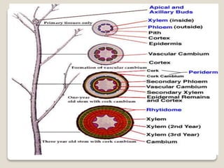
Periderm: This is formed during secondary growth replacing
primary epidermis.
Ground Tissue
This is inner to dermal tissue and is composed of simple tissues like
parenchyma.
Vascular tissue
Vascular tissue consists of conducting elements – xylem and
phloem.
TISSUE TYPES
In broad sense, tissues are classified as – meristematic
and permanent tissues.
Meristem (Meristos – divisible)
Initially all embryonic cells of an embryo have the
capacity to divide and multiply but as the embryo
develops into a plant body, this capacity for division
is restricted to certain parts of the plant body called
meristems which are active throughout the life of the
plant body (unlike that of an animal body).
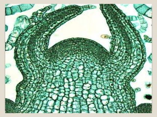
Meristems which occur at the apices of stem, root and other
branches are called apical meristems, which bring about
primary growth of the plants, hence also called as
primary meristems.
In many plants in addition to apical meristems, lateral
meristems like vascular cambium, cork cambium,
intercalary meristems are found.
Lateral meristems are arranged parallel to the sides of
organs in which they occur.
Intercalary Meristem: This is also a primary
meristem, found inserted between permanent tissues,
in the bases of internodes and leaf sheaths of grasses.
Sometimes, as in gynophores of groundnut, xylem may
be present in intercalary meristem.
Vascular cambium and cork cambium: These are
referred to as secondary meristems because they
produce secondary tissues, and increase the thickness
of the plant body.
This process is called secondary growth, seen in
dicotyledons and gymnosperms.
Permanent or Mature Tissues
In a strict sense only cells which have lost the power for
division must be regarded as permanent tissues, but in
a broad sense, cells derived from meristem that have
acquired a special function like photosynthesis,
secretion, storage are treated as part of matured tissue.
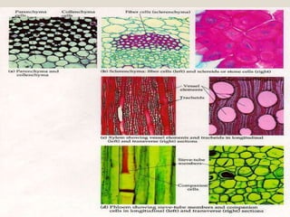
There are different types of mature tissues. Example:
Parenchyma, Collenchyma, Sclerenchyma, Xylem and
Phloem.
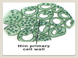
It is found in every part of the plant body like pith and
cortex of stem and root, mesophyll of leaves, flesh of
fruits, floral parts and even in xylem and Phloem.
Cells have thin primary walls.
Parenchymatous cells may also perform specialized
functions and are structurally modified.
The following are the different types of parenchyma.
Aerenchyma:
Intercellular spaces filled with air, are large in size and
many in number. Cells occupy a smaller area.
Though the cells are smaller, they provide the required
strength to the aquatic plants.
In these plants air spaces are common, helping in
aeration and buoyancy.
Air spaces are also seen in roots of grasses, petioles of
canna, aroids etc.
Armed Parenchyma: Cells are stellate in shape with
inward projections of cell wall as arms. Example:
Pinus leaf Mesophyll.
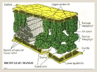
Chlorenchyma: Cells of photosynthetic parenchyma
contain numerous chloroplasts. These cells are
commonly seen in leaves, some times in young
shoots. Cells of Chlorenchyma are of two types –
1) Palisade cells that is elongated and compactly
arranged.
2) Spongy cells that are spaciously arranged and
irregularly shaped.
Prosenchyma (Pros-to): Cells are elongated, have
thick walls and serve as a supporting
tissue. Example: Endosperm cells of seeds.
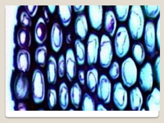
Collenchyma (Kolla – Glue)
Collenchyma is a simple, living tissue.
Cell walls are thickened due to deposition of pectin.
Collenchyma is the primary supporting tissue in stems,
leaves and floral parts of dicots, where as in stems
and leaves of monocots collenchyma is usually
absent, (instead, sclerenchyma is present in
monocots).
Intercellular spaces may or may not be present.
Three forms of collenchyma are recognized based on
the types of thickenings –
1. Thickening is on the tangential wall – lamellar
collenchyma.
2. Deposition of pectin is in the corners where several
cells meet-angular collenchyma.
3. Thickenings are around the intercellular spaces –
lacunar collenchyma.
Being plastic, collenchyma differs from elastic fibers
of sclerenchyma.
Sclerenchyma (Scleros-hard)
Cells of sclerenchyma are thick walled and are usually
lignified.
The thickness is due to formation of secondary wall.
Commonly sclerenchyma cells are classified into
fibers and sclereids.
The primary function is mechanical.
Fibers: These are usually long, spindle shaped
structures, with tapering or blunt ends.
Fibers are grouped into xylary fibers and extraxylary
fibers.
These are lignified or non lignified.
Commercial fibers like jute, flax, ramie are extraxylary
fibers.
Sclereids:
These are shorter than fibers.
Sclereids occur singly or in groups.
Sclereids are commonly found in fruit wall, seed coat,
epidermal scales, and occasionally found in cortex,
pith, mesophyll and petiole of submerged aquatics.
Vascular Tissue (complex tissue)
It is heterogeneous in nature with different types of
cells. (xylem, phloem and pericycle.)
Xylem (Wood)
It consists of living cells like parenchyma and dead cells
like tracheary elements. (fiber also present)
Xylem elements differentiating from an apical meristem
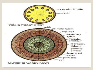
constitute primary xylem where as secondary xylem is
differentiated from vascular cambium.
Xylem is the most preserved tissue in fossils, due to the
development of lignified secondary walls.
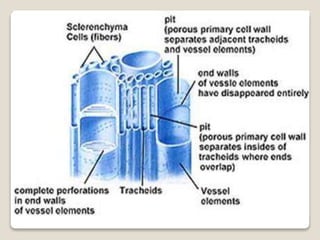
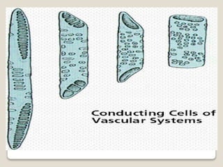
Tracheary elements (trachea – rough): These are of two
types – 1). Trachea or vessels 2) Tracheids, Vessels are
limited in their growth, are joined end to end to form
continuous tubular structures with perforations in their
cross walls.
Vessels are present in most of the Angiosperms and also in
certain lower plants like Gnetum, Marsilea and
Selaginella.
Tracheids are generally elongated and non-perforated.
Conduction of water and minerals is not as efficient as in
vessels
Primary Xylem: (differentiated into Protoxylem and
Metaxylem)
In stems, protoxylem is found nearest to central axis
(endarch xylem) and in roots away from centre (exarch
xylem).
Fibers are absent in protoxylem. Metaxylem appears after
protoxylem.
Metaxylem has only pitted secondary walls.
Metaxylem is more complex than protoxylem and its
elements are wider.
Secondary Xylem: The vascular cambium
(intrafascicular cambium interfascicular cambium)
producing secondary xylem consists of fusiform and
ray initials.
Here also the secondary xylem is also of two-system-
1) Axial or vertical system with living and nonliving
cells and 2) ray or horizontal system with living cells.
When compared with primary xylem, secondary
xylem is more complex and shows orderly
development
 Phloem (Phloos Bark) (food conducting tissue)
 It is composed of sieve elements, parenchyma cells, fibers
and sclereids.
 Phloem differentiated from procambium is called primary
phloem; secondary phloem is initiated from vascular
cambium.
 When xylem and phloem elements are found in the same line
the arrangement is called conjoint; if phloem is only external
to xylem it is called collateral as in many stems; if phloem is
both external and internal to xylem it is called bicollateral as
in leaves and a few stems; if xylem completely surrounds
phloem it is called amphivasal; if phloem surrounds xylem it
is called amphicribal; if xylem and phloem alternate with each
other as in roots, it is called radial.
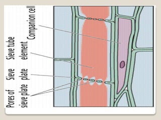
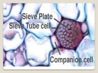
Sieve Elements:
These are classified into sieve cells and sieve tube
members.
Sieve cells are commonly long, slender, and taper at
both ends.
Sieve cells are found in Pteridophytes and
Gymnosperms Initially sieve tube members contain
uninucleated protoplast.
Sieve tube member is associated with one or more
specialized parenchyma cells called companion cells
are seen only in Angiosperms.
Both sieve tube member and companion cell are
derived from the same meristematic cell.
Companion cell is nucleated.
Probably these cells provide energy for food
conduction.
Primary Phloem: (Protophloem and Metaphloem. )
In protophloem, sieve tube members are without
companion cells.
Sieve tubes function for a brief period and soon they
get crushed by the surrounding pressure (disappear
crushed cell).
Metaphloem tissue survives for a longer period.
Usually fibers are absent in dicotyledons, where as in
monocotyledons and herbaceous dicots, parenchyma
cells are absent.
Secondary phloem:
Similar to secondary xylem, there are two systems of
arrangements in secondary phloem –
1) Axial system producing sieve elements, phloem
parenchyma and phloem fibers.
2) Transverse system producing ray parenchyma cells.
Pericycle (A Circle All Around)
Pericycle is the region, consisting of one – few layers
of cells, found external to central cylinder
(Stele). Example: Roots and Dicot stems.
Pericycle may be composed of parenchyma cells or
sclerenchyma cells or both. Lateral branches and
phellogen may arise from pericycle.
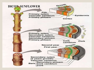
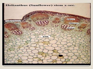
DICOT: SUNFLOWER
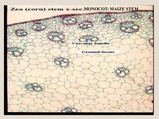
MONOCOT: MAIZE STEM
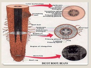
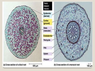
DICOT ROOT: BEANS
DICOT LEAF: MANGO
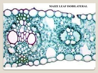
MAIZE LEAF ISOBILATERAL
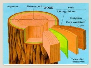
WOOD
THANK
YOU