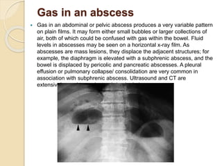
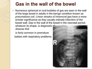
SlideShare a Scribd company logo
Plain Abdomen &
Gastrointestinal Tract
(OESOPHAGUS)
By: Khashayar
Cyrus
 The standard plain film of the abdomen is a supine
anteroposterior
 Free intraperitoneal air can be seen on an erect chest
radiograph (CXR), which is usually performed at the time of the
abdominal x-ray. Rarely, in patients who are unable to sit or
stand, a lateral decubitus view (i.e. an AP film with the patient
lying on his or her side) is performed using a horizontal x-ray
beam as a means of detecting free intraperitoneal air
 looking at a plain abdominal film:
• Analyze the intestinal gas pattern and identify any dilated
portion of the gastrointestinal tract.
• Look for gas outside the lumen of the bowel.
• Look for ascites and soft tissue masses in the abdomen and
pelvis.
• If there are any calcifications, try to locate exactly where they
lie.
• Assess the size of the liver and spleen.
Intestinal gas pattern:
 Relatively large amounts of gas are usually present in the
stomach and colon in a normal patient. The stomach can be
readily identified by its location above the transverse colon, by
the band-like shadows of the gastric rugae in the supine view.
The duodenum often contains air and there may be some gas
in the normal small bowel, but it is rarely sufficient to outline the
whole of a loop. If the bowel is dilated it is important to try and
decide which portion is involved.
 Dilatation of the bowel:
 Dilatation of the bowel is the cardinal plain film sign of intestinal
obstruction, and the pattern of dilatation is the key to the
radiological distinction between small and large bowel
obstruction. In small bowel obstruction, the small intestine is
dilated down to the point of obstruction and the bowel beyond
this point is either empty or of reduced calibre.
 In large bowel obstruction, the large bowel is dilated down to the
level of obstruction. If the ileocaecal valve, where the ileum joins
the colon, is incompetent, there will also be small bowel
dilatation. Making the distinction between large and small bowel
obstruction depends on the ability to recognize which portions of
bowel are dilated.Dilated small bowel usually lies in the centre
of the abdomen within the ‘frame’ of the large bowel (but the
sigmoid and transverse colon may be redundant and may also
lie in the centre of the abdomen, particularly when dilated).
When the proximal and mid small intestine are dilated, the
valvulae conniventes (plica circulares) can be identified. The
valvulae conniventes are always closer together and cross the
width of the bowel (the colonic haustra do not), often giving rise
to an appearance known as a ‘stack of coins’The distal small
intestine has a relatively smooth outline and it may be difficult to
distinguish the lower ileum and the sigmoid colon because both
may be smooth in outline. The radius of curvature of the loops is
sometimes helpful: the tighter the curve, the more likely the loop
is to be dilated small bowel.The colon is recognized by its
haustra, which usuallyform incomplete bands across the colonic
gas shadows.Haustra are always present in the ascending and
transverse colon, but may be absent distal to the splenic flexure.
A. B.
 If the cause or site of the obstruction is not evident from plain
films, and immediate exploratory surgery is not indicated,then
computed tomography (CT) or a contrast study(either a follow-
through for small bowel obstruction or instant enema for large
bowel obstruction) is helpful. CT can demonstrate the site of
obstruction by showing the location of the transition from dilated
to collapsed bowel, and can confirm or exclude a mass at the
site of obstruction
 Dilatation of the bowel occurs in a number of conditions. In
remembering causes of mechanical bowel obstruction it is often
easiest to think of conditions that: (i) obstruct the lumen (e.g.
gall stone ileus); (ii) affect the bowelwall and cause a narrowing
(e.g. Crohn’s disesase; or (iii)cause extrinsic compression of the
bowel (e.g. adhesions).Bowel dilatation, however, occurs in
conditions other than mechanical bowel obstruction, notably:
paralytic ileus,acute ischaemia and inflammatory bowel disease.
The radiologicaldiagnosis of these phenomena depends mainly
onthe pattern of distribution of the dilated loops
paralytic ileus Mechanical obstruction
of
the small bowel
Mechanical obstruction of Closed loop
obstruction
the large bowel
Toxic dilatation of the colon
Pneumoperitoneum
 The radiological diagnosis of perforation of the
gastrointestinal
 tract is based on recognizing free gas in the peritoneal cavity
(pneumoperitoneum) The most common cause of
spontaneous pneumoperitoneum is a perforated peptic ulcer
and two-thirds of such cases are recognizable radiologically.
The largest quantities of free gas are seen after colonic
perforation, and the smallest amounts with leakage from the
small bowel. A pneumoperitoneum is very rare in acute
appendicitis even if the appendix has perforated.Free
intraperitoneal air is a normal finding after laparotomy or
laparoscopy. In adults, all the air is usually absorbed within 7
days. In children, the air absorbs much faster, usually within
24 hours. An increase in the amount of airon successive films
indicates continuing leakage of air.
 Pneumoperitoneum under the right hemidiaphragm is usually
easy to recognize on an erect CXR as a curvilinearcollection
of gas between the line of the diaphragm and the opacity of
the liver. Free gas under the left hemidiaphragm is more
difficult to identify because of the overlapping gas shadows of
the stomach and the splenic flexure of the colon. Gas in these
organs may mimic free intraperitonealair when none is
present.
Gas in an abscess
 Gas in an abdominal or pelvic abscess produces a very variable pattern
on plain films. It may form either small bubbles or larger collections of
air, both of which could be confused with gas within the bowel. Fluid
levels in abscesses may be seen on a horizontal x-ray film. As
abscesses are mass lesions, they displace the adjacent structures; for
example, the diaphragm is elevated with a subphrenic abscess, and the
bowel is displaced by pericolic and pancreatic abscesses. A pleural
effusion or pulmonary collapse/ consolidation are very common in
association with subphrenic abscess. Ultrasound and CT are
extensively used to evaluate abdominal abscesses
Gas in the wall of the bowel
 Numerous spherical or oval bubbles of gas are seen in the wall
of the large bowel in adults in the benign condition known as
pneumatosis coli. Linear streaks of intramural gas have a more
sinister significance as they usually indicate infarction of the
bowel wall. Gas in the wall of the bowel in the neonatal period,
whatever its shape, is diagnostic of necrotizing enterocolitis a
disease that
is fairly common in premature
babies with respiratory problems.
Gas in the biliary system
 Gas in the biliary system is seen on plain films following
sphincterotomy or anastomosis of the common bile duct to the
bowel (Fig. 5.10). It is also seen with a fistula from erosion of a
gall stone into the duodenum or colon, or following penetration of
a duoden al ulcer into the common bile duct.
Gas may be seen, very
occasionally, in the wall or
lumen
of the gall bladder in acute
cholecystitis from gas-forming
organisms
Ascites
 Small amounts of ascites cannot be detected on plain films. Larger
quantities separate the loops of bowel from one another and
displace the ascending and descending colon from the fat stripes,
which indicate the position of the peritoneum along the lateral
abdominal walls The loops of small bowel float to the centre of the
abdomen In practice, plain films are of very limited value in the
diagnosis of ascites as the signs are so difficult to interpret
confidently except when a large amounts of ascites is present.
Ascites is readily recognized at ultrasound or CT
Abdominal calcification
 An attempt should always be made to determine the nature of
any abdominal calcification. The first essential is to localize the
calcification, as once the organ of origin is known the pattern or
shape of the calcification will usually limit the diagnosis to just
one or two choices The most common calcifications are of little
or no significance to the patient; most are phleboliths, calcified
lymph nodes, costal cartilages and arterial calcification.
Calcifications in the abdomen are likely to be one of the
following:
 Pelvic vein phleboliths:
Calcified mesenteric lymph nodes:
Vascular calcification:
Uterine fibroids
Malignant ovarian masses:
Adrenal calcification
Liver calcification
Splenic calcification
Pancreatic calcification
Faecoliths
Liver and spleen
 Substantial enlargement of the liver has to occur before it can be
recognized on a plain abdominal film. As the liver enlarges it
extends well below the costal margin, displacing the hepatic
flexure, transverse colon and right kidney downwards and
displacing the stomach to the left. The diaphragm may also be
elevated Occasionally, there is a tongue-like extension of the
right lobe into the right iliac fossa. This is a normal variant known
as a Reidl’s lobe and should not be confused with generalized
liver enlargement.As the spleen enlarges, the tip becomes visible
in the left
Abdominal and pelvic masses
 Attempting to diagnose the nature of an abdominal mass on a
plain film is notoriously difficult, and ultrasound, CT or magnetic
resonance imaging (MRI) are the appropriate imaging modalities.
The site of the mass, the displacemen of adjacent structures and
the hpresence of calcification are important diagnostic signs but
plain films are unable to distinguish between solid and cystic
masses.An enlarged bladder can be seen as a mass arising from
the pelvis, displacing loops of bowel. In females, uterine and
ovarian enlargements also appear as masses arising from the
pelvis. Ovarian cysts can become very large, almost filling the
abdomen and displacing the bowel to the sides of the abdomen
(Fig. 5.19). An ovarian dermoid cyst can be readily identified on a
plain film due to its content of low attenuation fat and often the
presence of other mesenchymal structures within it, such as
teeth.Retroperitoneal tumours and lymph nodes, when
large,become visible on plain films. Renal masses, especially cysts
and hydronephrosis, can become large and appear as masses in
the flank. With retroperitoneal masses the outline of the psoas
muscle may become invisible.
Gastrointestinal Tract
 Imaging techniques: general principles
 Contrast examinations
 Computed tomography
 Ultrasound examinations
 Magnetic resonance imaging
 Nuclear medicine
OESOPHAGUS
Plain films:
 Plain films do not normally show the oesophagus unless it is very dilated (e.g.
achalasia), but they are of use in demonstrating an opaque foreign body such as
a bone lodged in the oesophagus Plain films are also used to check the position
of a nasogastric tube, to ensure that thetube travels down through the
oesophagus and into the stomach, rather than down into one of the main bronchi
or an oesophageal pouch
Barium swallow examination
 The patient swallows a gas-producing agent to distend
theoesophagus, followed by barium, and its passage down the
oesophagus is observed on a television monitor. Films are taken
with the oesophagus both full of barium to show the outline, and
following the passage of the barium to show the mucosal
pattern.The oesophagus has a smooth outline when full of barium.
When empty and contracted, barium normally lies in between the
folds of mucosa, which appear as three or four long, straight,
parallel lines Peristaltic waves can be observed during
fluoroscopy.They move smoothly along the oesophagus to propel
the barium rapidly into the stomach. It is important not to confuse a
contraction wave with a true narrowing: a narrowing is constant
whereas a contraction wave is transitory.Sometimes the contraction
waves do not occur in an orderly fashion but are pronounced and
prolonged, giving the oesophagus an undulated appearance (Fig.
6.6). These socalled tertiary contractions usually occur in the
elderly, and in most instances they do not give rise to
symptoms.Occasionally, tertiary contractions cause
dysphagia.Endoscopy is the primary investigation in patients with
dysphagia.
Normal oesophagus barium has passed Tertiary
contractions
Computed tomograpphy
 Computed tomography is used in the staging of carcinoma of the
oesophagus. The primary function of CT is to detect distant
disease (e.g. lung, liver or bone metastases), and although it
does give information on local staging, this is more accurately
performed by endoscopic ultrasound
Fluorodeoxyglucose positron
emission tomography/
computed tomography
 In patients with oesophageal carcinoma who have potentially
curable disease, an FDG-PET/CT study is performed to identify
any occult metastases prior to undertaking either surgery or
definitive chemoradiotherapy.
Oesophageal abnormalities
 Strictures of the oesophagus
 Dilatation of the oesophagus
 Strictures of the oesophagus:
Oesophageal carcinoma
Peptic strictures
Achalasia
Corrosive strictures
Benign tumours
Other medistinal masses
Anomalous right subclavian artery
Dilatation of the oesophagus
 There are two main types of oesophageal dilatation –obstructive
and non-obstructive:
 Dilatation due to obstruction is associated with a visible
stricture. The patient with a carcinoma usually presents with
dysphagia before the oesophagus becomes very dilated.On the
other hand, a markedly dilated oesophagus indicates a very
longstanding condition, usually achalasiaor occasionally a
benign stricture.
 Dilatation without obstruction occurs in scleroderma.The
disease involves the oesophageal muscle, resulting in dilatation
of the oesophagus, which resembles an inert tube with no
peristaltic movement so that barium does not flow from the
oesophagus into the stomach unless the patient stands upright.
Other abnormalities of the oesophagus
 oesophageal web
Oesophageal diverticula
oesophageal atresia

More Related Content

What's hot

Abdominal xray - imaging and interpretation
Abdominal xray - imaging and interpretation Abdominal xray - imaging and interpretation
Abdominal xray - imaging and interpretation
ArushiGupta119
 
Radiology ----Classical Signs in GIT Dr. Muhammad Bin Zulfiqar
Radiology ----Classical Signs in GIT Dr. Muhammad Bin ZulfiqarRadiology ----Classical Signs in GIT Dr. Muhammad Bin Zulfiqar
Radiology ----Classical Signs in GIT Dr. Muhammad Bin Zulfiqar
Dr. Muhammad Bin Zulfiqar
 
radiology.Plain abd.(dr.kawa)
radiology.Plain abd.(dr.kawa)radiology.Plain abd.(dr.kawa)
radiology.Plain abd.(dr.kawa)student
 
Signs in pneumoperitoneum
Signs in pneumoperitoneumSigns in pneumoperitoneum
Signs in pneumoperitoneum
Vikram Patil
 
Imaging of Bowel Obstruction
Imaging of Bowel ObstructionImaging of Bowel Obstruction
Imaging of Bowel Obstruction
Rathachai Kaewlai
 
Radiological anatomy of liver segments
Radiological anatomy of liver segmentsRadiological anatomy of liver segments
Radiological anatomy of liver segments
Tarun Goyal
 
Pneumobilia vs portal vein gas
Pneumobilia vs portal vein gasPneumobilia vs portal vein gas
Pneumobilia vs portal vein gasairwave12
 
Presentation1, interpretation of x ray of the abdomen.
Presentation1, interpretation of x ray of the abdomen.Presentation1, interpretation of x ray of the abdomen.
Presentation1, interpretation of x ray of the abdomen.
Abdellah Nazeer
 
Imaging of stomach
Imaging of stomachImaging of stomach
Imaging of stomach
Rakesh Ca
 
USG in gall bladder pathologies
USG in gall bladder pathologiesUSG in gall bladder pathologies
USG in gall bladder pathologies
Pratibha Saran
 
Presentation1.pptx, radiological imaging of small bowel disease.
Presentation1.pptx, radiological imaging of small bowel disease.Presentation1.pptx, radiological imaging of small bowel disease.
Presentation1.pptx, radiological imaging of small bowel disease.Abdellah Nazeer
 
Imaging of acute abdomen
Imaging of acute abdomen Imaging of acute abdomen
Imaging of acute abdomen
Mohammed Bawazir
 
Presentation1.pptx, radiological imaging of intusussception.
Presentation1.pptx, radiological imaging of intusussception.Presentation1.pptx, radiological imaging of intusussception.
Presentation1.pptx, radiological imaging of intusussception.Abdellah Nazeer
 
Imaging anatomy of peritoneum
Imaging  anatomy of peritoneumImaging  anatomy of peritoneum
Imaging anatomy of peritoneum
Bajanagaraju
 
X ray signs of abdomen
X ray signs of abdomenX ray signs of abdomen
X ray signs of abdomen
Mathew Joseph
 
Imaging of abdominal trauma
Imaging of abdominal traumaImaging of abdominal trauma
Imaging of abdominal trauma
Dev Lakhera
 
Presentation2.pptx. radilogical imaging of intestinal obstruction.
Presentation2.pptx. radilogical imaging of intestinal obstruction.Presentation2.pptx. radilogical imaging of intestinal obstruction.
Presentation2.pptx. radilogical imaging of intestinal obstruction.Abdellah Nazeer
 
Abdominal X ray
Abdominal X rayAbdominal X ray
Abdominal X ray
Richin Koshy
 

What's hot (20)

Abdominal xray - imaging and interpretation
Abdominal xray - imaging and interpretation Abdominal xray - imaging and interpretation
Abdominal xray - imaging and interpretation
 
Ba.meal final
Ba.meal finalBa.meal final
Ba.meal final
 
Radiology ----Classical Signs in GIT Dr. Muhammad Bin Zulfiqar
Radiology ----Classical Signs in GIT Dr. Muhammad Bin ZulfiqarRadiology ----Classical Signs in GIT Dr. Muhammad Bin Zulfiqar
Radiology ----Classical Signs in GIT Dr. Muhammad Bin Zulfiqar
 
radiology.Plain abd.(dr.kawa)
radiology.Plain abd.(dr.kawa)radiology.Plain abd.(dr.kawa)
radiology.Plain abd.(dr.kawa)
 
Signs in pneumoperitoneum
Signs in pneumoperitoneumSigns in pneumoperitoneum
Signs in pneumoperitoneum
 
Imaging of Bowel Obstruction
Imaging of Bowel ObstructionImaging of Bowel Obstruction
Imaging of Bowel Obstruction
 
Radiological anatomy of liver segments
Radiological anatomy of liver segmentsRadiological anatomy of liver segments
Radiological anatomy of liver segments
 
Pneumobilia vs portal vein gas
Pneumobilia vs portal vein gasPneumobilia vs portal vein gas
Pneumobilia vs portal vein gas
 
Presentation1, interpretation of x ray of the abdomen.
Presentation1, interpretation of x ray of the abdomen.Presentation1, interpretation of x ray of the abdomen.
Presentation1, interpretation of x ray of the abdomen.
 
Imaging of stomach
Imaging of stomachImaging of stomach
Imaging of stomach
 
USG in gall bladder pathologies
USG in gall bladder pathologiesUSG in gall bladder pathologies
USG in gall bladder pathologies
 
Liver ultrasound
Liver ultrasoundLiver ultrasound
Liver ultrasound
 
Presentation1.pptx, radiological imaging of small bowel disease.
Presentation1.pptx, radiological imaging of small bowel disease.Presentation1.pptx, radiological imaging of small bowel disease.
Presentation1.pptx, radiological imaging of small bowel disease.
 
Imaging of acute abdomen
Imaging of acute abdomen Imaging of acute abdomen
Imaging of acute abdomen
 
Presentation1.pptx, radiological imaging of intusussception.
Presentation1.pptx, radiological imaging of intusussception.Presentation1.pptx, radiological imaging of intusussception.
Presentation1.pptx, radiological imaging of intusussception.
 
Imaging anatomy of peritoneum
Imaging  anatomy of peritoneumImaging  anatomy of peritoneum
Imaging anatomy of peritoneum
 
X ray signs of abdomen
X ray signs of abdomenX ray signs of abdomen
X ray signs of abdomen
 
Imaging of abdominal trauma
Imaging of abdominal traumaImaging of abdominal trauma
Imaging of abdominal trauma
 
Presentation2.pptx. radilogical imaging of intestinal obstruction.
Presentation2.pptx. radilogical imaging of intestinal obstruction.Presentation2.pptx. radilogical imaging of intestinal obstruction.
Presentation2.pptx. radilogical imaging of intestinal obstruction.
 
Abdominal X ray
Abdominal X rayAbdominal X ray
Abdominal X ray
 

Similar to Plain abdomen

Abdominal Plain.pptx
Abdominal Plain.pptxAbdominal Plain.pptx
Abdominal Plain.pptx
hussainAltaher
 
Abdominal Plain.pptx
Abdominal Plain.pptxAbdominal Plain.pptx
Abdominal Plain.pptx
hussainAltaher
 
ACUTE ABDOMEN
ACUTE ABDOMENACUTE ABDOMEN
ACUTE ABDOMEN
radiologyoffice
 
Presentation1aboutabdominalradiology.pptx
Presentation1aboutabdominalradiology.pptxPresentation1aboutabdominalradiology.pptx
Presentation1aboutabdominalradiology.pptx
ErumKhatri1
 
Chronic cholecystitis
Chronic cholecystitisChronic cholecystitis
Chronic cholecystitis
Allianze University
 
Pediatric High and Low intestinal Obstruction.pptx
Pediatric High and Low intestinal Obstruction.pptxPediatric High and Low intestinal Obstruction.pptx
Pediatric High and Low intestinal Obstruction.pptx
Dr Muhammad Tahir Javed
 
Git anomalies
Git anomaliesGit anomalies
Git anomalies
Dr Varun Bansal
 
Acute abdomen
Acute abdomenAcute abdomen
X ray abdomen
X ray abdomen X ray abdomen
X ray abdomen
Doha Rasheedy
 
Rapid review of radiology abdomen
Rapid review of radiology abdomenRapid review of radiology abdomen
Rapid review of radiology abdomen
Double M
 
Presentation1.pptx, radiological imaging of large bowel diseases
Presentation1.pptx, radiological imaging of large bowel diseasesPresentation1.pptx, radiological imaging of large bowel diseases
Presentation1.pptx, radiological imaging of large bowel diseasesAbdellah Nazeer
 
acute abdomen.pdf
acute abdomen.pdfacute abdomen.pdf
acute abdomen.pdf
DrMuhammadOmairChaud
 
Gall bladder & biliary tract anomalies and variants
Gall bladder & biliary tract  anomalies and variantsGall bladder & biliary tract  anomalies and variants
Gall bladder & biliary tract anomalies and variants
Sanal Kumar
 
Imaging of small bowel pathology
Imaging of small bowel pathologyImaging of small bowel pathology
Imaging of small bowel pathology
Sunil Kumar
 
Pathologyofthedigestivesystem
Pathologyofthedigestivesystem Pathologyofthedigestivesystem
Pathologyofthedigestivesystem UPM
 
quick abdomen radiology review .pptx
quick abdomen radiology review .pptxquick abdomen radiology review .pptx
quick abdomen radiology review .pptx
Mohammed Ali
 
VISCUS INJURY .ppt
VISCUS INJURY .pptVISCUS INJURY .ppt
VISCUS INJURY .ppt
farhanashafie3
 
Intestinal obstruction
Intestinal obstructionIntestinal obstruction
Intestinal obstruction
Dr. MD. Majedul Islam
 
Bohomolets Surgery 4th year Lecture #6
Bohomolets Surgery 4th year Lecture #6Bohomolets Surgery 4th year Lecture #6
Bohomolets Surgery 4th year Lecture #6
Dr. Rubz
 
Abdominal Inspection
Abdominal InspectionAbdominal Inspection
Abdominal Inspection
Arun Chhajer
 

Similar to Plain abdomen (20)

Abdominal Plain.pptx
Abdominal Plain.pptxAbdominal Plain.pptx
Abdominal Plain.pptx
 
Abdominal Plain.pptx
Abdominal Plain.pptxAbdominal Plain.pptx
Abdominal Plain.pptx
 
ACUTE ABDOMEN
ACUTE ABDOMENACUTE ABDOMEN
ACUTE ABDOMEN
 
Presentation1aboutabdominalradiology.pptx
Presentation1aboutabdominalradiology.pptxPresentation1aboutabdominalradiology.pptx
Presentation1aboutabdominalradiology.pptx
 
Chronic cholecystitis
Chronic cholecystitisChronic cholecystitis
Chronic cholecystitis
 
Pediatric High and Low intestinal Obstruction.pptx
Pediatric High and Low intestinal Obstruction.pptxPediatric High and Low intestinal Obstruction.pptx
Pediatric High and Low intestinal Obstruction.pptx
 
Git anomalies
Git anomaliesGit anomalies
Git anomalies
 
Acute abdomen
Acute abdomenAcute abdomen
Acute abdomen
 
X ray abdomen
X ray abdomen X ray abdomen
X ray abdomen
 
Rapid review of radiology abdomen
Rapid review of radiology abdomenRapid review of radiology abdomen
Rapid review of radiology abdomen
 
Presentation1.pptx, radiological imaging of large bowel diseases
Presentation1.pptx, radiological imaging of large bowel diseasesPresentation1.pptx, radiological imaging of large bowel diseases
Presentation1.pptx, radiological imaging of large bowel diseases
 
acute abdomen.pdf
acute abdomen.pdfacute abdomen.pdf
acute abdomen.pdf
 
Gall bladder & biliary tract anomalies and variants
Gall bladder & biliary tract  anomalies and variantsGall bladder & biliary tract  anomalies and variants
Gall bladder & biliary tract anomalies and variants
 
Imaging of small bowel pathology
Imaging of small bowel pathologyImaging of small bowel pathology
Imaging of small bowel pathology
 
Pathologyofthedigestivesystem
Pathologyofthedigestivesystem Pathologyofthedigestivesystem
Pathologyofthedigestivesystem
 
quick abdomen radiology review .pptx
quick abdomen radiology review .pptxquick abdomen radiology review .pptx
quick abdomen radiology review .pptx
 
VISCUS INJURY .ppt
VISCUS INJURY .pptVISCUS INJURY .ppt
VISCUS INJURY .ppt
 
Intestinal obstruction
Intestinal obstructionIntestinal obstruction
Intestinal obstruction
 
Bohomolets Surgery 4th year Lecture #6
Bohomolets Surgery 4th year Lecture #6Bohomolets Surgery 4th year Lecture #6
Bohomolets Surgery 4th year Lecture #6
 
Abdominal Inspection
Abdominal InspectionAbdominal Inspection
Abdominal Inspection
 

Recently uploaded

Ethnobotany and Ethnopharmacology ......
Ethnobotany and Ethnopharmacology ......Ethnobotany and Ethnopharmacology ......
Ethnobotany and Ethnopharmacology ......
Ashokrao Mane college of Pharmacy Peth-Vadgaon
 
special B.ed 2nd year old paper_20240531.pdf
special B.ed 2nd year old paper_20240531.pdfspecial B.ed 2nd year old paper_20240531.pdf
special B.ed 2nd year old paper_20240531.pdf
Special education needs
 
Model Attribute Check Company Auto Property
Model Attribute  Check Company Auto PropertyModel Attribute  Check Company Auto Property
Model Attribute Check Company Auto Property
Celine George
 
Introduction to Quality Improvement Essentials
Introduction to Quality Improvement EssentialsIntroduction to Quality Improvement Essentials
Introduction to Quality Improvement Essentials
Excellence Foundation for South Sudan
 
1.4 modern child centered education - mahatma gandhi-2.pptx
1.4 modern child centered education - mahatma gandhi-2.pptx1.4 modern child centered education - mahatma gandhi-2.pptx
1.4 modern child centered education - mahatma gandhi-2.pptx
JosvitaDsouza2
 
PART A. Introduction to Costumer Service
PART A. Introduction to Costumer ServicePART A. Introduction to Costumer Service
PART A. Introduction to Costumer Service
PedroFerreira53928
 
Thesis Statement for students diagnonsed withADHD.ppt
Thesis Statement for students diagnonsed withADHD.pptThesis Statement for students diagnonsed withADHD.ppt
Thesis Statement for students diagnonsed withADHD.ppt
EverAndrsGuerraGuerr
 
Cambridge International AS A Level Biology Coursebook - EBook (MaryFosbery J...
Cambridge International AS  A Level Biology Coursebook - EBook (MaryFosbery J...Cambridge International AS  A Level Biology Coursebook - EBook (MaryFosbery J...
Cambridge International AS A Level Biology Coursebook - EBook (MaryFosbery J...
AzmatAli747758
 
Mule 4.6 & Java 17 Upgrade | MuleSoft Mysore Meetup #46
Mule 4.6 & Java 17 Upgrade | MuleSoft Mysore Meetup #46Mule 4.6 & Java 17 Upgrade | MuleSoft Mysore Meetup #46
Mule 4.6 & Java 17 Upgrade | MuleSoft Mysore Meetup #46
MysoreMuleSoftMeetup
 
Overview on Edible Vaccine: Pros & Cons with Mechanism
Overview on Edible Vaccine: Pros & Cons with MechanismOverview on Edible Vaccine: Pros & Cons with Mechanism
Overview on Edible Vaccine: Pros & Cons with Mechanism
DeeptiGupta154
 
Template Jadual Bertugas Kelas (Boleh Edit)
Template Jadual Bertugas Kelas (Boleh Edit)Template Jadual Bertugas Kelas (Boleh Edit)
Template Jadual Bertugas Kelas (Boleh Edit)
rosedainty
 
Language Across the Curriculm LAC B.Ed.
Language Across the  Curriculm LAC B.Ed.Language Across the  Curriculm LAC B.Ed.
Language Across the Curriculm LAC B.Ed.
Atul Kumar Singh
 
The French Revolution Class 9 Study Material pdf free download
The French Revolution Class 9 Study Material pdf free downloadThe French Revolution Class 9 Study Material pdf free download
The French Revolution Class 9 Study Material pdf free download
Vivekanand Anglo Vedic Academy
 
The Roman Empire A Historical Colossus.pdf
The Roman Empire A Historical Colossus.pdfThe Roman Empire A Historical Colossus.pdf
The Roman Empire A Historical Colossus.pdf
kaushalkr1407
 
MARUTI SUZUKI- A Successful Joint Venture in India.pptx
MARUTI SUZUKI- A Successful Joint Venture in India.pptxMARUTI SUZUKI- A Successful Joint Venture in India.pptx
MARUTI SUZUKI- A Successful Joint Venture in India.pptx
bennyroshan06
 
Basic phrases for greeting and assisting costumers
Basic phrases for greeting and assisting costumersBasic phrases for greeting and assisting costumers
Basic phrases for greeting and assisting costumers
PedroFerreira53928
 
Fish and Chips - have they had their chips
Fish and Chips - have they had their chipsFish and Chips - have they had their chips
Fish and Chips - have they had their chips
GeoBlogs
 
Synthetic Fiber Construction in lab .pptx
Synthetic Fiber Construction in lab .pptxSynthetic Fiber Construction in lab .pptx
Synthetic Fiber Construction in lab .pptx
Pavel ( NSTU)
 
Sha'Carri Richardson Presentation 202345
Sha'Carri Richardson Presentation 202345Sha'Carri Richardson Presentation 202345
Sha'Carri Richardson Presentation 202345
beazzy04
 
Polish students' mobility in the Czech Republic
Polish students' mobility in the Czech RepublicPolish students' mobility in the Czech Republic
Polish students' mobility in the Czech Republic
Anna Sz.
 

Recently uploaded (20)

Ethnobotany and Ethnopharmacology ......
Ethnobotany and Ethnopharmacology ......Ethnobotany and Ethnopharmacology ......
Ethnobotany and Ethnopharmacology ......
 
special B.ed 2nd year old paper_20240531.pdf
special B.ed 2nd year old paper_20240531.pdfspecial B.ed 2nd year old paper_20240531.pdf
special B.ed 2nd year old paper_20240531.pdf
 
Model Attribute Check Company Auto Property
Model Attribute  Check Company Auto PropertyModel Attribute  Check Company Auto Property
Model Attribute Check Company Auto Property
 
Introduction to Quality Improvement Essentials
Introduction to Quality Improvement EssentialsIntroduction to Quality Improvement Essentials
Introduction to Quality Improvement Essentials
 
1.4 modern child centered education - mahatma gandhi-2.pptx
1.4 modern child centered education - mahatma gandhi-2.pptx1.4 modern child centered education - mahatma gandhi-2.pptx
1.4 modern child centered education - mahatma gandhi-2.pptx
 
PART A. Introduction to Costumer Service
PART A. Introduction to Costumer ServicePART A. Introduction to Costumer Service
PART A. Introduction to Costumer Service
 
Thesis Statement for students diagnonsed withADHD.ppt
Thesis Statement for students diagnonsed withADHD.pptThesis Statement for students diagnonsed withADHD.ppt
Thesis Statement for students diagnonsed withADHD.ppt
 
Cambridge International AS A Level Biology Coursebook - EBook (MaryFosbery J...
Cambridge International AS  A Level Biology Coursebook - EBook (MaryFosbery J...Cambridge International AS  A Level Biology Coursebook - EBook (MaryFosbery J...
Cambridge International AS A Level Biology Coursebook - EBook (MaryFosbery J...
 
Mule 4.6 & Java 17 Upgrade | MuleSoft Mysore Meetup #46
Mule 4.6 & Java 17 Upgrade | MuleSoft Mysore Meetup #46Mule 4.6 & Java 17 Upgrade | MuleSoft Mysore Meetup #46
Mule 4.6 & Java 17 Upgrade | MuleSoft Mysore Meetup #46
 
Overview on Edible Vaccine: Pros & Cons with Mechanism
Overview on Edible Vaccine: Pros & Cons with MechanismOverview on Edible Vaccine: Pros & Cons with Mechanism
Overview on Edible Vaccine: Pros & Cons with Mechanism
 
Template Jadual Bertugas Kelas (Boleh Edit)
Template Jadual Bertugas Kelas (Boleh Edit)Template Jadual Bertugas Kelas (Boleh Edit)
Template Jadual Bertugas Kelas (Boleh Edit)
 
Language Across the Curriculm LAC B.Ed.
Language Across the  Curriculm LAC B.Ed.Language Across the  Curriculm LAC B.Ed.
Language Across the Curriculm LAC B.Ed.
 
The French Revolution Class 9 Study Material pdf free download
The French Revolution Class 9 Study Material pdf free downloadThe French Revolution Class 9 Study Material pdf free download
The French Revolution Class 9 Study Material pdf free download
 
The Roman Empire A Historical Colossus.pdf
The Roman Empire A Historical Colossus.pdfThe Roman Empire A Historical Colossus.pdf
The Roman Empire A Historical Colossus.pdf
 
MARUTI SUZUKI- A Successful Joint Venture in India.pptx
MARUTI SUZUKI- A Successful Joint Venture in India.pptxMARUTI SUZUKI- A Successful Joint Venture in India.pptx
MARUTI SUZUKI- A Successful Joint Venture in India.pptx
 
Basic phrases for greeting and assisting costumers
Basic phrases for greeting and assisting costumersBasic phrases for greeting and assisting costumers
Basic phrases for greeting and assisting costumers
 
Fish and Chips - have they had their chips
Fish and Chips - have they had their chipsFish and Chips - have they had their chips
Fish and Chips - have they had their chips
 
Synthetic Fiber Construction in lab .pptx
Synthetic Fiber Construction in lab .pptxSynthetic Fiber Construction in lab .pptx
Synthetic Fiber Construction in lab .pptx
 
Sha'Carri Richardson Presentation 202345
Sha'Carri Richardson Presentation 202345Sha'Carri Richardson Presentation 202345
Sha'Carri Richardson Presentation 202345
 
Polish students' mobility in the Czech Republic
Polish students' mobility in the Czech RepublicPolish students' mobility in the Czech Republic
Polish students' mobility in the Czech Republic
 

Plain abdomen

  • 1. Plain Abdomen & Gastrointestinal Tract (OESOPHAGUS) By: Khashayar Cyrus
  • 2.  The standard plain film of the abdomen is a supine anteroposterior  Free intraperitoneal air can be seen on an erect chest radiograph (CXR), which is usually performed at the time of the abdominal x-ray. Rarely, in patients who are unable to sit or stand, a lateral decubitus view (i.e. an AP film with the patient lying on his or her side) is performed using a horizontal x-ray beam as a means of detecting free intraperitoneal air  looking at a plain abdominal film: • Analyze the intestinal gas pattern and identify any dilated portion of the gastrointestinal tract. • Look for gas outside the lumen of the bowel. • Look for ascites and soft tissue masses in the abdomen and pelvis. • If there are any calcifications, try to locate exactly where they lie. • Assess the size of the liver and spleen.
  • 3. Intestinal gas pattern:  Relatively large amounts of gas are usually present in the stomach and colon in a normal patient. The stomach can be readily identified by its location above the transverse colon, by the band-like shadows of the gastric rugae in the supine view. The duodenum often contains air and there may be some gas in the normal small bowel, but it is rarely sufficient to outline the whole of a loop. If the bowel is dilated it is important to try and decide which portion is involved.  Dilatation of the bowel:  Dilatation of the bowel is the cardinal plain film sign of intestinal obstruction, and the pattern of dilatation is the key to the radiological distinction between small and large bowel obstruction. In small bowel obstruction, the small intestine is dilated down to the point of obstruction and the bowel beyond this point is either empty or of reduced calibre.
  • 4.  In large bowel obstruction, the large bowel is dilated down to the level of obstruction. If the ileocaecal valve, where the ileum joins the colon, is incompetent, there will also be small bowel dilatation. Making the distinction between large and small bowel obstruction depends on the ability to recognize which portions of bowel are dilated.Dilated small bowel usually lies in the centre of the abdomen within the ‘frame’ of the large bowel (but the sigmoid and transverse colon may be redundant and may also lie in the centre of the abdomen, particularly when dilated). When the proximal and mid small intestine are dilated, the valvulae conniventes (plica circulares) can be identified. The valvulae conniventes are always closer together and cross the width of the bowel (the colonic haustra do not), often giving rise to an appearance known as a ‘stack of coins’The distal small intestine has a relatively smooth outline and it may be difficult to distinguish the lower ileum and the sigmoid colon because both may be smooth in outline. The radius of curvature of the loops is sometimes helpful: the tighter the curve, the more likely the loop is to be dilated small bowel.The colon is recognized by its haustra, which usuallyform incomplete bands across the colonic gas shadows.Haustra are always present in the ascending and transverse colon, but may be absent distal to the splenic flexure.
  • 6.  If the cause or site of the obstruction is not evident from plain films, and immediate exploratory surgery is not indicated,then computed tomography (CT) or a contrast study(either a follow- through for small bowel obstruction or instant enema for large bowel obstruction) is helpful. CT can demonstrate the site of obstruction by showing the location of the transition from dilated to collapsed bowel, and can confirm or exclude a mass at the site of obstruction  Dilatation of the bowel occurs in a number of conditions. In remembering causes of mechanical bowel obstruction it is often easiest to think of conditions that: (i) obstruct the lumen (e.g. gall stone ileus); (ii) affect the bowelwall and cause a narrowing (e.g. Crohn’s disesase; or (iii)cause extrinsic compression of the bowel (e.g. adhesions).Bowel dilatation, however, occurs in conditions other than mechanical bowel obstruction, notably: paralytic ileus,acute ischaemia and inflammatory bowel disease. The radiologicaldiagnosis of these phenomena depends mainly onthe pattern of distribution of the dilated loops
  • 7. paralytic ileus Mechanical obstruction of the small bowel
  • 8. Mechanical obstruction of Closed loop obstruction the large bowel
  • 10. Pneumoperitoneum  The radiological diagnosis of perforation of the gastrointestinal  tract is based on recognizing free gas in the peritoneal cavity (pneumoperitoneum) The most common cause of spontaneous pneumoperitoneum is a perforated peptic ulcer and two-thirds of such cases are recognizable radiologically. The largest quantities of free gas are seen after colonic perforation, and the smallest amounts with leakage from the small bowel. A pneumoperitoneum is very rare in acute appendicitis even if the appendix has perforated.Free intraperitoneal air is a normal finding after laparotomy or laparoscopy. In adults, all the air is usually absorbed within 7 days. In children, the air absorbs much faster, usually within 24 hours. An increase in the amount of airon successive films indicates continuing leakage of air.
  • 11.  Pneumoperitoneum under the right hemidiaphragm is usually easy to recognize on an erect CXR as a curvilinearcollection of gas between the line of the diaphragm and the opacity of the liver. Free gas under the left hemidiaphragm is more difficult to identify because of the overlapping gas shadows of the stomach and the splenic flexure of the colon. Gas in these organs may mimic free intraperitonealair when none is present.
  • 12. Gas in an abscess  Gas in an abdominal or pelvic abscess produces a very variable pattern on plain films. It may form either small bubbles or larger collections of air, both of which could be confused with gas within the bowel. Fluid levels in abscesses may be seen on a horizontal x-ray film. As abscesses are mass lesions, they displace the adjacent structures; for example, the diaphragm is elevated with a subphrenic abscess, and the bowel is displaced by pericolic and pancreatic abscesses. A pleural effusion or pulmonary collapse/ consolidation are very common in association with subphrenic abscess. Ultrasound and CT are extensively used to evaluate abdominal abscesses
  • 13. Gas in the wall of the bowel  Numerous spherical or oval bubbles of gas are seen in the wall of the large bowel in adults in the benign condition known as pneumatosis coli. Linear streaks of intramural gas have a more sinister significance as they usually indicate infarction of the bowel wall. Gas in the wall of the bowel in the neonatal period, whatever its shape, is diagnostic of necrotizing enterocolitis a disease that is fairly common in premature babies with respiratory problems.
  • 14. Gas in the biliary system  Gas in the biliary system is seen on plain films following sphincterotomy or anastomosis of the common bile duct to the bowel (Fig. 5.10). It is also seen with a fistula from erosion of a gall stone into the duodenum or colon, or following penetration of a duoden al ulcer into the common bile duct. Gas may be seen, very occasionally, in the wall or lumen of the gall bladder in acute cholecystitis from gas-forming organisms
  • 15. Ascites  Small amounts of ascites cannot be detected on plain films. Larger quantities separate the loops of bowel from one another and displace the ascending and descending colon from the fat stripes, which indicate the position of the peritoneum along the lateral abdominal walls The loops of small bowel float to the centre of the abdomen In practice, plain films are of very limited value in the diagnosis of ascites as the signs are so difficult to interpret confidently except when a large amounts of ascites is present. Ascites is readily recognized at ultrasound or CT
  • 16. Abdominal calcification  An attempt should always be made to determine the nature of any abdominal calcification. The first essential is to localize the calcification, as once the organ of origin is known the pattern or shape of the calcification will usually limit the diagnosis to just one or two choices The most common calcifications are of little or no significance to the patient; most are phleboliths, calcified lymph nodes, costal cartilages and arterial calcification. Calcifications in the abdomen are likely to be one of the following:  Pelvic vein phleboliths:
  • 17. Calcified mesenteric lymph nodes: Vascular calcification:
  • 21. Liver and spleen  Substantial enlargement of the liver has to occur before it can be recognized on a plain abdominal film. As the liver enlarges it extends well below the costal margin, displacing the hepatic flexure, transverse colon and right kidney downwards and displacing the stomach to the left. The diaphragm may also be elevated Occasionally, there is a tongue-like extension of the right lobe into the right iliac fossa. This is a normal variant known as a Reidl’s lobe and should not be confused with generalized liver enlargement.As the spleen enlarges, the tip becomes visible in the left
  • 22. Abdominal and pelvic masses  Attempting to diagnose the nature of an abdominal mass on a plain film is notoriously difficult, and ultrasound, CT or magnetic resonance imaging (MRI) are the appropriate imaging modalities. The site of the mass, the displacemen of adjacent structures and the hpresence of calcification are important diagnostic signs but plain films are unable to distinguish between solid and cystic masses.An enlarged bladder can be seen as a mass arising from the pelvis, displacing loops of bowel. In females, uterine and ovarian enlargements also appear as masses arising from the pelvis. Ovarian cysts can become very large, almost filling the abdomen and displacing the bowel to the sides of the abdomen (Fig. 5.19). An ovarian dermoid cyst can be readily identified on a plain film due to its content of low attenuation fat and often the presence of other mesenchymal structures within it, such as teeth.Retroperitoneal tumours and lymph nodes, when large,become visible on plain films. Renal masses, especially cysts and hydronephrosis, can become large and appear as masses in the flank. With retroperitoneal masses the outline of the psoas muscle may become invisible.
  • 23. Gastrointestinal Tract  Imaging techniques: general principles  Contrast examinations  Computed tomography  Ultrasound examinations  Magnetic resonance imaging  Nuclear medicine
  • 24. OESOPHAGUS Plain films:  Plain films do not normally show the oesophagus unless it is very dilated (e.g. achalasia), but they are of use in demonstrating an opaque foreign body such as a bone lodged in the oesophagus Plain films are also used to check the position of a nasogastric tube, to ensure that thetube travels down through the oesophagus and into the stomach, rather than down into one of the main bronchi or an oesophageal pouch
  • 25. Barium swallow examination  The patient swallows a gas-producing agent to distend theoesophagus, followed by barium, and its passage down the oesophagus is observed on a television monitor. Films are taken with the oesophagus both full of barium to show the outline, and following the passage of the barium to show the mucosal pattern.The oesophagus has a smooth outline when full of barium. When empty and contracted, barium normally lies in between the folds of mucosa, which appear as three or four long, straight, parallel lines Peristaltic waves can be observed during fluoroscopy.They move smoothly along the oesophagus to propel the barium rapidly into the stomach. It is important not to confuse a contraction wave with a true narrowing: a narrowing is constant whereas a contraction wave is transitory.Sometimes the contraction waves do not occur in an orderly fashion but are pronounced and prolonged, giving the oesophagus an undulated appearance (Fig. 6.6). These socalled tertiary contractions usually occur in the elderly, and in most instances they do not give rise to symptoms.Occasionally, tertiary contractions cause dysphagia.Endoscopy is the primary investigation in patients with dysphagia.
  • 26. Normal oesophagus barium has passed Tertiary contractions
  • 27. Computed tomograpphy  Computed tomography is used in the staging of carcinoma of the oesophagus. The primary function of CT is to detect distant disease (e.g. lung, liver or bone metastases), and although it does give information on local staging, this is more accurately performed by endoscopic ultrasound
  • 28. Fluorodeoxyglucose positron emission tomography/ computed tomography  In patients with oesophageal carcinoma who have potentially curable disease, an FDG-PET/CT study is performed to identify any occult metastases prior to undertaking either surgery or definitive chemoradiotherapy.
  • 29. Oesophageal abnormalities  Strictures of the oesophagus  Dilatation of the oesophagus  Strictures of the oesophagus: Oesophageal carcinoma
  • 33. Benign tumours Other medistinal masses Anomalous right subclavian artery
  • 34. Dilatation of the oesophagus  There are two main types of oesophageal dilatation –obstructive and non-obstructive:  Dilatation due to obstruction is associated with a visible stricture. The patient with a carcinoma usually presents with dysphagia before the oesophagus becomes very dilated.On the other hand, a markedly dilated oesophagus indicates a very longstanding condition, usually achalasiaor occasionally a benign stricture.  Dilatation without obstruction occurs in scleroderma.The disease involves the oesophageal muscle, resulting in dilatation of the oesophagus, which resembles an inert tube with no peristaltic movement so that barium does not flow from the oesophagus into the stomach unless the patient stands upright.
  • 35. Other abnormalities of the oesophagus  oesophageal web