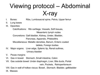
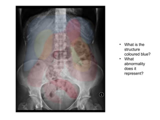
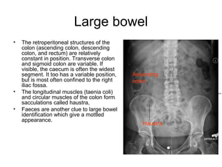
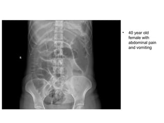
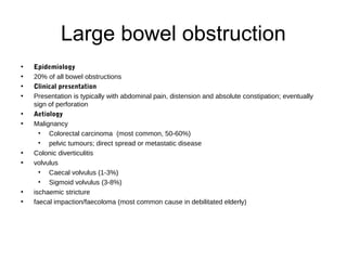
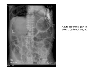
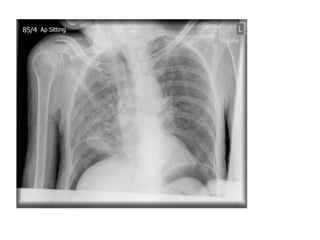
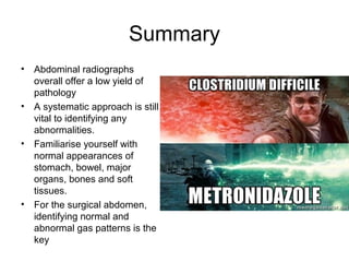
Introduction to abdominal X-
ray interpretation
Dr G Abraham
Technical Aspects
• Check that it is
standard supine AP
projection.
• Other projections
important in surgical
abdomen include
decubitus radiograph
or erect chest
radiograph.
• Field of exposure –
ideally diaphragm to
hernial orifices and
left to right of
abdomen.
• Penetration
Viewing protocol – Abdominal
X-ray
I. Bones: Ribs, Lumbosacral spine, Pelvis, Upper femur
II. Lung bases
III. Opacities:
Calcifications: Rib cartilage, Vessels, Soft tissues,
Mesenteric lymph nodes
Concretions: Gall bladder, Kidney, Ureter, Bladder,
Pancreas, Appendix, Phleboliths
Miscellaneous: Metallic densities, Barium, Enteric coated
tablets, Foreign bodies
IV. Major organs: Liver edge, Splenic tip, Renal outlines,
Urinary bladder
V. Psoas margins
VI. Gas in bowel: Stomach, Small intestine, Colon
VII. Gas outside bowel: Under diaphragm, Liver, Bile ducts, Portal
vein, Pockets, Retroperitoneum
VIII. Gas in wall of hollow viscus: Bowel , Stomach. Bladder, gallbladder
IX. Masses
Bony review
Soft tissues/organs
• Liver
• Lung bases
• Psoas muscles
• Kidneys
• Spleen
• Bladder
• What is the
structure
coloured blue?
• What
abnormality
does it
represent?
Bowel Gas Pattern
• Gas is of low density and forms a natural
contrast against surrounding denser soft
tissues. It is often difficult to differentiate
between normal small and large bowel.
• The upper limit of normal diameter of the
bowel is generally accepted as 3cm for the
small bowel, 6cm for the colon and 9cm
for the caecum (‘3/6/9 rule’).
Stomach
• If the stomach
contains air it
may be visible in
the left upper
quadrant of the
abdomen. The
lowest part of the
stomach may
cross the midline.
Stomach
Small bowel – duodenum to
terminal ileum
• Generally the small
bowel lies centrally
within the abdomen.
• The valvulae
coniventes are
circumferential folds of
mucosa and are seen
on an X-ray to pass
across the full width of
the lumen.
Large bowel
• The retroperitoneal structures of the
colon (ascending colon, descending
colon, and rectum) are relatively
constant in position. Transverse colon
and sigmoid colon are variable. If
visible, the caecum is often the widest
segment. It too has a variable position,
but is most often confined to the right
iliac fossa.
• The longitudinal muscles (taenia coli)
and circular muscles of the colon form
sacculations called haustra,
• Faeces are another clue to large bowel
identification which give a mottled
appearance.
Ascending
colon
Haustra
• 61 year old
female
• Previous
laparotomy
and incisional
hernia repair.
Distended
abdomen and
vomiting.
Small Bowel Obstruction
• epidemiology 1
• 80% of all mechanical bowel obstruction
• average age: 64 years
• females comprise 60% of patients
• presentation
• abdominal distension, nausea and vomiting
• the level will determine the acuity of presentation
• high obstruction presents early, possibly with bilious vomiting
• lower obstruction presents late and may have faeculent vomiting
• pathology
• may be complete or incomplete
• causes
• adhesional SBO: occurs almost exclusively from prior surgery
• herniae (often femoral or inguinal, but incisional occur)
• foreign bodies or other masses, e.g. gallstones
• rare: small bowel tumours causing intussusception
• 40 year old
female with
abdominal pain
and vomiting
Large bowel obstruction
• Epidemiology
• 20% of all bowel obstructions
• Clinical presentation
• Presentation is typically with abdominal pain, distension and absolute constipation; eventually 
sign of perforation
• Aetiology
• Malignancy
• Colorectal carcinoma  (most common, 50-60%)
• pelvic tumours; direct spread or metastatic disease
• Colonic diverticulitis
• volvulus
• Caecal volvulus (1-3%)
• Sigmoid volvulus (3-8%)
• ischaemic stricture 
• faecal impaction/faecoloma (most common cause in debilitated elderly)
Acute abdominal pain in
an ICU patient, male, 65.
Bowel perforation
Clinical Features
•severe and generalised abdominal pain (upper)
•gradual and localised pain (lower)
•anorexia, nausea and vomiting
•rigid abdomen and generalised tenderness
•guarding and rebound
•bowel sounds range from quiet to absent
Aetiology
•gastric and duodenal ulceration
•infection (diverticulitis, appendicitis), ischaemia and cancer
•blunt and penetrating trauma
•ingestion of corrosive materials
•iatrogenic causes (ERCP, colonoscopy, laparotomy, biopsy)
• Rigler’s sign LUQ
• Free
intraperitoneal
gas is widespread
with moderate
dilatation of small
bowel in particular
and there may
also be intramural
gas.
Are these urinary
tract calculi?
• phleboliths
73 year old female
with abdominal pain
and distension
• 85 year old man
from a Nursing
home with
advanced
Parkinson’s
disease.
• 35 year old patient
with Crohn’s
disease, 3 days of
nausea and vomiting
• Erect film
• Supine abdo film
of same patient
• Residual CT
contrast
showing
‘thumb-
printing’
suggestive of
mucosal
oedema
Summary
• Abdominal radiographs
overall offer a low yield of
pathology
• A systematic approach is still
vital to identifying any
abnormalities.
• Familiarise yourself with
normal appearances of
stomach, bowel, major
organs, bones and soft
tissues.
• For the surgical abdomen,
identifying normal and
abnormal gas patterns is the
key
References
• Dr Samuel Withey for cases
• https://radiopaedia.org
• http://www.radiologymasterclass.co.uk/

Introduction to abdominal x ray interpretation

  • 1.
    Introduction to abdominalX- ray interpretation Dr G Abraham
  • 2.
    Technical Aspects • Checkthat it is standard supine AP projection. • Other projections important in surgical abdomen include decubitus radiograph or erect chest radiograph. • Field of exposure – ideally diaphragm to hernial orifices and left to right of abdomen. • Penetration
  • 3.
    Viewing protocol –Abdominal X-ray I. Bones: Ribs, Lumbosacral spine, Pelvis, Upper femur II. Lung bases III. Opacities: Calcifications: Rib cartilage, Vessels, Soft tissues, Mesenteric lymph nodes Concretions: Gall bladder, Kidney, Ureter, Bladder, Pancreas, Appendix, Phleboliths Miscellaneous: Metallic densities, Barium, Enteric coated tablets, Foreign bodies IV. Major organs: Liver edge, Splenic tip, Renal outlines, Urinary bladder V. Psoas margins VI. Gas in bowel: Stomach, Small intestine, Colon VII. Gas outside bowel: Under diaphragm, Liver, Bile ducts, Portal vein, Pockets, Retroperitoneum VIII. Gas in wall of hollow viscus: Bowel , Stomach. Bladder, gallbladder IX. Masses
  • 4.
  • 5.
    Soft tissues/organs • Liver •Lung bases • Psoas muscles • Kidneys • Spleen • Bladder
  • 6.
    • What isthe structure coloured blue? • What abnormality does it represent?
  • 7.
    Bowel Gas Pattern •Gas is of low density and forms a natural contrast against surrounding denser soft tissues. It is often difficult to differentiate between normal small and large bowel. • The upper limit of normal diameter of the bowel is generally accepted as 3cm for the small bowel, 6cm for the colon and 9cm for the caecum (‘3/6/9 rule’).
  • 8.
    Stomach • If thestomach contains air it may be visible in the left upper quadrant of the abdomen. The lowest part of the stomach may cross the midline. Stomach
  • 9.
    Small bowel –duodenum to terminal ileum • Generally the small bowel lies centrally within the abdomen. • The valvulae coniventes are circumferential folds of mucosa and are seen on an X-ray to pass across the full width of the lumen.
  • 10.
    Large bowel • Theretroperitoneal structures of the colon (ascending colon, descending colon, and rectum) are relatively constant in position. Transverse colon and sigmoid colon are variable. If visible, the caecum is often the widest segment. It too has a variable position, but is most often confined to the right iliac fossa. • The longitudinal muscles (taenia coli) and circular muscles of the colon form sacculations called haustra, • Faeces are another clue to large bowel identification which give a mottled appearance. Ascending colon Haustra
  • 12.
    • 61 yearold female • Previous laparotomy and incisional hernia repair. Distended abdomen and vomiting.
  • 13.
    Small Bowel Obstruction •epidemiology 1 • 80% of all mechanical bowel obstruction • average age: 64 years • females comprise 60% of patients • presentation • abdominal distension, nausea and vomiting • the level will determine the acuity of presentation • high obstruction presents early, possibly with bilious vomiting • lower obstruction presents late and may have faeculent vomiting • pathology • may be complete or incomplete • causes • adhesional SBO: occurs almost exclusively from prior surgery • herniae (often femoral or inguinal, but incisional occur) • foreign bodies or other masses, e.g. gallstones • rare: small bowel tumours causing intussusception
  • 14.
    • 40 yearold female with abdominal pain and vomiting
  • 15.
    Large bowel obstruction •Epidemiology • 20% of all bowel obstructions • Clinical presentation • Presentation is typically with abdominal pain, distension and absolute constipation; eventually  sign of perforation • Aetiology • Malignancy • Colorectal carcinoma  (most common, 50-60%) • pelvic tumours; direct spread or metastatic disease • Colonic diverticulitis • volvulus • Caecal volvulus (1-3%) • Sigmoid volvulus (3-8%) • ischaemic stricture  • faecal impaction/faecoloma (most common cause in debilitated elderly)
  • 16.
    Acute abdominal painin an ICU patient, male, 65.
  • 18.
    Bowel perforation Clinical Features •severeand generalised abdominal pain (upper) •gradual and localised pain (lower) •anorexia, nausea and vomiting •rigid abdomen and generalised tenderness •guarding and rebound •bowel sounds range from quiet to absent Aetiology •gastric and duodenal ulceration •infection (diverticulitis, appendicitis), ischaemia and cancer •blunt and penetrating trauma •ingestion of corrosive materials •iatrogenic causes (ERCP, colonoscopy, laparotomy, biopsy)
  • 19.
    • Rigler’s signLUQ • Free intraperitoneal gas is widespread with moderate dilatation of small bowel in particular and there may also be intramural gas.
  • 23.
    Are these urinary tractcalculi? • phleboliths
  • 24.
    73 year oldfemale with abdominal pain and distension
  • 25.
    • 85 yearold man from a Nursing home with advanced Parkinson’s disease.
  • 26.
    • 35 yearold patient with Crohn’s disease, 3 days of nausea and vomiting • Erect film
  • 27.
    • Supine abdofilm of same patient • Residual CT contrast showing ‘thumb- printing’ suggestive of mucosal oedema
  • 28.
    Summary • Abdominal radiographs overalloffer a low yield of pathology • A systematic approach is still vital to identifying any abnormalities. • Familiarise yourself with normal appearances of stomach, bowel, major organs, bones and soft tissues. • For the surgical abdomen, identifying normal and abnormal gas patterns is the key
  • 29.
    References • Dr SamuelWithey for cases • https://radiopaedia.org • http://www.radiologymasterclass.co.uk/