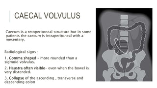
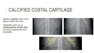
everythng about abdominal radiograph is discussed from views to obstruction to foreign body.
definetly u r not going to get bored
read and share with your peers.
Ultrasound renal stone differential diagnosis .AHMED ESAWY
renal sinus ultrasound
stone location in calyx
(not in medulla,not in cortex)
echogenic foci
acoustic shadowing
twinkle artifact on colour Doppler
color comet tail artifact
Staghorn calculi
Ultrasound beam-stone angle
Difference between kidney gravel & stone
RENAL ECHOCONCRETION
Vascular Reflectors (Normal or calcified renal vessels are the most
notable and common causes of intrarenal
bright echo reflectors)
Segmental Arteries
Arcuate Arteries
Sinus Vessels
renal vein thrombosis calcification
Calcifications of the branches of the renal artery
Nonvascular Reflectors: Prominent Papillae
Reflectors Within the Renal Parenchyma
Milk of Calcium Cysts
Renal Cortical Calcification
Junctional Parenchymal Line
Angiomyolipomas
Foreign Bodies
Bright echoes within the Renal Parenchyma
medullary nephrocalcinosis
focal pyelonephritis.
Echogenic tips of the renal pyramids apex
Transient pyramidal echogenicity
echogenic neonate renal pyramids (Tamm-Horsfall protein)
fungal balls
blood clots
Renal calcification in infants with Furosemid therapy
Rapid review of radiology text book, abdominal imaging, contrast imaging, CT , plain x ray, IVU , power point of abdominal pathological cases and description of diagnosis , differential diagnosis of diagnosis
various congenital gastrointestinal diseases manifesting in childhood or even in adults, their radiographic findings on various imaging modalities such as radiograph, barium, ultrasound etc.
This presentation explains in detail the definition, pathophysiology, signs & symptoms, management, and prognosis of intestinal obstruction, ileus, and volvulus.
Similar to Abdominal xray - imaging and interpretation (20)
Ethanol (CH3CH2OH), or beverage alcohol, is a two-carbon alcohol
that is rapidly distributed in the body and brain. Ethanol alters many
neurochemical systems and has rewarding and addictive properties. It
is the oldest recreational drug and likely contributes to more morbidity,
mortality, and public health costs than all illicit drugs combined. The
5th edition of the Diagnostic and Statistical Manual of Mental Disorders
(DSM-5) integrates alcohol abuse and alcohol dependence into a single
disorder called alcohol use disorder (AUD), with mild, moderate,
and severe subclassifications (American Psychiatric Association, 2013).
In the DSM-5, all types of substance abuse and dependence have been
combined into a single substance use disorder (SUD) on a continuum
from mild to severe. A diagnosis of AUD requires that at least two of
the 11 DSM-5 behaviors be present within a 12-month period (mild
AUD: 2–3 criteria; moderate AUD: 4–5 criteria; severe AUD: 6–11 criteria).
The four main behavioral effects of AUD are impaired control over
drinking, negative social consequences, risky use, and altered physiological
effects (tolerance, withdrawal). This chapter presents an overview
of the prevalence and harmful consequences of AUD in the U.S.,
the systemic nature of the disease, neurocircuitry and stages of AUD,
comorbidities, fetal alcohol spectrum disorders, genetic risk factors, and
pharmacotherapies for AUD.
HOT NEW PRODUCT! BIG SALES FAST SHIPPING NOW FROM CHINA!! EU KU DB BK substit...GL Anaacs
Contact us if you are interested:
Email / Skype : kefaya1771@gmail.com
Threema: PXHY5PDH
New BATCH Ku !!! MUCH IN DEMAND FAST SALE EVERY BATCH HAPPY GOOD EFFECT BIG BATCH !
Contact me on Threema or skype to start big business!!
Hot-sale products:
NEW HOT EUTYLONE WHITE CRYSTAL!!
5cl-adba precursor (semi finished )
5cl-adba raw materials
ADBB precursor (semi finished )
ADBB raw materials
APVP powder
5fadb/4f-adb
Jwh018 / Jwh210
Eutylone crystal
Protonitazene (hydrochloride) CAS: 119276-01-6
Flubrotizolam CAS: 57801-95-3
Metonitazene CAS: 14680-51-4
Payment terms: Western Union,MoneyGram,Bitcoin or USDT.
Deliver Time: Usually 7-15days
Shipping method: FedEx, TNT, DHL,UPS etc.Our deliveries are 100% safe, fast, reliable and discreet.
Samples will be sent for your evaluation!If you are interested in, please contact me, let's talk details.
We specializes in exporting high quality Research chemical, medical intermediate, Pharmaceutical chemicals and so on. Products are exported to USA, Canada, France, Korea, Japan,Russia, Southeast Asia and other countries.
Ozempic: Preoperative Management of Patients on GLP-1 Receptor Agonists Saeid Safari
Preoperative Management of Patients on GLP-1 Receptor Agonists like Ozempic and Semiglutide
ASA GUIDELINE
NYSORA Guideline
2 Case Reports of Gastric Ultrasound
Basavarajeeyam is an important text for ayurvedic physician belonging to andhra pradehs. It is a popular compendium in various parts of our country as well as in andhra pradesh. The content of the text was presented in sanskrit and telugu language (Bilingual). One of the most famous book in ayurvedic pharmaceutics and therapeutics. This book contains 25 chapters called as prakaranas. Many rasaoushadis were explained, pioneer of dhatu druti, nadi pareeksha, mutra pareeksha etc. Belongs to the period of 15-16 century. New diseases like upadamsha, phiranga rogas are explained.
263778731218 Abortion Clinic /Pills In Harare ,sisternakatoto
263778731218 Abortion Clinic /Pills In Harare ,ABORTION WOMEN’S CLINIC +27730423979 IN women clinic we believe that every woman should be able to make choices in her pregnancy. Our job is to provide compassionate care, safety,affordable and confidential services. That’s why we have won the trust from all generations of women all over the world. we use non surgical method(Abortion pills) to terminate…Dr.LISA +27730423979women Clinic is committed to providing the highest quality of obstetrical and gynecological care to women of all ages. Our dedicated staff aim to treat each patient and her health concerns with compassion and respect.Our dedicated group ABORTION WOMEN’S CLINIC +27730423979 IN women clinic we believe that every woman should be able to make choices in her pregnancy. Our job is to provide compassionate care, safety,affordable and confidential services. That’s why we have won the trust from all generations of women all over the world. we use non surgical method(Abortion pills) to terminate…Dr.LISA +27730423979women Clinic is committed to providing the highest quality of obstetrical and gynecological care to women of all ages. Our dedicated staff aim to treat each patient and her health concerns with compassion and respect.Our dedicated group of receptionists, nurses, and physicians have worked together as a teamof receptionists, nurses, and physicians have worked together as a team wwww.lisywomensclinic.co.za/
Lung Cancer: Artificial Intelligence, Synergetics, Complex System Analysis, S...Oleg Kshivets
RESULTS: Overall life span (LS) was 2252.1±1742.5 days and cumulative 5-year survival (5YS) reached 73.2%, 10 years – 64.8%, 20 years – 42.5%. 513 LCP lived more than 5 years (LS=3124.6±1525.6 days), 148 LCP – more than 10 years (LS=5054.4±1504.1 days).199 LCP died because of LC (LS=562.7±374.5 days). 5YS of LCP after bi/lobectomies was significantly superior in comparison with LCP after pneumonectomies (78.1% vs.63.7%, P=0.00001 by log-rank test). AT significantly improved 5YS (66.3% vs. 34.8%) (P=0.00000 by log-rank test) only for LCP with N1-2. Cox modeling displayed that 5YS of LCP significantly depended on: phase transition (PT) early-invasive LC in terms of synergetics, PT N0—N12, cell ratio factors (ratio between cancer cells- CC and blood cells subpopulations), G1-3, histology, glucose, AT, blood cell circuit, prothrombin index, heparin tolerance, recalcification time (P=0.000-0.038). Neural networks, genetic algorithm selection and bootstrap simulation revealed relationships between 5YS and PT early-invasive LC (rank=1), PT N0—N12 (rank=2), thrombocytes/CC (3), erythrocytes/CC (4), eosinophils/CC (5), healthy cells/CC (6), lymphocytes/CC (7), segmented neutrophils/CC (8), stick neutrophils/CC (9), monocytes/CC (10); leucocytes/CC (11). Correct prediction of 5YS was 100% by neural networks computing (area under ROC curve=1.0; error=0.0).
CONCLUSIONS: 5YS of LCP after radical procedures significantly depended on: 1) PT early-invasive cancer; 2) PT N0--N12; 3) cell ratio factors; 4) blood cell circuit; 5) biochemical factors; 6) hemostasis system; 7) AT; 8) LC characteristics; 9) LC cell dynamics; 10) surgery type: lobectomy/pneumonectomy; 11) anthropometric data. Optimal diagnosis and treatment strategies for LC are: 1) screening and early detection of LC; 2) availability of experienced thoracic surgeons because of complexity of radical procedures; 3) aggressive en block surgery and adequate lymph node dissection for completeness; 4) precise prediction; 5) adjuvant chemoimmunoradiotherapy for LCP with unfavorable prognosis.
Flu Vaccine Alert in Bangalore Karnatakaaddon Scans
As flu season approaches, health officials in Bangalore, Karnataka, are urging residents to get their flu vaccinations. The seasonal flu, while common, can lead to severe health complications, particularly for vulnerable populations such as young children, the elderly, and those with underlying health conditions.
Dr. Vidisha Kumari, a leading epidemiologist in Bangalore, emphasizes the importance of getting vaccinated. "The flu vaccine is our best defense against the influenza virus. It not only protects individuals but also helps prevent the spread of the virus in our communities," he says.
This year, the flu season is expected to coincide with a potential increase in other respiratory illnesses. The Karnataka Health Department has launched an awareness campaign highlighting the significance of flu vaccinations. They have set up multiple vaccination centers across Bangalore, making it convenient for residents to receive their shots.
To encourage widespread vaccination, the government is also collaborating with local schools, workplaces, and community centers to facilitate vaccination drives. Special attention is being given to ensuring that the vaccine is accessible to all, including marginalized communities who may have limited access to healthcare.
Residents are reminded that the flu vaccine is safe and effective. Common side effects are mild and may include soreness at the injection site, mild fever, or muscle aches. These side effects are generally short-lived and far less severe than the flu itself.
Healthcare providers are also stressing the importance of continuing COVID-19 precautions. Wearing masks, practicing good hand hygiene, and maintaining social distancing are still crucial, especially in crowded places.
Protect yourself and your loved ones by getting vaccinated. Together, we can help keep Bangalore healthy and safe this flu season. For more information on vaccination centers and schedules, residents can visit the Karnataka Health Department’s official website or follow their social media pages.
Stay informed, stay safe, and get your flu shot today!
Title: Sense of Taste
Presenter: Dr. Faiza, Assistant Professor of Physiology
Qualifications:
MBBS (Best Graduate, AIMC Lahore)
FCPS Physiology
ICMT, CHPE, DHPE (STMU)
MPH (GC University, Faisalabad)
MBA (Virtual University of Pakistan)
Learning Objectives:
Describe the structure and function of taste buds.
Describe the relationship between the taste threshold and taste index of common substances.
Explain the chemical basis and signal transduction of taste perception for each type of primary taste sensation.
Recognize different abnormalities of taste perception and their causes.
Key Topics:
Significance of Taste Sensation:
Differentiation between pleasant and harmful food
Influence on behavior
Selection of food based on metabolic needs
Receptors of Taste:
Taste buds on the tongue
Influence of sense of smell, texture of food, and pain stimulation (e.g., by pepper)
Primary and Secondary Taste Sensations:
Primary taste sensations: Sweet, Sour, Salty, Bitter, Umami
Chemical basis and signal transduction mechanisms for each taste
Taste Threshold and Index:
Taste threshold values for Sweet (sucrose), Salty (NaCl), Sour (HCl), and Bitter (Quinine)
Taste index relationship: Inversely proportional to taste threshold
Taste Blindness:
Inability to taste certain substances, particularly thiourea compounds
Example: Phenylthiocarbamide
Structure and Function of Taste Buds:
Composition: Epithelial cells, Sustentacular/Supporting cells, Taste cells, Basal cells
Features: Taste pores, Taste hairs/microvilli, and Taste nerve fibers
Location of Taste Buds:
Found in papillae of the tongue (Fungiform, Circumvallate, Foliate)
Also present on the palate, tonsillar pillars, epiglottis, and proximal esophagus
Mechanism of Taste Stimulation:
Interaction of taste substances with receptors on microvilli
Signal transduction pathways for Umami, Sweet, Bitter, Sour, and Salty tastes
Taste Sensitivity and Adaptation:
Decrease in sensitivity with age
Rapid adaptation of taste sensation
Role of Saliva in Taste:
Dissolution of tastants to reach receptors
Washing away the stimulus
Taste Preferences and Aversions:
Mechanisms behind taste preference and aversion
Influence of receptors and neural pathways
Impact of Sensory Nerve Damage:
Degeneration of taste buds if the sensory nerve fiber is cut
Abnormalities of Taste Detection:
Conditions: Ageusia, Hypogeusia, Dysgeusia (parageusia)
Causes: Nerve damage, neurological disorders, infections, poor oral hygiene, adverse drug effects, deficiencies, aging, tobacco use, altered neurotransmitter levels
Neurotransmitters and Taste Threshold:
Effects of serotonin (5-HT) and norepinephrine (NE) on taste sensitivity
Supertasters:
25% of the population with heightened sensitivity to taste, especially bitterness
Increased number of fungiform papillae
2. INTRODUCTION
Patients with acute abdomen comprise the largest group of people
presenting as a general surgical emergency.
Following history and clinical examination , plain film radiograph
have tradionally been one of the first and most useful method of
further investigation..
3. INDICATIONS
Suspected bowel obstruction
Suspected perforation
Moderate to severe
undifferentiated abdominal pain
Suspected foreign body
Renal tract calculi follow up
For other clinical situations,
abdominal xray is not
recommended:
Abdominal trauma
Right upper quadrant pain
Suspected intra-abdominal
collection
Acute upper GIT bleed
Suspected intra-abdominal
malignancy
Constipation
4. VIEWS
The standard view is an anterior-
posterior (AP) supine abdominal
xray (AXR).
You should assume that an
abdominal radiograph is taken
AP supine unless otherwise
stated.
5. AP SUPINE ABDOMINAL X-RAY
•Patient lies supine
•Xray pass in AP direction
•Patient asked to hold breath.
•Easier for patients (ill or post op)
•Allows distribution of gas and the caliber of
bowel to be determined and may show
displacement of bowel by soft tissue
masses.
•Obliteration of fat lines normally visualized
eg psoas outline , may indicate fluid or
inflammatory exudate.
6. ERECT XRAY
CHEST XRAY
•Xray beam passing tangentially
to the free gas.
•Better exposure
•chest diseases such as
pneumonia, aortic dissection,
myocardial infarction present
with abdominal symptoms.
•Abd diseases get complicated eg
pancreatitis with pleural effusion
•valuable baseline for post-op
studies
ABDOMEN XRAY
•Xray beam passes at an oblique
angle and diverges.
•Exposure unfavorable (more
black and more radiation dose)
•assess number and length of any
air fluid levels . unreliable and
misleading.
It is essential that patients should be in a position for 10 min prior to allow free gas to rise
to the highest point.
7. LEFT LATERAL DECUBITUS AXR
•Small amounts of free gas can be detected
•Patient lies on left side with the xray beam
horizontal
•Free gas seen trapped between the edge of the
liver and lateral abdominal wall.
•Eg a gas filled duodenal loop , one of the
commonest signs of acute pancreatitis , is best
shown in this view..
8. AIR FLUID LEVELS
•Three to five air fluid levels less than 2.5cm are normal mostly in
RLQ.
•However , more than 2 AFL in dilated small bowel ( calibre >2.5cm)
is abnormal .
•Doesnot help in differentiating intestinal obstruction from paralytic
ileus.
•The transverse colon normally measures less than 5.5 cm across.
•Caecum is highly distensible , 9cm is a critical dimension beyond
which danger of perforation exists.
•Significance of AFL is often overstated.
9. SOME CAUSES OF SMALL BOWEL
AFL
1. Small bowel obstruction
2. Large bowel obstruction
3. Paralytic ileus
4. Gastroenteritis
5. Hypokalemia
6. Uraemia
7. Jejunal diverticulosis
8. Mesentric thrombosis
9. Saline cathartics
10. Peritoneal metastases
11. Cleansing enema
12. Normal (<2.5cm)
10. RADIOGRAPH QUALITY
INCLUSION
From hemidiaphragm to the
pubis symphysis.
The superior aspect of liver ,
spleen
The lateral abdominal wall
The pubic symphysis
11. EXPOSURE
Under or over exposure.
Check the spine should be clearly visualised
12. NORMAL ANATOMY ON AN
ABDOMINAL XRAY
1) RIGHT AND LEFT
Left side of the image is patients right
side.
Always describe findings according to
the patients side.
R
L
18. 7) BOWEL
Normally most of the
bowel contains fluid/
faeces (light grey ) and
hence not visualized.
Colon most likely to
contain gas than the
small bowel , hence
easier to identify.
Stomach is visible if it
contains air .
20. PRESENTING AN ABDOMINAL
RADIOGRAPH
Give the type of radiograph
Give the patient’s name.
Assess the radiograph quality
Run through the ABCDE of abdominal radiograph
Short summary at the end
21. A IS FOR AIR IN THE WRONG
PLACE
• Look for pneumoperitoneum and pneumoretroperitoneum
• Look for gas in the biliary tree and portal vein
22. B IS FOR BOWEL
• Look for dilated small and large bowel
• Look for volvulus
• Look for a distended stomach
• Look for a hernia
• Look for evidence of bowel wall thickening
• Faecal loading
• Faecal impaction
23. C IS FOR CALCIFICATION
• clinically significant calcified
structures eg. Calcified gall
stones , renal stones ,
pancreatic calcifications,
abdominal aortic aneurysm
25. D IS FOR DISABILITY ( BONES AND
SOLID ORGANS)
•Bony skeleton for fracture pelvis – 3 rings test
•Sclerotic/lytic lesions
•Spine for vertebral body height, alignment, pedicles
•Solid organ enlargement
26. E IS FOR EVERYTHING ELSE
•Evidence of previous surgery or devices :
surgical clips, urinary catheter , supra-pubic catheter , NG and NJ tube
, flatus tube , surgical drain , nephrostomy catheter , peritoneal
dialysis catheter , gastric band device , syoma bag , stents , IVC filter ,
IUCD , pessary
•Foreign bodies:
Retained surgical swabs, Swallowed objects , Clothing artefacts,
Piercing , Body packer
•Lung bases.
27. PNEUMOPERITONEUM
•Free gas in peritoneal cavity
•Indicates bowel perforation
•Seen upto 3 weeks after abdominal surgery and in trauma
•Main causes :
Perforated peptic ulcer
Perforated appendix/bowel diverticulum
Post-surgery
Trauma
28. CAUSES OF PNEUMOPERITONEUM
WITHOUT PERITONITIS
1. Silent perforation of a viscus that has sealed itself , in aged ,
patient on steroid , unconscious patients or patients on ventilator.
2. Post-operative
3. Peritoneal dialysis
4. Perforated jejunal diverticulosis
5. Tracking down from pneumomediastinum
6. Leakage through a distended stomach (endoscopy)
7. Vaginal tube entry of air
29. An abdominal radiograph
and an erect chest
radiograph are requested
when looking for
pneumoperitoneum.
Erect radiograph is very
sensitive. Can detect 2-3
ml free gas .
32. Sometimes ,
two loops of
bowel lying
next to each
other may
mimic this
sign. This
can be
identified by
haustra or
valvulae.
33. 2) GAS OUTLINING THE
LIVER
Normally liver(light
grey) is surrounded by
peritoneal fat(dark
grey).
If pneumoperitoneum ,
liver is outlined by
gas(black) giving
better contrast.
34. 3) FALCIFORM
LIGAMENT SIGN
Is a ligament
attaching the liver
to the anterior
abdominal wall
(remnant of
umbilical vein).
Normally not
visible.
Visible if outlined
by free gas either
side in a supine
patient.
35. PNEUMORETROPERITONEUM
Rarely seen but always abnormal.
Contains kidneys, ureter, adrenal, aorta, IVC, most pancreas,
duodenum , ascending and descending colon.
Main causes :
1. bowel perforation:
Posterior duodenal perforation – PUD / post-op
Ascending and descending colon perforation
Rectal perforation
2. Post surgical ( residual air)
36. It may look similar to
pneumoperitoneum . The
key to identify is gas
surrounding all or part of
the kidneys.
Impossible to have both
pneumoperitoneum and
pneumoretroperitoneum at
the same time.
37.
38. PNEUMOBILIA
Branching dark lines within the centre of
the liver , more prominent towards the
hilum.
Main causes :
1. Recent ERCP/ incompetent sphincter
of Oddi (post sphincterotomy)
2. External biliary drain insertion/
biliary stent insertion
3. Biliary-enteric connection
Surgical anastomoses (eg whipple’s
procedure)
Spontaneous (eg Gallstone ileus)
4. Infection(rare) – emphysematous
39.
40. PORTAL VENOUS GAS
Branching dark lines within the periphery of the liver.
High mortality rate.
Main causes:
1. ischaemic bowel (most common)
2. necrotising enterocolitis (NEC) – most common in infants.
3. severe intra-abdominal sepsis
41.
42. DILATED SMALL BOWEL
Is a sign of mechanical obstruction or ileus.
In normal individual the small bowel is not seen because it is
collapsed or contains fluid.
Two main processes :
1. Mechanical obstruction
Physical obstruction.
Proximal bowel is dilated.
More distal the occlusion , more loops are visible.
43.
44. 2. Ileus – disruption of the normal propulsive ability of the GIT.
Causes :
•Post –operative
•Intra abdominal infection or inflammation
•Anti-cholinergic drugs.
Mechanical obstruction and ileum appears identical and underlying
cause cannot be determined on a xray.
45. Radiological signs :
1. dilatation >3cm - for quick
comparison the adult vertebral
body measures 4cm.
2. central location
3. valvulae conniventes-
musical folds of the SI. Thin ,
closely spaced and classically
seen as a continuous thin line
across the entire width of the
bowel .
48. SPECIAL CASE: GALLSTONE ILEUS
Recurrent episodes of
cholecystitis cause adhesion of
the gallbladder to the bowel
(usually duodenum) and
eventually a fistula.
A large gallstone then enters the
bowel and cause obstruction ,
typically at the ilececal valve.
Rigler’s triad:
1. pneumobilia
2. small bowel obstruction
3. gallstone ( usually in RIF – only
in 30%)
49. DILATED LARGE BOWEL
Bowel proximal is dilated and distal is collapsed.
Causes:
1. malignancy ( colorectal carcinoma- MCC in adults)
2. diverticular stricture
3. faecal impaction
4. volvulus.
50. Radiographic appearance:
1. dilatation >5.5cm – caecum is allowed to reach
9cm before being called dilated.
2. circumferential location- peripherally located .
Exception is transverse colon – loops down towards
pelvis and can cross centre of the radiograph.
3. haustra : small pouches in the wall of LI. Taenia
coli (ribbons of smooth muscle which run along the
length of the colon) are shorter than the colon itself,
therefore the colon becomes sacculated between the
taenia coli forming the haustra.
Lines between the haustra are called haustral folds
and do not cross the entire width of the bowel
If the bowel is grossly distended, then the haustra
may not be seen.
51.
52.
53. COMPARISON :
DILATED SMALL
BOWEL
DIALTED LARGE
BOWEL
SIZE >3cm >5.5cm
>9cm for caecum
POSITION Central Peripheral
MUCOSAL/WALL
PATTERN
Valvulae conniventes Haustral folds
PRESENCE OF SOLID
FAECES IS THE ONLY
RELIABLE SIGN
54. VOLVULUS
Twisting of the bowel on its mesentry.
Causes partial or complete bowel obstruction.
Two commonest types : sigmoid and caecal volvulus.
Symptoms due to :
1. bowel obstruction – ‘closed-loop’ obstruction.
2. bowel ischaemia – and later necrosis.
55. SIGMOID VOLVULUS
Caused when the sigmoid colon twists on
its mesentery.
elderly/ institutionalised patients.
Radiological signs :
1. coffee bean sign – closed loop of colon
2. general lack of haustra.
3. distension of the ascending , transverse
and descending colon.
56.
57. CAECAL VOLVULUS
Caecum is a retoperitoneal structure but in some
patients the caecum is intraperitoneal with a
mesentery.
Radiological signs :
1. Comma shaped – more rounded than a
sigmoid volvulus.
2. Haustra often visible- even when the bowel is
very distended.
3. Collapse of the ascending , transverse and
descending colon
58.
59. DILATED STOMACH
If filled with gas or fluid.
Causes of gas filled stomach :
1. bowel obstruction (malignancy or scarring in the duodenum)
2. aerophagia (distressed patients or side effect of non-invasive ventilation)
Causes of fluid – filled stomach:
1. bowel obstruction
2. Chronic gastro paresis (autonomic neuropathy from poorly controlled
diabetes)
60. Radiological appearances:
Large stomach shaped gas
filled(dark) or fluid filled (light
grey) loop in the upper abdomen
(LUQ). If very large can extend
inferiorly over the centre of the
abdomen. Normal rugae may not
be seen.
Wall of the greater curvature
convex caudally and the pyloric
antrum pointing cranially.
61.
62. HERNIA
Protrusion of an organ through the wall
of the cavity.
Only hernia seen on abdominal
radiograph is inguinal hernia –
containing a loop of bowel which
contains gas. If the bowel loop contains
fluid – will not be easily seen.
Radiological appearance :
1. loops of gas-filled bowel seen below
the level of inguinal ligament.
Quick way to see if above or below the
obturator foramen.
2. soft tissue swelling on the side of
hernia : loop of bowel (black) and soft
tissue (light grey) . This is due to
herniated mesenteric fat/ associated
oedema
63. BOWEL WALL INFLAMATION
Most commonly seen in large bowel – colitis.
Main causes :
1. IBD
2. Ischaemic bowel
3. infection (e.g. pseudomembranous colitis from clostridium difficile)
Impossible to differentiate the different causes of colitis on a xray.
UC – only large bowel
Crohns and infection – anywhere along the GIT
Ischaemic – along a specific vascular territory ( SMA- midgut , IMA – hindgut)
64. Radiological signs of bowel wall
inflammation:
1. Bowel wall thickening: inflammation
causes mucosal edema and later
thickening.
Thickened wall outlined by gas within
the lumen and peritoneal fat outside.
Thumb printing : mucosal edema cause
severe thickening of the haustral folds
of the colon , such that the folds appear
as ‘thumb-shaped’ projections into the
lumen.
Featureless bowel : chronic bowel
thickening causes complete loss of the
normal hausral markings. The colon
appears smooth walled. In chronic UC –
lead pipe appearance.
2. loss of formed faecal matter in the
65.
66. SPECIAL CASE : TOXIC
MEGACOLON
Acute form of bowel dilatation
occurring as a complication of IBD
or infection.
Rapid dilatation of colon and signs
of septic shock.
1. large bowel dilatation to >6cm
diameter
2. inflammatory pseudopoylps
(mucosal islands) – lobulated
opacities in the bowel wall from the
areas of raised mucosal tissue
surrounded by areas of ulceration.
3. thumb printing and mucosal
67. FAECAL LOADING
Large volume of faecal matter in the
colon of any consistency.
Usually result of chronic
constipation.
Hardened faecal matter has a
characterstic appearance:
1. rounded masses
2. mottled or granular texture
Hardened faecal matter within the
right side of the colon – highly
suggestive of faecal loading as the
faecal material here normally of fluid
consistency.
68. FAECAL IMPACTION
More severe than faecal loading.
Solid, immobile bulk of faeces in
rectum
Result of chronic constipation –
elderly , immobile or
institutionalized
Appearance is fairly
characteristic : large bulk of
faeces in the rectum .
In severe cases – can extend into
sigmoid and even rest of large
bowel.
69. CHOLELITHIASIS
Only 15% are radio-opaque.
Seen over RUQ along the lower lobe
of the liver.
Appearances may be variable :
1. large or small
2. single or multiple
3. radio-opaque outline with a lucent
centre
4. polygonal shape (flat surface – due
to stones abutting one another)
5. laminated (concentric ring)
70. SPECIAL CASES :
1) Milk of calcium or ‘Limey’
Bile
Dense fluid containing
calcium carbonate.
RO gall bladder lumen
Gallstone outlined by bile.
2) Porcelain gallbladder
Heavily calcified walls.
Increased risk of GB
malignancy.
Rim of calcification outlining
the GB. Edge appearing more
71. UROLITHIASIS
90% renal calculi are radio-
opaque.
Radiological signs :
1. calcific density projected
over the kidney
2. calcific density projected
over the course of ureter :
medial aspect of the kidney
and inferiorly along the tips
of the transverse processes.
3. staghorn calculus.
72.
73. BLADDER STONES
Main causes:
1. urinary stasis (most common)
- BOO
- bladder diverticulum
- neurogenic bladder
2. UTI
3. Migrated renal calculus
4. foreign material left for long
- long term urinary catheterisation
74. Appear as rounded or
oval shaped opacities
projected over the
lower pelvis near the
midline.
Often large and may
be multiple.
Some may have a
laminated appearance.
75. NEPHROCALCINOSIS
Abnormal deposition of calcium in the kidney parenchyma.
Medulla is far more commonly affected.
Usually associated with metabolic disorders.
1. hyperparathyroidism.
2. medullary sponge kidney
3. renal tubular acidosis.
Appears as :
Generalised Calcium deposition
Little clusters corresponding to the medullary pyramids.
76. PANCREATIC CALCIFICATION
Most commonly a sign
of chronic pancreatitis.
Most common
underlying cause is
alcohol abuse.
Not visualized in
normal patients ,
retroperitoneal and
crosses midline.
Appears as irregular
clusters or foci of
calcification crossing
the midline in the mid
abdomen. Can take the
78. ABDOMINAL AORTIC ANEURYSM
CALCIFICATION
An AAA pronounced ‘triple-a’ is
abnormal dilatation of the abdominal
aorta to >3cm diameter.
Normally <2.5cm.
When AAA grows to >5.5cm the risk of
rupture increases and surgery is
recommended.
AAA are only seen when :
1. calcification in the wall of the aorta.
2. both sides of the aortic wall must be
visualized to diagnose AAA. If only one
side is seen bulging , can be tortous.
3. Most (>90%) are infra-renal.
79. FETUS
11 weeks to birth .
Very rare since radiation
should be avoided in
pregancy
Look for skeleton within the
abdomen.
Large circular opacity and a
linear array of smaller
opacities are seen.
80. CALCIFIED COSTAL CARTILAGE
Appear patchy and more
dense than the ribs.
Typically seen as a
continuation of the ribs ,
curving superiorly and
medially.
82. CALCIFIED MESENTERIC LYMPH
NODE
Incidental findings. Most in
elderly.
Secondary to a previous
granulomatous infection such
as TB.
Appear as oval , mottled areas
of calcification , usually 5-
15mm in size often in RLQ or
central abdomen.
Normally appear in clusters of
two or more.
NOTE : distinguish from renal
calculi.
1. mottled appearance
83. CALCIFIED UTERINE FIBROID
Leiomyomas are
benign tumor of
myometrial origin.
Long standing
fibroid may
calcify.
Appear as
rounded calcified
structure with
‘splatter like
calcification’
85. ABDOMINAL AORTIC
CALCIFICATION
With normal caliber.
In elderly and diabetic .
Indicative of atheromatous
processes in the wall of
vessel.
Linear areas of calcification
projected over the midline.
86. SPLENIC ARTERY CALCIFICATION
Seen projected over the
left upper quadrant and
has a distinctive tourtour
‘Chinese dragon’ like
appearance.
87. PELVIC FRACTURE
Three rings :
Pelvic ring – paired ilium ,
ischium and pubic bones along
with sacrum
Ring of bone surrounding the
right obturator foramen
Ring of bone surrounding the
left obturator foramen
If you see a fracture in one part
of the ring , look for the second
fracture.
It is impossible to have a
significant fracture in one place
88. SCLEROTIC AND LUCENT BONE
LESIONS
Sclerotic –
increased
density. Many
causes
including
malignancy.
Prostate
metastases.
Lucent –
abnormal area
of reduced
density. Many
causes
including mets.
91. SOLID ORGAN ENLARGEMENT
Either by increase in the overall size of one of the solid organs or by a large
tumor in the abdomen.
Incidental finding as first IOC – ultrasound.
Common causes :
1)Solid organ
•Hepatomegaly – Riedel’s lobe : inferior , tongue like projection of the right
lobe of the liver. Normal variant in 17% population.
•Splenomegaly
2) Tumor
•Renal masses
•Pelvic masses
92. RADIOLOGICAL SIGNS :
•Large soft tissue density (light grey)mass
•Loops of bowel often displaced by the mass
•Location gives a clue
RUQ – liver , right kidney
LUQ – spleen ,left kidney , fluid filled stomach
Lower abdomen – ovaries,uterus , distended urinary bladder.
93.
94.
95. CHOLECYSTECTOMY CLIPS
Surgical clips , stapes and anastomoses are common findings on an
abdominal radiograph and it is important to recognize the differences
between them
101. URINARY CATHETER
Classical position in the
lower pelvis with their tip
projected over the position
of the urinary bladder.
Radio-opaque line along the
length of the catheter.
The inflated urinary catheter
balloon is not visualized as
it contains water , which is
the same density as the
urine.
102. SUPRA-PUBIC CATHETER
Hollow flexible tube
inserted into the urinary
bladder via an incision in
the anterior abdominal
wall.
Catheter is seen to enter
the bladder from above
rather than below.
103. NASO-GASTRIC TUBE
Plastic tube inserted
through the nose ,
down the oesphagus
and into the stomach.
Can be used for
short-term feeding ,
drug administration
and aspiration of
stomach contents.
104. NASO-JEJUNAL TUBE
A plastic tube
similar to NG Tube
passes through
stomach and
duodenum into the
jejunum.
Used inpatients
unable to tolerate
feeding into the
stomach.
105. FLATUS TUBE
Long soft tube
usually inserted into
the sigmoid colon
with the help of a
rigid or flexible
sigmoidoscope. It is
used to decompress
a sigmoid volvulus.
106. SURGICAL DRAIN
Used to drain /
prevent the
accumulation of
fluid/ blood/ pus
from the area of
surgery.
108. PERITONEAL DIALYSIS
Placed in the
peritoneal cavity and
used to introduce and
remove dialysate fluid
for patients
undergoing
peritoneal dialysis.
Easily recognized by
its large coiled tip.
109. GASTRIC BAND DEVICE
Inflatable ring inserted
surgically around the
top portion of the
stomach.
Used as a treatment for
obesity by creating a
smaller stomach pouch
to limit the amount of
food that can be
consumed at one sitting.
The inflatable ring is
attached to a small
access port placed just
under the skin to allow
re-adjustment of the
110. PEG/ RIG
A gastrostomy is a
tube passed into the
patients stomach
through the
abdominal wall.
Used to provide
feeding when oral
intake is not
adequate or safe.
A PEG – endoscopic
guidance
A RIG – radiological
guidance
111. STOMA BAG
A stoma is a surgically
created opening in the
abdomen to connect
bowel with the outside
environment.
Three main types –
ileostomy ,colostomy ,
urostomy.
On a radiograph you
may see a dense ring ,
which is the site of
attachment of the
stoma bag to the skin
around the stoma.
112. BILIARY STENT
Stent is a tube inserted into
a natural passage in the
body to improve flow or
prevent blockage.
Placed between common
bile duct and/or hepatic
duct and are usually
projected just to the right of
the midline in the upper
abdomen.
116. ENDOVASCULAR ANEURYSM
REPAIR STENT GRAFT
Used to treat
AAA.
A stent graft
is placed in
the lumen of
the aorta to
allow blood
to flow and
reduce
pressure in
the aneurysm
, preventing
rupture.
117. IVC FILTER
Umbrella like device
placed in IVC to reduce
the risk of large
pulmonary emboli.
Wires allow blood to
flow past and prevent
large clots from
reaching pulmonary
arteries.
Often used when anti-
coagulation is
contraindicated.
118. IUCD
Small often T
shaped device
which is
inserted into
the uterus.
Long-acting
reversible
contraception
119. PESSARY
Medical device
inserted into the
vagina to provide
structural support or
to deliver medication.
Come in all shapes
and sizes but ring
pessary is the most
common.
120. FOREIGN BODIES
• range and number of different items are limitless.
• don’t be surprised by some of the things people do.
125. BODY PACKER
•Smuggle illicit drugs by concealing
drugs in their GIT.
•Drugs are packed into a balloon,
latex glove or condom and
swallowed.
•Vary from a few to more than 200.
•Radiologically : multiple oval or
tubular soft tissue opacities ,
sometimes surrounded by a rim of
halo.
•Earlier managed by surgical retrieval
but high mortality.
•Nowadays , conservatively managed
by bowel irrigation.