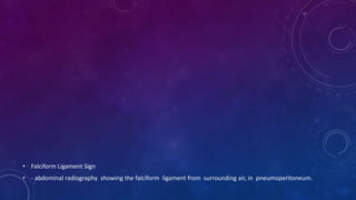
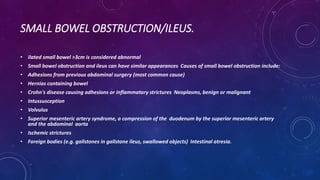
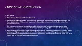
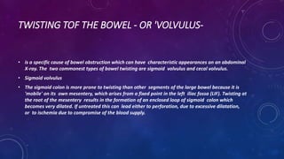
INTERPRETATION OF ABNORMAL X-RAY OF THE
ABDOMEN.
PATHOLOGY
• ABCDE approach:
• A-Air in a wrong place. B-Bowel loops.
• C-Calcifications.
• D-Dense structures like soft tissue and bones densities.
• E-Everything as foreign bodies.
LOOKING FOR AIR IN A WRONG PLACE
1-pneumoretroperitoneal(air at the
• retroperitoneal space).
• Pneumatosis intestinalis(Air along the bowel wall).
• Pneumobilia(air at the biliary tree).
• Portal venoupneumoperiton
• eum(air at peritoneal cavity). 2-Ps air(air at the portal vein).
• Pneumoperitoneum (abnormal presence of air or other gas) in the peritoneal cavity, a potential space
within the abdominal cavity.
• Causes
• Perforated duodenal ulcer – The most common cause of rupture in the abdomen. Perforated peptic
ulcer
• Bowel obstruction Ruptured diverticulum Penetrating trauma
• Ruptured inflammatory bowel disease (e.g. megacolon) Necrotizing enterocolitis/Pneumatosis coli.
• Bowel cancer Ischemic bowel Steroids
• After laparotomy and laparoscopy Breakdown of a surgical anastomosis Bowel injury after endoscopy
• Crescent Sign: plain film showing appearance of a sliver of air usually beneath the both
hemidiaphragms in pneumoperitoneum. b) Chilaiditi’s Sign: plain film showing interposition of bowel
gas between the liver and the right hemidiaphragm.
• Crescent Sign: abdominal radiography showing air beneath both hemidiaphragms, in relation with
pneumoperitoneum.
• Cupola sign:
• abdominal radiography showing free intraperitoneal air under the central diaphragmatic tendon.
• Falciform Ligament Sign
• - abdominal radiography showing the falciform ligament from surrounding air, in pneumoperitoneum.
Football sign (pneumoperitoneum)
• Plain X-ray of the chest and upper abdomen displaying obvious Chilaiditi's sign, or presence of gas in
the right colic angle between the liver and right.
• Rigler sign -
• abdominal radiography
showing free air outlining
the small bowel
wall, indicating pneumoperit
oneum.
PNEUMORETROPERITONEUM.
• Clinical
• The most common cause of pneumoretroperitoneum is perforation of the second, third, or fourth
portion of the duodenum or retroperitoneal colon secondary to trauma, diverticulitis, or ulceration.
• Post-surgical(Post-urology or adrenal surgery).
• Radiological findings
• Pneumoretroperitoneum is most often seen on the right side where the air can outline the right
kidney and the undersurface of the liver. In contrast to pneumoperitoneum, air in the
retroperitoneum does not move freely with change in position. The gas can extend up into the
mediastinum or neck because there is no barrier between them.
• massive retroperitoneal air.
• Pneumoretroperitoneum
• - presence of gas
within the
retroperitoneal space.
Typically the air outlines
structures like the kidneys,
psoas muscles
and retroperitoneal
portions of the bowel
PNEUOMBILIA
• Clinical
• Gas in the biliary tree is most commonly secondary to surgical procedures such as choledochoenterostomy
or sphincterotmy of the sphincter of Oddi. It may also arise in the setting of trauma, infection by gas
producing organisms (i.e. emphysematous cholecystitis), fistulas connecting the biliary system and the
intestinal tract (i.e. from dudodenal ulcers, or gallstones), malignant involvement of the ampulla of Vater,
or as a congenital anomaly.
• Radiological findings
• Ultrasound is the modality of choice for visualizing gas in the biliary tree. Air in the bile ducts generally
causes bright linear or globular reflections with shadowing and ring-down artifacts. The gas will move with
patient positioning. On plain film, biliary gas may be seen outlining the bile ducts but it is important to be
cautious in making this diagnosis as normal periductal fat that surrounds and parallels the course of the
bile ducts may give the appearance known as pseudopneumobilia.
BOWEL LOOPS PATHOLOGY.
• Dilated stomach.
• Dilated small and large bowel loops. Volvulus.
• Inflammatory bowel disease. Hernia.
• Constipation with fecal impaction.
• CT scout image showing massive gastric dilation.
• Double Bubble Sign:
abdominal radiography showin
g two air- filled structures
in the upper abdomen, with
no air distally. The proximal
bubble in the left side
filled with air represents the
stomach and the second
bubble to the right of
the midline represents the
proximal duodenum.
SMALL BOWEL OBSTRUCTION/ILEUS.
• ilated small bowel >3cm is considered abnormal
• Small bowel obstruction and ileus can have similar appearances Causes of small bowel obstruction include:
• Adhesions from previous abdominal surgery (most common cause)
• Hernias containing bowel
• Crohn's disease causing adhesions or inflammatory strictures Neoplasms, benign or malignant
• Intussusception
• Volvulus
• Superior mesenteric artery syndrome, a compression of the duodenum by the superior mesenteric artery
and the abdominal aorta
• Ischemic strictures
• Foreign bodies (e.g. gallstones in gallstone ileus, swallowed objects) Intestinal atresia.
• String of Beads
Sign - abdominal
radiography showi
ng linearly
arranged small
pockets of air in
a fluid-dilated
small bowel loop.
• Upright abdominal X-
ray demonstrating
a small
bowel obstruction.
Note multiple air fluid
levels.
LARGE BOWEL OBSTRUCTION
• Key points
• Dilatation of the caecum >9cm is abnormal
• Dilatation of any other part of the colon >6cm is abnormal Abdominal X-ray may demonstrate the
level of obstruction Abdominal X-ray cannot reliably differentiate mechanical obstruction from
pseudo-obstruction
• The most common causes of large bowel obstruction are colorectal carcinoma and diverticular
strictures. Less common causes are hernias or volvulus (twisting of the bowel on its mesentery).
• Adhesions do not commonly cause large bowel obstruction. Radiological appearances of large bowel
obstruction differ from those of small bowel obstruction, however, with large bowel obstruction
there is often co-existing small bowel dilatation proximally. Dilatation of the caecum >9cm, and >6cm
for the rest of the colon is considered abnormal.
• Toxic Megacolon
Abdominal X-ray -
large bowel
obstruction.
TWISTING TOF THE BOWEL - OR 'VOLVULUS-
• is a specific cause of bowel obstruction which can have characteristic appearances on an abdominal
X-ray. The two commonest types of bowel twisting are sigmoid volvulus and cecal volvulus.
• Sigmoid volvulus
• The sigmoid colon is more prone to twisting than other segments of the large bowel because it is
'mobile' on its own mesentery, which arises from a fixed point in the left iliac fossa (LIF). Twisting at
the root of the mesentery results in the formation of an enclosed loop of sigmoid colon which
becomes very dilated. If untreated this can lead either to perforation, due to excessive dilatation,
or to ischemia due to compromise of the blood supply.
• Cecal volvulus: The caecum is
most frequently a
retroperitoneal structure,
and therefore not susceptible
to twisting. However, in up to
20% of individuals there
is congenital incomplete
peritoneal covering of the
caecum with formation of
a 'mobile' caecum on a
mesentery, such that it no
longer lies in the right iliac
fossa.
BOWEL WALL INFLAMMATION
• Abdominal X-rays sometimes demonstrate signs of bowel inflammation such as mucosal thickening
'thumb-printing' or a featureless colon 'lead pipe' colon.
• Occasionally, abdominal X-rays show signs of inflammation in patients with inflammatory bowel
disease. Abnormalities may relate to either acute or chronic stages of disease
• thumbprinting – abdominal
radiography showing
• ‘thumbprinting’(arrows). The
normal haustral folds are replaced
by wide transverse
thickened bands.
• Ttt
INTRA-ABDOMINAL CALCIFICATION IS COMMON AND THE
CAUSES MAY
BE CLASSIFIED INTO FOUR BROAD GROUPS BASED ON
MORPHOLOGY:
• Stones: renal stones, ureteric stones, bladder stones, gallstones pancreatic ductal calcification
• nodal calcification: most commonly from treated lymphoma, tuberculosis or
• histoplasmosis
• Phlebolith, appendicolith, calcified granuloma failed renal transplant
• encapsulating peritoneal sclerosis
• Conduit calcification
• Calcification within the walls of any fluid-filled hollow tube:
• abdominal aorta, pancreatic duct, ductus deferens, large veins
• Cystic calcification
• Calcification in the wall of a mass such as a cyst, pseudocyst or aneurysm. simple serous cysts, Aneurysms, echinococcal cysts, hematoma, 'porcelain' gallbladder,
calcified appendiceal mucocele
• Solid mass calcification
• mesenteric nodes, adrenal calcifications, uterine fibroids Primary tumours, e.g. ovarian dermoid, metastases, adenoma Spleen (autosplenectomy in sickle cell disease)
• Renal tuberculosis with autonephrectomy
• A and B, Two patients with pancreatic calculi and
chronic pancreatitis in a patient with alcoholism.
This is the typical appearance of numerous dense,
discrete opacities that cross the midline at the
level of L1 to L2 (arrow). The normal pancreas is
not visible on abdominal plain films.
• Bladder calculi. Three homogeneously
dense bladder stones with a continuous
rim of calcification typical of concretions.
Incidentally noted is a phlebolith with the
diagnostic concentric lucency that should
not be mistaken for a ureteral stone
(arrow).
DENSITY(D).
• Bones
• Lots of bones are visible on an AXR and it’s important that you can identify each and screen for any
pathology (which may be expected or unexpected). In addition, bones on the AXR provide
useful landmarks for where you might expect to see a soft tissue structure (e.g. ischial spines are the
usual level of the vesicoureteric junction). Bones commonly visible on AXR include:
• Ribs(Look for increased or decreased density).
• Lumbar vertebrae( look for alignment, vertebral height and pedicle Sacrum and Coccyx(Look for lytic
or sclerotic lesion).
• Pelvis(look for fracture, lytic or sclerotic lesion).
• Proximal femurs(Looks for sclerotic, lytic lesion or fractures).
• Scoliosis of
the lumbar spine.
• Bowel loops are
displaced inferiorly by an
enlarged liver. No evidence
of obstruction
or pneumoperitoneum.
No fecal loading. Multiple lytic
lesions demonstrated through
bony pelvis and proximal
femurs. Upper abdominal
surgical clips.
• Intussusception with the 'target' and
'meniscus' signs. Supine view shows a
round soft-tissue mass in the right
upper quadrant. The mass contains
a ring-like area of lucency representing
the 'target' sign (small arrows). The
mass protrudes into the gas-
filled transverse colon representing the
'meniscus' sign (long arrows).
•Thank You.

Presentation1aboutabdominalradiology.pptx

  • 1.
    INTERPRETATION OF ABNORMALX-RAY OF THE ABDOMEN.
  • 2.
    PATHOLOGY • ABCDE approach: •A-Air in a wrong place. B-Bowel loops. • C-Calcifications. • D-Dense structures like soft tissue and bones densities. • E-Everything as foreign bodies.
  • 3.
    LOOKING FOR AIRIN A WRONG PLACE 1-pneumoretroperitoneal(air at the • retroperitoneal space). • Pneumatosis intestinalis(Air along the bowel wall). • Pneumobilia(air at the biliary tree). • Portal venoupneumoperiton • eum(air at peritoneal cavity). 2-Ps air(air at the portal vein).
  • 4.
    • Pneumoperitoneum (abnormalpresence of air or other gas) in the peritoneal cavity, a potential space within the abdominal cavity. • Causes • Perforated duodenal ulcer – The most common cause of rupture in the abdomen. Perforated peptic ulcer • Bowel obstruction Ruptured diverticulum Penetrating trauma • Ruptured inflammatory bowel disease (e.g. megacolon) Necrotizing enterocolitis/Pneumatosis coli. • Bowel cancer Ischemic bowel Steroids • After laparotomy and laparoscopy Breakdown of a surgical anastomosis Bowel injury after endoscopy
  • 5.
    • Crescent Sign:plain film showing appearance of a sliver of air usually beneath the both hemidiaphragms in pneumoperitoneum. b) Chilaiditi’s Sign: plain film showing interposition of bowel gas between the liver and the right hemidiaphragm.
  • 6.
    • Crescent Sign:abdominal radiography showing air beneath both hemidiaphragms, in relation with pneumoperitoneum.
  • 7.
    • Cupola sign: •abdominal radiography showing free intraperitoneal air under the central diaphragmatic tendon.
  • 8.
    • Falciform LigamentSign • - abdominal radiography showing the falciform ligament from surrounding air, in pneumoperitoneum.
  • 9.
  • 10.
    • Plain X-rayof the chest and upper abdomen displaying obvious Chilaiditi's sign, or presence of gas in the right colic angle between the liver and right.
  • 11.
    • Rigler sign- • abdominal radiography showing free air outlining the small bowel wall, indicating pneumoperit oneum.
  • 12.
    PNEUMORETROPERITONEUM. • Clinical • Themost common cause of pneumoretroperitoneum is perforation of the second, third, or fourth portion of the duodenum or retroperitoneal colon secondary to trauma, diverticulitis, or ulceration. • Post-surgical(Post-urology or adrenal surgery). • Radiological findings • Pneumoretroperitoneum is most often seen on the right side where the air can outline the right kidney and the undersurface of the liver. In contrast to pneumoperitoneum, air in the retroperitoneum does not move freely with change in position. The gas can extend up into the mediastinum or neck because there is no barrier between them.
  • 13.
  • 14.
    • Pneumoretroperitoneum • -presence of gas within the retroperitoneal space. Typically the air outlines structures like the kidneys, psoas muscles and retroperitoneal portions of the bowel
  • 15.
    PNEUOMBILIA • Clinical • Gasin the biliary tree is most commonly secondary to surgical procedures such as choledochoenterostomy or sphincterotmy of the sphincter of Oddi. It may also arise in the setting of trauma, infection by gas producing organisms (i.e. emphysematous cholecystitis), fistulas connecting the biliary system and the intestinal tract (i.e. from dudodenal ulcers, or gallstones), malignant involvement of the ampulla of Vater, or as a congenital anomaly. • Radiological findings • Ultrasound is the modality of choice for visualizing gas in the biliary tree. Air in the bile ducts generally causes bright linear or globular reflections with shadowing and ring-down artifacts. The gas will move with patient positioning. On plain film, biliary gas may be seen outlining the bile ducts but it is important to be cautious in making this diagnosis as normal periductal fat that surrounds and parallels the course of the bile ducts may give the appearance known as pseudopneumobilia.
  • 16.
    BOWEL LOOPS PATHOLOGY. •Dilated stomach. • Dilated small and large bowel loops. Volvulus. • Inflammatory bowel disease. Hernia. • Constipation with fecal impaction.
  • 17.
    • CT scoutimage showing massive gastric dilation.
  • 18.
    • Double BubbleSign: abdominal radiography showin g two air- filled structures in the upper abdomen, with no air distally. The proximal bubble in the left side filled with air represents the stomach and the second bubble to the right of the midline represents the proximal duodenum.
  • 19.
    SMALL BOWEL OBSTRUCTION/ILEUS. •ilated small bowel >3cm is considered abnormal • Small bowel obstruction and ileus can have similar appearances Causes of small bowel obstruction include: • Adhesions from previous abdominal surgery (most common cause) • Hernias containing bowel • Crohn's disease causing adhesions or inflammatory strictures Neoplasms, benign or malignant • Intussusception • Volvulus • Superior mesenteric artery syndrome, a compression of the duodenum by the superior mesenteric artery and the abdominal aorta • Ischemic strictures • Foreign bodies (e.g. gallstones in gallstone ileus, swallowed objects) Intestinal atresia.
  • 20.
    • String ofBeads Sign - abdominal radiography showi ng linearly arranged small pockets of air in a fluid-dilated small bowel loop.
  • 21.
    • Upright abdominalX- ray demonstrating a small bowel obstruction. Note multiple air fluid levels.
  • 22.
    LARGE BOWEL OBSTRUCTION •Key points • Dilatation of the caecum >9cm is abnormal • Dilatation of any other part of the colon >6cm is abnormal Abdominal X-ray may demonstrate the level of obstruction Abdominal X-ray cannot reliably differentiate mechanical obstruction from pseudo-obstruction • The most common causes of large bowel obstruction are colorectal carcinoma and diverticular strictures. Less common causes are hernias or volvulus (twisting of the bowel on its mesentery). • Adhesions do not commonly cause large bowel obstruction. Radiological appearances of large bowel obstruction differ from those of small bowel obstruction, however, with large bowel obstruction there is often co-existing small bowel dilatation proximally. Dilatation of the caecum >9cm, and >6cm for the rest of the colon is considered abnormal.
  • 23.
    • Toxic Megacolon AbdominalX-ray - large bowel obstruction.
  • 24.
    TWISTING TOF THEBOWEL - OR 'VOLVULUS- • is a specific cause of bowel obstruction which can have characteristic appearances on an abdominal X-ray. The two commonest types of bowel twisting are sigmoid volvulus and cecal volvulus. • Sigmoid volvulus • The sigmoid colon is more prone to twisting than other segments of the large bowel because it is 'mobile' on its own mesentery, which arises from a fixed point in the left iliac fossa (LIF). Twisting at the root of the mesentery results in the formation of an enclosed loop of sigmoid colon which becomes very dilated. If untreated this can lead either to perforation, due to excessive dilatation, or to ischemia due to compromise of the blood supply.
  • 25.
    • Cecal volvulus:The caecum is most frequently a retroperitoneal structure, and therefore not susceptible to twisting. However, in up to 20% of individuals there is congenital incomplete peritoneal covering of the caecum with formation of a 'mobile' caecum on a mesentery, such that it no longer lies in the right iliac fossa.
  • 26.
    BOWEL WALL INFLAMMATION •Abdominal X-rays sometimes demonstrate signs of bowel inflammation such as mucosal thickening 'thumb-printing' or a featureless colon 'lead pipe' colon. • Occasionally, abdominal X-rays show signs of inflammation in patients with inflammatory bowel disease. Abnormalities may relate to either acute or chronic stages of disease
  • 27.
    • thumbprinting –abdominal radiography showing • ‘thumbprinting’(arrows). The normal haustral folds are replaced by wide transverse thickened bands. • Ttt
  • 28.
    INTRA-ABDOMINAL CALCIFICATION ISCOMMON AND THE CAUSES MAY BE CLASSIFIED INTO FOUR BROAD GROUPS BASED ON MORPHOLOGY: • Stones: renal stones, ureteric stones, bladder stones, gallstones pancreatic ductal calcification • nodal calcification: most commonly from treated lymphoma, tuberculosis or • histoplasmosis • Phlebolith, appendicolith, calcified granuloma failed renal transplant • encapsulating peritoneal sclerosis • Conduit calcification • Calcification within the walls of any fluid-filled hollow tube: • abdominal aorta, pancreatic duct, ductus deferens, large veins • Cystic calcification • Calcification in the wall of a mass such as a cyst, pseudocyst or aneurysm. simple serous cysts, Aneurysms, echinococcal cysts, hematoma, 'porcelain' gallbladder, calcified appendiceal mucocele • Solid mass calcification • mesenteric nodes, adrenal calcifications, uterine fibroids Primary tumours, e.g. ovarian dermoid, metastases, adenoma Spleen (autosplenectomy in sickle cell disease) • Renal tuberculosis with autonephrectomy
  • 29.
    • A andB, Two patients with pancreatic calculi and chronic pancreatitis in a patient with alcoholism. This is the typical appearance of numerous dense, discrete opacities that cross the midline at the level of L1 to L2 (arrow). The normal pancreas is not visible on abdominal plain films.
  • 30.
    • Bladder calculi.Three homogeneously dense bladder stones with a continuous rim of calcification typical of concretions. Incidentally noted is a phlebolith with the diagnostic concentric lucency that should not be mistaken for a ureteral stone (arrow).
  • 31.
    DENSITY(D). • Bones • Lotsof bones are visible on an AXR and it’s important that you can identify each and screen for any pathology (which may be expected or unexpected). In addition, bones on the AXR provide useful landmarks for where you might expect to see a soft tissue structure (e.g. ischial spines are the usual level of the vesicoureteric junction). Bones commonly visible on AXR include: • Ribs(Look for increased or decreased density). • Lumbar vertebrae( look for alignment, vertebral height and pedicle Sacrum and Coccyx(Look for lytic or sclerotic lesion). • Pelvis(look for fracture, lytic or sclerotic lesion). • Proximal femurs(Looks for sclerotic, lytic lesion or fractures).
  • 32.
    • Scoliosis of thelumbar spine.
  • 33.
    • Bowel loopsare displaced inferiorly by an enlarged liver. No evidence of obstruction or pneumoperitoneum. No fecal loading. Multiple lytic lesions demonstrated through bony pelvis and proximal femurs. Upper abdominal surgical clips.
  • 34.
    • Intussusception withthe 'target' and 'meniscus' signs. Supine view shows a round soft-tissue mass in the right upper quadrant. The mass contains a ring-like area of lucency representing the 'target' sign (small arrows). The mass protrudes into the gas- filled transverse colon representing the 'meniscus' sign (long arrows).
  • 35.