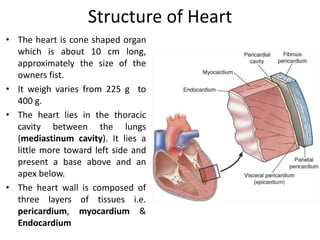
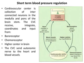
The cardiovascular system consists of the heart and blood vessels. The heart pumps blood through a network of arteries, capillaries and veins. The heart has four chambers and is composed of three layers. It is located in the chest cavity slightly left of center. The heart's natural pacemaker, the sinoatrial node, initiates electrical impulses that cause coordinated contractions. Blood pressure, cardiac output and peripheral resistance determine blood flow. Diseases can disrupt blood flow and oxygen delivery, like atherosclerosis, heart failure and ischemic heart disease including angina and myocardial infarction.
The lymphatic system consists of lymph, lymph vessels, lymph nodes, and lymph organs such as the spleen and thymus. Lymph is a clear fluid that transports plasma proteins from tissues back to the bloodstream and carries away debris from damaged tissues. Lymph vessels are thin-walled and valve-lined, transporting lymph towards the heart. Lymph nodes filter lymph and proliferate lymphocytes. The spleen and thymus are major lymph organs - the spleen filters blood and stores lymphocytes, while the thymus develops T-lymphocytes. Mucosa-associated lymphoid tissues such as tonsils and Peyer's patches intercept antigens from the mouth, throat, and intestines.
Blood and lymph are the two main body fluids. Blood consists of plasma, red blood cells, white blood cells, and platelets. Lymph circulates within lymphatic vessels. The document discusses the composition, functions, and formation of blood cells through hematopoiesis. Hematopoietic stem cells in bone marrow give rise to myeloid cells like red blood cells and platelets, and lymphoid cells like lymphocytes. The stages of erythropoiesis and hemoglobin synthesis are described. Anemia results from low red blood cell count, hemoglobin, or hematocrit, and can be classified by cell morphology or etiology.
The document provides information on the structure and function of the digestive system. It begins with an overview of the basic components of the alimentary canal including the mouth, salivary glands, pharynx, esophagus, stomach, small intestine, large intestine, rectum, anal canal, and accessory organs like the liver, pancreas, and bile duct. It then describes the layers of the alimentary canal including the peritoneum, muscle layers, submucosa, and mucosa. Specific structures like the tongue, teeth, stomach, small intestine and large intestine are examined in more detail. The functions of digestion and absorption in the mouth, stomach, small intestine, large intestine and accessory organs are
The respiratory system provides oxygen to the body's cells through respiration and removes carbon dioxide. It includes the nose, pharynx, larynx, trachea, bronchi, lungs, and muscles of respiration. Oxygen and carbon dioxide are exchanged between the alveoli and blood in the lungs through diffusion. Respiration is regulated by the respiratory center in the brainstem and chemoreceptors that detect changes in blood gases. Artificial respiration can prevent deaths by reopening the airway and exchanging gases until natural breathing resumes.
The urinary system consists of the kidneys, ureters, urinary bladder, and urethra. The kidneys filter waste from the blood to produce urine. The urine passes through the ureters to the bladder, where it is stored until excretion through the urethra. The kidneys play a vital role in regulating water balance and electrolyte levels in the body by selectively reabsorbing needed substances and secreting waste through urine production. The urinary system works to maintain homeostasis in the body.
7. Male and Female reproductive system.pptxAbhiDabra
The document provides an overview of the male and female reproductive systems. It describes the key organs involved in both systems, including their structure and functions. For the male system it covers the testes, spermatic cord, seminal vesicles, prostate gland, and urethra. For the female system it discusses the ovaries, fallopian tubes, uterus, vagina, and external genitalia. It also explains processes like the menstrual cycle, fertilization, pregnancy, and menopause.
The skin is the largest organ of the body and has three main layers - the epidermis, dermis and hypodermis. The epidermis is made of stratified squamous epithelium and provides a protective barrier. It has five layers including the stratum corneum. The dermis contains collagen, elastic fibers, blood vessels, nerves and skin appendages. The hypodermis is a subcutaneous layer containing fat and lobules. Skin has several functions like protection, sensation, temperature regulation and immunity. It also contains appendages like hair, nails, sweat and sebaceous glands.
The document classifies joints into three main types - fibrous, cartilaginous, and synovial - based on the type of tissue that connects the bones and the amount of movement allowed. It describes the characteristics and examples of each joint type. Common disorders of joints like dislocation, sprain, bursitis, and arthritis are also outlined.
The urinary system regulates blood composition and excretes waste through the kidneys, ureters, bladder and urethra. The kidneys filter blood to form urine and regulate electrolyte and fluid levels. Urine passes from the kidneys through the ureters to the bladder, where it is temporarily stored until urination. During urination, urine exits the body through the urethra under control of sphincter muscles. The urinary system helps maintain homeostasis by regulating water balance and removing toxins and wastes from the bloodstream.
BP201T. Human Anatomy And Physiology-II
Unit-III: - Urinary System.
Anatomy of urinary tract with special reference to anatomy of kidney and
nephrons, functions of kidney and urinary tract, physiology of urine formation,
micturition reflex and role of kidneys in acid base balance, role of RAS in kidney
and disorders of kidney.
Cardiovascular system- Human Anatomy & Physiology 1st.RAHUL PAL
The document discusses the anatomy and functions of the heart. It describes the heart's location in the thoracic cavity and layers (epicardium, myocardium, endocardium). It details the heart valves (tricuspid, bicuspid, pulmonary, aortic semi-lunar), blood vessels (arteries, veins, capillaries), conduction system (SA node, AV node, bundle of His), cardiac cycle and output. Common heart disorders are also listed.
The urinary system functions to regulate homeostasis by controlling water, electrolyte, and acid-base balance and eliminating waste from the body. The major organs of the urinary system include the kidneys, ureters, urinary bladder, and urethra. The kidneys filter waste from the blood to form urine and are made up of nephrons, the functional units that filter blood and remove waste. Urine is formed through the processes of filtration, reabsorption, and secretion in the nephrons and is temporarily stored in the bladder before being excreted through the urethra.
Unit-III, chapter-2- Lymphatic System,
Functions of Lymphatic System,
Major Parts of Lymphatic System,
Composition of Lymph,
Lymph and Lymphatic Capillaries,
Structure of lymph node,
Mechanisms of Lymph Flow,
Functions of Lymph Node,
Mucosa-Associated Lymphoid Tissue (MALT),
As per PCI syllabus,
B. Pharm. First Year,
Human Anatomy and Physiology-I.
The urinary system helps remove waste from the body through urine. It includes the kidneys, ureters, urinary bladder, and urethra. The kidneys filter waste from the blood to form urine, which travels through the ureters to the bladder. In the bladder, urine is stored until emptying through the urethra. The kidneys contain nephrons, which filter blood and reabsorb useful components back into blood while secreting waste to form urine.
The respiratory system allows for oxygen intake and carbon dioxide removal. It consists of the nose, pharynx, larynx, trachea, bronchi, lungs and muscles. The nose and mouth allow air intake and filter it. The pharynx and larynx direct air to the proper passages. The trachea and bronchi form the conducting airways to the lungs. In the lungs, gas exchange occurs between the air in alveoli and blood in capillaries, transferring oxygen to blood and carbon dioxide out of blood.
Cardiovascular System, Heart, Blood Vessel, ECG, Hypertension, Arrhythmia Audumbar Mali
Cardiovascular System,
Human Anatomy and Physiology-I,
The Blood Vessels,
The Heart,
The Electrocardiogram,
The Vascular Pathways,
As per PCI syllabus,
Atherosclerosis,
Coronary bypass operation,
Heart Transplants and Artificial Hearts
The urinary system consists of the kidneys, ureters, urinary bladder, and urethra. The kidneys filter waste from the blood and regulate fluid and electrolyte balance. Urine travels from the kidneys down the ureters into the muscular urinary bladder, which stores urine until emptying through the urethra. The urinary system works to eliminate waste from the body and maintain homeostasis.
The digestive system begins at the mouth and includes the esophagus, stomach, and small and large intestines. Food is ingested and broken down through both mechanical and chemical digestion. Mechanical digestion begins with chewing in the mouth. Chemical digestion involves enzymes that break down carbohydrates, proteins, and fats in specific areas of the digestive tract. Digested nutrients are then absorbed through the walls of the tract and transported throughout the body.
Unit I, chapter-2 Cellular level of organization.Audumbar Mali
The document provides an overview of the structure and function of eukaryotic cells. It discusses the key parts of the cell including the plasma membrane, cytoplasm, organelles like the nucleus, mitochondria and Golgi apparatus. It also describes cellular transport mechanisms like passive diffusion and active transport. The summary is as follows:
The document discusses the basic structure and functions of eukaryotic cells. It describes the key parts of the cell including the plasma membrane, cytoplasm and organelles. It also explains cellular transport mechanisms such as passive and active transport that allow movement of substances into and out of cells.
The kidneys filter blood to produce urine and are located on either side of the spine in the posterior abdominal wall. Each kidney contains around 1-2 million functional filtering units called nephrons. The nephron filters blood in the glomerulus to form an initial filtrate. Most of the filtrate is reabsorbed back into the bloodstream, with only around 1% excreted as urine. Through selective reabsorption and secretion, the kidneys regulate water and electrolyte balance and remove nitrogenous wastes from the body.
Reproductive system
Anatomy of male and female reproductive system, Functions of male and female
reproductive system, sex hormones, physiology of menstruation, fertilization,
spermatogenesis, oogenesis, pregnancy and parturition
The urinary system has several important functions:
1. It filters waste and excess water from the blood and removes it from the body through urine.
2. It maintains the chemical consistency of the blood.
3. Its main organs - the kidneys, ureters, urinary bladder, and urethra - work together to produce, transport, store, and eliminate urine from the body.
There are three kinds of blood vessels: arteries, veins, and capillaries.
Each of these plays a very specific role in the circulation process/blood circulation.
Arteries transport blood away from the heart.
Veins return blood back toward the heart.
Capillaries surround body cells and tissues to deliver and absorb oxygen, nutrients, and other substances.
The capillaries also connect the branches of arteries and to the branches of veins.
The walls of most blood vessels have three distinct layers: the tunica externa, the tunica media, and the tunica intima.
Learning Objectives:
Compare and contrast the structure and function
of
Arteries
Veins
Capillaries
ulatory
system
Arteries
Arterioles
Capillaries
Venules
Veins
3 tunics
Lume
The Vessels
Functions:
Distribution of blood
Exchange of materials with tissues
Return of blood to the heart
Structure:
Most have the same basic structure:
– 3 layers surrounding a hollow lumen
Renal physiology, nephron structure, function,jga. hussein f. sakrHussein Sakr
The document provides an overview of renal physiology, including the anatomy, structure, function, and blood supply of the kidney. It describes the nephron as the functional unit of the kidney, composed of the renal corpuscle and renal tubules. There are two main types of nephrons - superficial cortical nephrons and juxtamedullary nephrons. The document also discusses the juxtaglomerular apparatus, which regulates renin release in response to changes in sodium concentration or blood pressure. Renin triggers the renin-angiotensin-aldosterone system, which functions to retain salt and water and constrict blood vessels.
The urinary system consists of the kidneys, ureters, bladder, and urethra. The kidneys filter the blood to remove waste and regulate electrolyte and fluid levels. The nephrons are the functional units of the kidneys that filter blood to form urine. Urine travels from the kidneys through the ureters to the bladder, where it is stored until excreted through the urethra. The urinary system regulates blood volume and pressure and removes waste from the body.
✓Heart
✓Anatomy of heart
✓Blood circulation
✓Blood Vessels
✓Structure and function of artery, vein and capillaries
✓Elements of conduction system of heart and heart beat
✓Its regulation by nervous system
✓Cardiac output
✓Cardiac cycle
✓Regulation of bood pressure
✓Pulse
✓Electrocardiogram
✓Disorder of heart
The circulatory system consists of the heart, blood vessels, and blood. The heart pumps blood through two circuits - pulmonary circulation to the lungs and systemic circulation to the entire body. It has four chambers and four valves that ensure one-way blood flow. The cardiac cycle involves repeated heart contraction and relaxation to pump blood. Key components like arteries, veins, and capillaries form blood vessels that deliver oxygen and nutrients throughout the body.
Cardiovascular physiology for university studentsItsOnyii
A detailed pdf document on cardiovascular physiology for university students including structure and functions of heart, Electrocardiogram, echocardiography, chest and limb leads, Diseases and disorders of the heart.
The cardiovascular system delivers oxygen and nutrients to tissues and removes waste. It has three main components:
1. The heart, which is a muscular pump located in the chest cavity with four chambers that drive blood circulation.
2. Arteries, which carry oxygenated blood away from the heart. Capillaries allow for gas and nutrient exchange before veins return deoxygenated blood to the heart.
3. Valves ensure one-way blood flow, with the tricuspid and bicuspid valves located between the heart's upper and lower chambers.
The cardiovascular system consists of the heart and blood vessels. The heart is hollow and located in the thorax, protected by the pericardium. It pumps blood through the circulatory system via contraction and relaxation in a cardiac cycle. Blood flow is regulated by arteries, veins and capillaries which allow for gas and nutrient exchange. Blood pressure and flow are influenced by many factors including the heart, blood properties and vessel dynamics.
The cardiovascular system consists of the heart and blood vessels. The heart is hollow and located in the thorax, protected by the pericardium. It pumps blood through the circulatory system via contraction and relaxation. Blood flows from the heart through arteries, then capillaries where gas exchange occurs, and returns to the heart through veins, aided by valves. The heart's rhythm is regulated by electrical conduction pathways and nerves.
This document provides an overview of the cardiovascular system, including:
- The heart's structure, chambers, valves, and conduction system.
- Blood flow through the heart and the events of the cardiac cycle.
- Types of blood vessels and circulation (pulmonary, systemic, coronary, hepatic portal).
- Functions of the cardiovascular system like oxygen transport and nutrient delivery.
- Key terms like stroke volume and cardiac output.
The document summarizes the structure and function of blood vessels. It describes the three main types of blood vessels - arteries, capillaries, and veins. It explains the layers (tunics) that make up blood vessel walls, and how they differ in each vessel type. It discusses the roles of elastic and muscular arteries in circulation. It also outlines neural, chemical, and renal mechanisms that regulate blood pressure through controlling vessel diameter and cardiac output.
1) Mammals require transport systems like the circulatory system to efficiently deliver oxygen and nutrients to cells throughout the body since diffusion is only effective over short distances. The circulatory system can be open, closed, or a double circulatory system.
2) In a closed circulatory system, blood remains within blood vessels and nutrients/gases are exchanged through vessel walls. A double circulatory system has two circuits - pulmonary and systemic - allowing blood to pass through the heart twice.
3) Diseases can occur if cholesterol builds up in artery walls, restricting blood flow and oxygen delivery. This can cause heart attacks, strokes, aneurysms, and high blood pressure. Proper diet, exercise, and managing
This system has three main components: the heart, the blood vessel and the blood itself. The heart is the system's pump and the blood vessels are like the delivery routes. Blood can be thought of as a fluid which contains the oxygen and nutrients the body needs and carries the wastes which need to be removed.
The heart has four chambers. The two superior receiving chambers are the atria (= entry halls or chambers), and the two inferior pumping chambers are the ventricles (= little bellies).
On the anterior surface of each atrium is a wrinkled pouchlike structure called an auricle
The document summarizes the anatomy and physiology of the human heart. It describes the heart's location in the thoracic cavity. It then details the three layers of the heart - pericardium, myocardium, and endocardium. It explains the interior structures of the heart including the atria, ventricles, and valves. The conducting system and cardiac cycle are then summarized. Finally, it briefly discusses cardiac output, factors affecting stroke volume and heart rate, and the baroreceptor reflex.
The document discusses cardiovascular physiology, including:
- The location of the heart in the thoracic cavity between the lungs.
- The three layers of the heart - pericardium, endocardium, and myocardium - and its four chambers - two atria and two ventricles.
- The heart pumps oxygenated blood to the body and deoxygenated blood returns to the heart through arteries, veins and capillaries.
- The electrical conduction system generates heartbeats and the electrocardiogram can be used to measure heart rate and rhythm.
The circulatory system is divided into the heart and blood vessels. The heart pumps blood into the pulmonary and systemic circulations. The right side pumps blood to the lungs and the left side pumps oxygenated blood to the rest of the body. The heart has four chambers, valves to ensure one-way blood flow, and a conducting system to coordinate contractions. Arteries carry blood away from the heart while veins return blood to the heart.
The document discusses the structure and function of the circulatory system. It describes the heart as a hollow muscular organ that pumps blood through arteries, capillaries, and veins. The heart has four chambers and a conduction system that generates electrical signals to coordinate contractions. Blood vessels branch throughout the body to deliver oxygen, nutrients and remove waste. Arteries carry blood away from the heart, veins carry blood back to the heart, and capillaries facilitate exchange of materials between blood and tissues.
The document summarizes the cardiovascular system, including:
1. The anatomy and physiology of the heart, including its chambers and layers. The heart pumps deoxygenated blood to the lungs and oxygenated blood to the body.
2. The three main types of blood circulation - pulmonary circulation between the heart and lungs, coronary circulation supplying the heart, and systemic circulation supplying the body. Blood vessels have three layers and blood flows through arteries, arterioles, capillaries and veins.
3. Key aspects of cardiac cycle including the phases of atrial and ventricular systole and diastole. Heart sounds and electrocardiograms are also summarized.
The cardiovascular system transports blood throughout the body using the heart, arteries, veins and capillaries. The heart has four chambers - two atria that receive blood and two ventricles that pump blood out. It is surrounded by membranes and protected in the chest cavity. Arteries carry oxygenated blood away from the heart while veins return deoxygenated blood back to the heart. The smallest blood vessels, capillaries, allow for nutrient and gas exchange with tissues.
The document summarizes the cardiovascular system, including the circulatory and lymphatic systems. It describes the heart, blood vessels, blood flow through pulmonary and systemic circulation. It also discusses common diseases like atherosclerosis, heart attack, angina, and their diagnosis and treatment. The circulatory system functions to transport blood throughout the body, while the lymphatic system transports lymph and maintains fluid balance.
A powerpoint designed for the South African Life Sciences syllabus for grade 11. Includes information about blood and it's transportation, the human heart, the lymph system etc. Hope it helps :)
This presentation was provided by Racquel Jemison, Ph.D., Christina MacLaughlin, Ph.D., and Paulomi Majumder. Ph.D., all of the American Chemical Society, for the second session of NISO's 2024 Training Series "DEIA in the Scholarly Landscape." Session Two: 'Expanding Pathways to Publishing Careers,' was held June 13, 2024.
How to Setup Warehouse & Location in Odoo 17 InventoryCeline George
In this slide, we'll explore how to set up warehouses and locations in Odoo 17 Inventory. This will help us manage our stock effectively, track inventory levels, and streamline warehouse operations.
ISO/IEC 27001, ISO/IEC 42001, and GDPR: Best Practices for Implementation and...PECB
Denis is a dynamic and results-driven Chief Information Officer (CIO) with a distinguished career spanning information systems analysis and technical project management. With a proven track record of spearheading the design and delivery of cutting-edge Information Management solutions, he has consistently elevated business operations, streamlined reporting functions, and maximized process efficiency.
Certified as an ISO/IEC 27001: Information Security Management Systems (ISMS) Lead Implementer, Data Protection Officer, and Cyber Risks Analyst, Denis brings a heightened focus on data security, privacy, and cyber resilience to every endeavor.
His expertise extends across a diverse spectrum of reporting, database, and web development applications, underpinned by an exceptional grasp of data storage and virtualization technologies. His proficiency in application testing, database administration, and data cleansing ensures seamless execution of complex projects.
What sets Denis apart is his comprehensive understanding of Business and Systems Analysis technologies, honed through involvement in all phases of the Software Development Lifecycle (SDLC). From meticulous requirements gathering to precise analysis, innovative design, rigorous development, thorough testing, and successful implementation, he has consistently delivered exceptional results.
Throughout his career, he has taken on multifaceted roles, from leading technical project management teams to owning solutions that drive operational excellence. His conscientious and proactive approach is unwavering, whether he is working independently or collaboratively within a team. His ability to connect with colleagues on a personal level underscores his commitment to fostering a harmonious and productive workplace environment.
Date: May 29, 2024
Tags: Information Security, ISO/IEC 27001, ISO/IEC 42001, Artificial Intelligence, GDPR
-------------------------------------------------------------------------------
Find out more about ISO training and certification services
Training: ISO/IEC 27001 Information Security Management System - EN | PECB
ISO/IEC 42001 Artificial Intelligence Management System - EN | PECB
General Data Protection Regulation (GDPR) - Training Courses - EN | PECB
Webinars: https://pecb.com/webinars
Article: https://pecb.com/article
-------------------------------------------------------------------------------
For more information about PECB:
Website: https://pecb.com/
LinkedIn: https://www.linkedin.com/company/pecb/
Facebook: https://www.facebook.com/PECBInternational/
Slideshare: http://www.slideshare.net/PECBCERTIFICATION
Philippine Edukasyong Pantahanan at Pangkabuhayan (EPP) CurriculumMJDuyan
(𝐓𝐋𝐄 𝟏𝟎𝟎) (𝐋𝐞𝐬𝐬𝐨𝐧 𝟏)-𝐏𝐫𝐞𝐥𝐢𝐦𝐬
𝐃𝐢𝐬𝐜𝐮𝐬𝐬 𝐭𝐡𝐞 𝐄𝐏𝐏 𝐂𝐮𝐫𝐫𝐢𝐜𝐮𝐥𝐮𝐦 𝐢𝐧 𝐭𝐡𝐞 𝐏𝐡𝐢𝐥𝐢𝐩𝐩𝐢𝐧𝐞𝐬:
- Understand the goals and objectives of the Edukasyong Pantahanan at Pangkabuhayan (EPP) curriculum, recognizing its importance in fostering practical life skills and values among students. Students will also be able to identify the key components and subjects covered, such as agriculture, home economics, industrial arts, and information and communication technology.
𝐄𝐱𝐩𝐥𝐚𝐢𝐧 𝐭𝐡𝐞 𝐍𝐚𝐭𝐮𝐫𝐞 𝐚𝐧𝐝 𝐒𝐜𝐨𝐩𝐞 𝐨𝐟 𝐚𝐧 𝐄𝐧𝐭𝐫𝐞𝐩𝐫𝐞𝐧𝐞𝐮𝐫:
-Define entrepreneurship, distinguishing it from general business activities by emphasizing its focus on innovation, risk-taking, and value creation. Students will describe the characteristics and traits of successful entrepreneurs, including their roles and responsibilities, and discuss the broader economic and social impacts of entrepreneurial activities on both local and global scales.
Chapter wise All Notes of First year Basic Civil Engineering.pptxDenish Jangid
Chapter wise All Notes of First year Basic Civil Engineering
Syllabus
Chapter-1
Introduction to objective, scope and outcome the subject
Chapter 2
Introduction: Scope and Specialization of Civil Engineering, Role of civil Engineer in Society, Impact of infrastructural development on economy of country.
Chapter 3
Surveying: Object Principles & Types of Surveying; Site Plans, Plans & Maps; Scales & Unit of different Measurements.
Linear Measurements: Instruments used. Linear Measurement by Tape, Ranging out Survey Lines and overcoming Obstructions; Measurements on sloping ground; Tape corrections, conventional symbols. Angular Measurements: Instruments used; Introduction to Compass Surveying, Bearings and Longitude & Latitude of a Line, Introduction to total station.
Levelling: Instrument used Object of levelling, Methods of levelling in brief, and Contour maps.
Chapter 4
Buildings: Selection of site for Buildings, Layout of Building Plan, Types of buildings, Plinth area, carpet area, floor space index, Introduction to building byelaws, concept of sun light & ventilation. Components of Buildings & their functions, Basic concept of R.C.C., Introduction to types of foundation
Chapter 5
Transportation: Introduction to Transportation Engineering; Traffic and Road Safety: Types and Characteristics of Various Modes of Transportation; Various Road Traffic Signs, Causes of Accidents and Road Safety Measures.
Chapter 6
Environmental Engineering: Environmental Pollution, Environmental Acts and Regulations, Functional Concepts of Ecology, Basics of Species, Biodiversity, Ecosystem, Hydrological Cycle; Chemical Cycles: Carbon, Nitrogen & Phosphorus; Energy Flow in Ecosystems.
Water Pollution: Water Quality standards, Introduction to Treatment & Disposal of Waste Water. Reuse and Saving of Water, Rain Water Harvesting. Solid Waste Management: Classification of Solid Waste, Collection, Transportation and Disposal of Solid. Recycling of Solid Waste: Energy Recovery, Sanitary Landfill, On-Site Sanitation. Air & Noise Pollution: Primary and Secondary air pollutants, Harmful effects of Air Pollution, Control of Air Pollution. . Noise Pollution Harmful Effects of noise pollution, control of noise pollution, Global warming & Climate Change, Ozone depletion, Greenhouse effect
Text Books:
1. Palancharmy, Basic Civil Engineering, McGraw Hill publishers.
2. Satheesh Gopi, Basic Civil Engineering, Pearson Publishers.
3. Ketki Rangwala Dalal, Essentials of Civil Engineering, Charotar Publishing House.
4. BCP, Surveying volume 1
LAND USE LAND COVER AND NDVI OF MIRZAPUR DISTRICT, UPRAHUL
This Dissertation explores the particular circumstances of Mirzapur, a region located in the
core of India. Mirzapur, with its varied terrains and abundant biodiversity, offers an optimal
environment for investigating the changes in vegetation cover dynamics. Our study utilizes
advanced technologies such as GIS (Geographic Information Systems) and Remote sensing to
analyze the transformations that have taken place over the course of a decade.
The complex relationship between human activities and the environment has been the focus
of extensive research and worry. As the global community grapples with swift urbanization,
population expansion, and economic progress, the effects on natural ecosystems are becoming
more evident. A crucial element of this impact is the alteration of vegetation cover, which plays a
significant role in maintaining the ecological equilibrium of our planet.Land serves as the foundation for all human activities and provides the necessary materials for
these activities. As the most crucial natural resource, its utilization by humans results in different
'Land uses,' which are determined by both human activities and the physical characteristics of the
land.
The utilization of land is impacted by human needs and environmental factors. In countries
like India, rapid population growth and the emphasis on extensive resource exploitation can lead
to significant land degradation, adversely affecting the region's land cover.
Therefore, human intervention has significantly influenced land use patterns over many
centuries, evolving its structure over time and space. In the present era, these changes have
accelerated due to factors such as agriculture and urbanization. Information regarding land use and
cover is essential for various planning and management tasks related to the Earth's surface,
providing crucial environmental data for scientific, resource management, policy purposes, and
diverse human activities.
Accurate understanding of land use and cover is imperative for the development planning
of any area. Consequently, a wide range of professionals, including earth system scientists, land
and water managers, and urban planners, are interested in obtaining data on land use and cover
changes, conversion trends, and other related patterns. The spatial dimensions of land use and
cover support policymakers and scientists in making well-informed decisions, as alterations in
these patterns indicate shifts in economic and social conditions. Monitoring such changes with the
help of Advanced technologies like Remote Sensing and Geographic Information Systems is
crucial for coordinated efforts across different administrative levels. Advanced technologies like
Remote Sensing and Geographic Information Systems
9
Changes in vegetation cover refer to variations in the distribution, composition, and overall
structure of plant communities across different temporal and spatial scales. These changes can
occur natural.
2. Cardiovascular system
This system divided into two main
parts
• Cardio (Heart): whose pumping
actin ensure constant circulation
of blood
• Vascular (Blood vessels): which
form a lengthy network
throughout the body through
which blood flows.
Lymphatic system is closely
associated with cardiovascular
system both structurally and
functionally.
3. Structure of Heart
• The heart is cone shaped organ
which is about 10 cm long,
approximately the size of the
owners fist.
• It weigh varies from 225 g to
400 g.
• The heart lies in the thoracic
cavity between the lungs
(mediastinum cavity). It lies a
little more toward left side and
present a base above and an
apex below.
• The heart wall is composed of
three layers of tissues i.e.
pericardium, myocardium &
Endocardium
4. Pericardium
• It is the outermost layer made up of two sacs i.e. Fibrous
pericardium (outer layer) and serous pericardium (Inner layer).
The inelastic fibrous nature of this layer prevents overdistension
of the heart.
• Serous pericardium has two layers:
i. Parietal pericardium; attached with fibrous percardium
ii. Visceral pericardium; is adhered to heart muscle.
Serous pericardium consist of flattened epithelial cells, secretes
serous fluid called pericardial fluid (present between parietal an
visceral pericardium)
5. Myocardium
• It composed of specialized cardiac muscle found only in heart. It
is striated like skeletal muscle but an involuntary muscle.
• The end of the cell and their branches are very close contact with
the ends and branches of adjacent cells.
• Microscopically these joint or intercalated disc are thicker, darker
lines than the striations. Due to this arrangement cardiac muscle
appear as a sheet rather than a very large number of individual
cells.
• Because of end to end continuity of the fibers, each one does not
need to have a separate nerve supply.
• When an impulse is initiated it spreads from cell to cell via the
branches and intercalated discs over the whole ‘sheet’ of muscle,
causing contraction
6. Myocardium contd.
• The sheet arrangement of myocardium enables the atria and
ventricle to contact in a coordinated and efficient manner.
• Apex of myocardium is thick while base is thin comparatively.
7. Endocardium
• This lines the chamber and valve of the heart. It is thin smooth
membrane to ensure smooth flow of blood throughout the heart
• Consist of flattened epithelial cells and is continuous with the
endothelium lining the blood vessels.
10. Structure and function of Blood vessels
(artery, vein and capillaries)
• Blood vessels vary in structure, size and function and there are
several types: Arteries, arterioles, capillaries, venules and veins
Arteries and arterioles:
These blood vessels transport blood away from the heart. They
vary considerably in size and their walls consist of three layers of
tissue
• Tunica adventitia (Outer fibrous tissue layer)
• Tunica media (Middle layer of smooth muscle and elastic tissue)
• Tunica intima (Inner most layer of squamous epithelium known
as endothelium)
11. Capillaries and sinusoids
• The smallest arteries breakup into a number of minute
vessels called capillaries. Capillary wall consist of a single layer
of endothelial cells sitting on a very thin basement
membrane, through which water and other small molecule
can pass.
• Blood cells and large molecule (plasma proteins) do not
normally pass through capillary walls.
• The capillary form a vast network of tiny vessels that link the
smallest arterioles to the smallest venules.
• The capillary bed is the site of exchange of substance
between the blood and tissue fluid which bathes the body
cells and with the exception of those on the skin surface and
in the cornea of the eye, every body cells lies close to a
capillary.
12. Capillaries and sinusoids contd.
• Entry to capillary bed is guarded by smooth muscle (Capillary
sphincter) that direct blood flow.
• In certain places, including the liver and bone marrow, the
capillaries are significantly wider and leakier than normal and
are known as sinusoids.
• Due to their incomplete wall and larger lumen, blood flow
with less pressure and can come directly in contact with the
cells outside the sinusoid wall
• This allows much faster exchange of substances between the
blood and the tissues.
13. Veins and venules
• Veins returns blood at low pressure to heart. Walls of vein are
thinner than arteries but have the same three layers of tissue.
• They are thin because there is less muscle and elastic tissue in
the tunica media, as vein carry low blood pressure than arteries.
When cut, the veins collapse while the thicker walled arteries
remain open and blood spurts at high pressure while a steady
flow of blood escapes from a vein.
• Some veins possess valve ( valve cusp) which prevent back flow
of blood and ensure the flow of blood towards heart. Cusp are
semilunar in shape.
14. Anastomoses and End-arteries
• Anastomoses are the arteries that form a link between main
arteries supplying an area e.g. palms of hands, soles of feet, the
brain and joint, if any artery supplying the area is occluded
anastomotic arteries provide a co-lateral circulation just to provide
an adequate time to the main artery to dilate.
• An End-artery is an artery that is the sole source of blood to a tissue
e.g. branches from the circulus arterious in the brain and central
artery to the retina of eye. When an end-artery is occluded, the
tissue it supplies die because there is no alternative blood supply.
15. Element of conduction system of heart
• The heart possesses the property of auto-rhythmicity which
means it generate its own electrical impulse and beats
independently of nervous or hormonal control i.e. it is not reliant
on external mechanism to each heart beat.
• However it is supplied with both sympathetic and
parasympathetic nerve fibers which increase or decreases
respectively the intrinsic heart rate.
• In addition the heart respond to a number of circulating
hormones, including adrenaline and thyroxine.
• Small group of specialized neuromuscular cells in the
myocardium initiate and conduct impulses, causing coordinated
and synchronized contraction of heart muscle.
16. Sino-Atrial node (SA node)
• Small mass of specialized cells
lies in the wall of right atrium
near the opening of superior
vena cava.
• Sino-atrial cells generate
regular impulses because they
are electrically unstable. This
instability leads them to
discharge (depolarize) regularly
(usually 60 to 80 times a
minute).
• This depolarization is followed
by recovery (repolarization) but
almost immediately their
instability leads them to
discharge again.
17. Atrio-ventricular (AV) node
• This small mass of neuromuscular tissue is situated in the wall of
the atrial septum near atrio-ventricular valve.
• The AV node merely transmit the electrical signal from the atria
into the ventricles. There is a delay here; the electrical signal takes
0.1 of a second to pass through into the ventricles. This allow the
atria to finish contracting before the ventricle starts.
• It acts as secondary pacemaker
Bundle of His
• The mass of specialized fibers originates from the AV node
18. The Cardiac cycle
• Normal heart beat
60 to 80 b. p. m.
• During each heart
beat / cardiac
cycle, the heart
contract (Systole)
and then relaxes
(diastole).
Stages of cardiac cycle:
• Atrial systole:
Contraction of the
atria
• Ventricular systole:
Contraction of
ventricles
• Complete cardiac
diastole: Relaxation
of atria and
ventricles
19. Cardiac output
• The cardiac output is the amount of blood ejected from each
ventricle every minute. The amount expelled by each contraction of
each ventricle is the stroke volume.
• Cardiac output is expressed in liter per minute (L/min). And can be
calculated by multiplying the stroke volume by the heart rate
(measured in beats per minute).
Cardiac output = Stroke volume X Heart rate
• Normal stroke volume 72 ml and heart rate is 72, so accordingly the
cardiac output is 5 L/min
20. Regulation of blood pressure
• Blood pressure is the force or pressure that the blood exerts on the
walls of blood vessels. There are two type of blood pressure
• Systolic blood pressure: When the left ventricle contracts and
pushes blood into the aorta, the pressure produced within the
arterial system is called the systolic blood pressure. In adults it is
about 120 mmHg or 16kPa.
• Diastolic blood pressure: In complete cardiac diastole when the
heart is resting following the ejection of blood, the pressure within
the arteries is much lower and is called diastolic blood pressure.
BP = 120/80 mmHg or BP= 16/11 kPa
21. Factors determining blood pressure
• Cardiac output
• Peripheral or arteriolar resistance
• Auto-regulation
Control of blood pressure:
Blood pressure is controlled in two way:
• Short term control, on a moment to moment basis, which mainly
involves the baroreceptor reflex, chemoreceptor and circulating
hormone
• Long term control, involves regulation of blood volume by kidney
and the renin-angiotensin-aldosterone system
22. Short term blood pressure regulation
• Cardiovascular center is
collection of inter
connected neurons in the
medulla and pons of the
brain stem. The CVC
receive, integrate,
coordinates and input
from:
• Baroreceptor
• Chemoreceptor
• Higher center in brain
• The CVC send autonomic
nerve to the heart and
blood vessels
23.
24.
25.
26. Electrical changes in the heart
• Body tissue and fluid conduct electricity well so electrical activity of the heart
can be recorded on the skin surface using electrode positioned on the limbs and
chest.
• This recording is called as electrocardiogram (ECG). It shows the spread of
electrical signal generated by the SA node as it travels through the atria, AV
node and the ventricles.
• Normal ECG tracing shows five wave which have been named P, Q, R, S and T.
• The P wave arises when the impulse from the SA node sweeps overs the atria
(atrial depolarization).
• The QRS complex represents the very rapid spread of the impulse from the AV
node through the AV bundle and the purkinje fibers and the electrical activity of
the ventricular muscle (ventricular depolarization).
• A delay between the completion of P wave and onset of QRS complex
represents the conduction of the impulse through the AV node which is much
slower the conduction elsewhere in the heart, and allow atrial contraction to
finish completely before ventricular contraction starts.
• The T wave represents the relaxation of the ventricular muscle (ventricular
repolarization). Atrial repolarization occurs during ventricular contraction is not
seen because of larger QRS complex.
27.
28. Disorder of heart
1. Shock:
Shock occurs when the metabolic needs of cells are not being met
because of inadequate blood flow. In effect, there is a reduction in
circulating blood volume, in blood pressure and in cardiac output.
This causes tissue hypoxia, an inadequate supply of nutrients and
the accumulation of waste products.
i. Hypovolaemic shock: This occurs when the blood volume is
reduced by 15 to 25%. Reduced venous return and in turn cardiac
output may occur following:
i. Hypovolaemic
ii. Cardiogenic
iii. Septic
iv. Neurogenic
v. Anaphylactic
29. a) severe hemorrhage — whole blood is lost
b) extensive superficial burns — serum is lost and blood cells at the
site of the burn are destroyed
c) severe vomiting and diarrhoea — water and electrolytes are lost
ii. Cardiogenic shock:
This occurs in acute heart disease when the damaged heart muscle
cannot maintain an adequate cardiac output, e.g. in myocardial
infarction
iii. Septic shock:
This is caused by severe infections in which endotoxins are
released into the circulation from dead Gram-negative bacteria,
e.g. Enterobacteria, Pseudomonas.
They cause an apparent reduction in the blood volume because of
vasodilatation and pooling of blood in the large veins. This reduces
the venous return to the heart and the cardiac output
30. iv. Neurogenic shock (vasovagal attack, fainting):
The causes include sudden acute pain, severe emotional experience,
spinal anesthesia and spinal cord damage. Parasympathetic nerve
impulses reduce the heart rate, and in turn, the cardiac output.
The venous return may also be reduced by the pooling of blood in
dilated veins. These changes effectively reduce the blood supply to the
brain, causing fainting. The period of unconsciousness is usually of short
duration.
v. Anaphylactic shock:
In allergic reactions an antigen interacts with an antibody and a variety
of responses can occur. In severe cases, the chemicals released, e.g.
histamine, bradykinin, produce widespread vasodilatation and
constriction of bronchiolar smooth muscle (bronchospasm).
The vasodilatation profoundly reduces the venous return and cardiac
output resulting in tissue hypoxia. Bronchospasm reduces the amount of
air entering the lungs, increasing tissue hypoxia.
31. 2. Disease of blood vessels:
Patchy changes (atheromatous plaques) develop in the tunica
intima of large and medium-sized arteries. These consist of
accumulations of cholesterol and other lipid compounds, excess
smooth muscle and fat-filled monocytes (foam cells). The plaque is
covered with a fibrous cap. As plaques grow they spread along the
artery wall forming swellings that protrude into the lumen.
3. Arteriosclerosis:
This is a progressive degeneration of arterial walls, associated with
ageing and accompanied by hypertension.
i) Large and medium arteries
The tunica media is infiltrated with fibrous tissue and calcium. This
causes the vessels to lose their elasticity. The lumen dilates and
they become tortuous Loss of elasticity increases systolic blood
pressure.
32. ii) Small arteries and arterioles:
Hyaline thickening of the tunica media and tunica intima causes
narrowing of the lumen and they become tortuous. These arteries
are the main determinants of peripheral resistance (p. 80) and
narrowing of their lumens increases peripheral resistance and
blood pressure.
Ischaemia of tissues supplied by affected arteries may occur. In the
limbs, the resultant ischaemia predisposes to gangrene which is
particularly serious in people with diabetes mellitus.
3. Cardiac failure
The heart is described as failing when the cardiac output is unable
to maintain the circulation of sufficient blood to meet the needs of
the body. In mild cases, cardiac output is adequate at rest and
becomes inadequate only when increased cardiac output is
required, e.g. in exercise.
33. 4. Ischaemic Heart Disease:
Ischaemic heart disease is due to the effects of atheroma,
causing narrowing or occlusion of one or more branches of the
coronary arteries.
The narrowing is caused by atheromatous plaques. Occlusion
may be by plaques alone, or plaques complicated by
thrombosis.
The overall effect depends on the size of the coronary artery
involved and whether it is narrowed or occluded. Narrowing of
an artery leads to angina pectoris, and occlusion to myocardial
infarction, i.e. an area of dead tissue.
a) Angina pectoris:
This is sometimes called angina of effort because increased cardiac
output required during extra physical effort causes severe
ischaemic pain in the chest.
34. The pain may also radiate to the arms, neck and jaw. Other factors
which may precipitate angina include:
• Cold weather
• exercising after a heavy meal
• strong emotions.
b) Myocardial infarction
An infarct is an area of tissue that has died because of lack of
oxygenated blood. The myocardium is affected when a branch
of a coronary artery is occluded.
The commonest cause is an atheromatous plaque complicated
by thrombosis.
The extent of myocardial damage depends on the size of the
blood vessel and site of the infarct.
The damage is permanent because cardiac muscle cannot
regenerate and the dead tissue is replaced with non-functional
fibrous tissue.
35. Speedy restoration of blood flow through the blocked artery
using clot-dissolving (thrombolytic) drugs can greatly reduce the
extent of the permanent damage and improve prognosis, but
treatment must be started within a few hours of the infarction
occurring.
The effects and complications are greatest when the left
ventricle is involved.