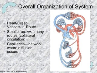
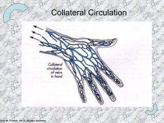
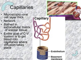
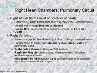
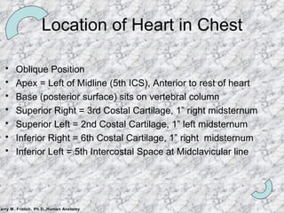
CARDIO-VASCULAR SYSTEM Principles Structures Two circuits Pulmonary Systemic Fetal Circulation Heart Details Other details
What Does C-V System do? Circulate blood throughout entire body for Transport of oxygen to cells Transport of CO2 away from cells Transport of nutrients (glucose) to cells Movement of immune system components (cells, antibodies) Transport of endocrine gland secretions
How does it do it? Heart is pump Arteries and veins are main tubes (plumbing) A rteries  A way from Heart Veins to Heart Diffusion happens in capillaries (oxygen, CO2, glucose diffuse in or out of blood)
Heart/Great Vessels--1 Route Smaller aa. vv.--many routes (collateral circulation) Capillaries—network where diffusion occurs Overall Organization of System
Collateral Circulation
Walls of Arteries and Veins Tunica externa Outermost layer CT w/elastin and collagen Strengthens, Anchors Tunica media Middle layer Circular Smooth Muscle Vaso-constriction/dilation Tunica intima Innermost layer Endothelium Minimize friction Lumen pg 546
Artery/Vein differences Lower Higher Pressure Valves Lumen Walls Direction of flow Valves (see next) No valves Larger Smaller THINNER:  Tunica externa thicker than tunica media THICKER:  Tunica media thicker than tunica externa Blood to Heart Blood Away from Heart Veins (vv.) Arteries (aa.)
 
Capillaries Microscopic--one cell layer thick Network  Bathed in extracellular matrix  of areolar tissue Entire goal of C-V system is to get blood into capillaries where diffusion takes place
Two circulatory paths Pulmonary Systemic
GREAT VESSELS Aorta IVC, SVC Pulmonary Trunk Pulmonary Veins
Heart Chambers and Valves
Right Heart Chambers: Pulmonary Circuit Right Atrium (forms most of posterior of heart) Receives O 2 -poor  blood from body via IVC, SVC, Coronary sinus Ventral wall = rough  Pectinate muscle Fossa Ovalis - on interatrial septum, remnant of  Foramen Ovale Right Ventricle Receives O 2 -poor  blood from right atrium through tricuspid valve Pumps blood to lungs via  Pulmonary Semilunar Valve  in pulmonary trunk Trabeculae Carnae  along ventral surface Papillary Muscle -cone-shaped muscle to which chordae tendinae are anchored Moderator Band- muscular band connecting anterior papillary muscle to interventricular septum
Left Heart Chambers: Systemic Circuit Left Atrium Receives O 2 -rich blood from 4 Pulmonary Veins Pectinate Muscles  line only auricle Left Ventricle (forms apex of heart) Receives blood from Left Atrium via bicuspid valve Pumps blood into aorta via  Aortic Semilunar Valve  to body Same structures as Rt Ventricle: Trabeculae carnae, Papillary muscles, Chordae tendinae No Moderator Band
Heart Valves: Lub*-Dub** *Tricuspid Valve: Right AV valve   3 Cusps (flaps) made of endocardium and CT Cusps anchored in Rt. Ventricle by  Chordae Tendinae Chordae Tendinae prevent inversion of cusps into atrium Flow of blood pushes cusps open When ventricle in diastole (relaxed), cusps hang limp in ventricle Ventricular contraction increases pressure and forces cusps closed *Bicuspid (Mitral) Valve: Left AV valve 2 cusps anchored in Lft. Ventricle by chordae tendinae Functions same as Rt. AV valve **Semilunar valves : prevents backflow in large arteries Pulmonary Semilunar Valve:  Rt Ventricle and Pulmonary Trunk Aortic Semilunar Valve:  Left Ventricle and Aorta 3 cusps: blood rushes past they’re flattened, as it settles they’re pushed down (valve closed)
Another View
Location of Heart in Thorax pg 523
Location of Heart in Chest Oblique Position Apex = Left of Midline (5th ICS), Anterior to rest of heart Base (posterior surface) sits on vertebral column Superior Right = 3rd Costal Cartilage, 1” right midsternum Superior Left = 2nd Costal Cartilage, 1” left midsternum Inferior Right = 6th Costal Cartilage, 1” right  midsternum Inferior Left = 5th Intercostal Space at Midclavicular line
Heart Wall   Epicardium (most superficial) –  Visceral pleura Myocardium (middle layer) Cardiac muscle Contracts  Endocardium (inner) Endothelium on CT Lines the heart  Creates the valves pg 524 How does heart muscle get blood supply?
Blood supply to heart wall Rt and Lft Coronary Arteries Branch from Ascending Aorta Have multiple branches along heart Sit  in Coronary Sulcus Coronary Heart Disease Cardiac Veins Coronary Sinus (largest) Many branches feed into sinus Sit  in Coronary Sulcus
Heart Innervation Heart receives visceral motor innervation Sympathetic (speeds up) Parasympathetic (slows down) p. 534
Fetal Circulation No circulation to lungs Foramen ovale Ductus arteriosum Circulation must go to placenta Umbilical aa., vv.
Adult remnants of fetal circulation Umbilical cord (leaving fetus) Medial umbilical ligament Ductus venosus Ligamentum venosum Umbilical v.(within fetus) Round ligament (ligamentum teres) of liver Umbilical aa.(within fetus) Medial umbilical ligaments Ductus arteriosus Ligamentum arteriosum Foramen ovale Fossa ovale Fetus Adult
Hepatic Portal System
Lymphatic System…The Players: Lymph- clear fluid from loose areolar CT around capillaries Lymphatic capillaries (near blood capillaries)   Lymph collecting vessels (small, 3 tunicas, valves)  Lymph nodes (sit along collecting vessels)-clean lymph of pathogens, they are NOT glands Lymphatic trunks   (convergence large collecting vessels) Lumbar, intestinal, bronchomediastinal, subclavian, jugular Lymphatic ducts    empty into veins of neck
Lymphatic System--Function Function: to collect excess tissue fluid collecting at arteriole end  and return leaked blood proteins to blood (maintain osmotic pressure needed to take up water into bloodstream) Lymph moved through vessels Pulse of nearby arteries Contraction of surrounding skeletal muscle Regular movement of body (wiggling legs) Muscle in Tunica Media Lacteals-lymphatic capillaries w/unique function In mucosa of small intestine, receive digested fat from intestine Fatty lymph becomes milky =  Chyle Chyle  goes to bloodstream

Cardiovascular system

  • 1.
    CARDIO-VASCULAR SYSTEM PrinciplesStructures Two circuits Pulmonary Systemic Fetal Circulation Heart Details Other details
  • 2.
    What Does C-VSystem do? Circulate blood throughout entire body for Transport of oxygen to cells Transport of CO2 away from cells Transport of nutrients (glucose) to cells Movement of immune system components (cells, antibodies) Transport of endocrine gland secretions
  • 3.
    How does itdo it? Heart is pump Arteries and veins are main tubes (plumbing) A rteries A way from Heart Veins to Heart Diffusion happens in capillaries (oxygen, CO2, glucose diffuse in or out of blood)
  • 4.
    Heart/Great Vessels--1 RouteSmaller aa. vv.--many routes (collateral circulation) Capillaries—network where diffusion occurs Overall Organization of System
  • 5.
  • 6.
    Walls of Arteriesand Veins Tunica externa Outermost layer CT w/elastin and collagen Strengthens, Anchors Tunica media Middle layer Circular Smooth Muscle Vaso-constriction/dilation Tunica intima Innermost layer Endothelium Minimize friction Lumen pg 546
  • 7.
    Artery/Vein differences LowerHigher Pressure Valves Lumen Walls Direction of flow Valves (see next) No valves Larger Smaller THINNER: Tunica externa thicker than tunica media THICKER: Tunica media thicker than tunica externa Blood to Heart Blood Away from Heart Veins (vv.) Arteries (aa.)
  • 8.
  • 9.
    Capillaries Microscopic--one celllayer thick Network Bathed in extracellular matrix of areolar tissue Entire goal of C-V system is to get blood into capillaries where diffusion takes place
  • 10.
    Two circulatory pathsPulmonary Systemic
  • 11.
    GREAT VESSELS AortaIVC, SVC Pulmonary Trunk Pulmonary Veins
  • 12.
  • 13.
    Right Heart Chambers:Pulmonary Circuit Right Atrium (forms most of posterior of heart) Receives O 2 -poor blood from body via IVC, SVC, Coronary sinus Ventral wall = rough Pectinate muscle Fossa Ovalis - on interatrial septum, remnant of Foramen Ovale Right Ventricle Receives O 2 -poor blood from right atrium through tricuspid valve Pumps blood to lungs via Pulmonary Semilunar Valve in pulmonary trunk Trabeculae Carnae along ventral surface Papillary Muscle -cone-shaped muscle to which chordae tendinae are anchored Moderator Band- muscular band connecting anterior papillary muscle to interventricular septum
  • 14.
    Left Heart Chambers:Systemic Circuit Left Atrium Receives O 2 -rich blood from 4 Pulmonary Veins Pectinate Muscles line only auricle Left Ventricle (forms apex of heart) Receives blood from Left Atrium via bicuspid valve Pumps blood into aorta via Aortic Semilunar Valve to body Same structures as Rt Ventricle: Trabeculae carnae, Papillary muscles, Chordae tendinae No Moderator Band
  • 15.
    Heart Valves: Lub*-Dub***Tricuspid Valve: Right AV valve 3 Cusps (flaps) made of endocardium and CT Cusps anchored in Rt. Ventricle by Chordae Tendinae Chordae Tendinae prevent inversion of cusps into atrium Flow of blood pushes cusps open When ventricle in diastole (relaxed), cusps hang limp in ventricle Ventricular contraction increases pressure and forces cusps closed *Bicuspid (Mitral) Valve: Left AV valve 2 cusps anchored in Lft. Ventricle by chordae tendinae Functions same as Rt. AV valve **Semilunar valves : prevents backflow in large arteries Pulmonary Semilunar Valve: Rt Ventricle and Pulmonary Trunk Aortic Semilunar Valve: Left Ventricle and Aorta 3 cusps: blood rushes past they’re flattened, as it settles they’re pushed down (valve closed)
  • 16.
  • 17.
    Location of Heartin Thorax pg 523
  • 18.
    Location of Heartin Chest Oblique Position Apex = Left of Midline (5th ICS), Anterior to rest of heart Base (posterior surface) sits on vertebral column Superior Right = 3rd Costal Cartilage, 1” right midsternum Superior Left = 2nd Costal Cartilage, 1” left midsternum Inferior Right = 6th Costal Cartilage, 1” right midsternum Inferior Left = 5th Intercostal Space at Midclavicular line
  • 19.
    Heart Wall Epicardium (most superficial) – Visceral pleura Myocardium (middle layer) Cardiac muscle Contracts Endocardium (inner) Endothelium on CT Lines the heart Creates the valves pg 524 How does heart muscle get blood supply?
  • 20.
    Blood supply toheart wall Rt and Lft Coronary Arteries Branch from Ascending Aorta Have multiple branches along heart Sit in Coronary Sulcus Coronary Heart Disease Cardiac Veins Coronary Sinus (largest) Many branches feed into sinus Sit in Coronary Sulcus
  • 21.
    Heart Innervation Heartreceives visceral motor innervation Sympathetic (speeds up) Parasympathetic (slows down) p. 534
  • 22.
    Fetal Circulation Nocirculation to lungs Foramen ovale Ductus arteriosum Circulation must go to placenta Umbilical aa., vv.
  • 23.
    Adult remnants offetal circulation Umbilical cord (leaving fetus) Medial umbilical ligament Ductus venosus Ligamentum venosum Umbilical v.(within fetus) Round ligament (ligamentum teres) of liver Umbilical aa.(within fetus) Medial umbilical ligaments Ductus arteriosus Ligamentum arteriosum Foramen ovale Fossa ovale Fetus Adult
  • 24.
  • 25.
    Lymphatic System…The Players:Lymph- clear fluid from loose areolar CT around capillaries Lymphatic capillaries (near blood capillaries)  Lymph collecting vessels (small, 3 tunicas, valves)  Lymph nodes (sit along collecting vessels)-clean lymph of pathogens, they are NOT glands Lymphatic trunks  (convergence large collecting vessels) Lumbar, intestinal, bronchomediastinal, subclavian, jugular Lymphatic ducts  empty into veins of neck
  • 26.
    Lymphatic System--Function Function:to collect excess tissue fluid collecting at arteriole end and return leaked blood proteins to blood (maintain osmotic pressure needed to take up water into bloodstream) Lymph moved through vessels Pulse of nearby arteries Contraction of surrounding skeletal muscle Regular movement of body (wiggling legs) Muscle in Tunica Media Lacteals-lymphatic capillaries w/unique function In mucosa of small intestine, receive digested fat from intestine Fatty lymph becomes milky = Chyle Chyle goes to bloodstream