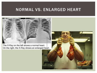
The document summarizes key aspects of the cardiovascular system, including:
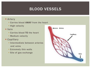
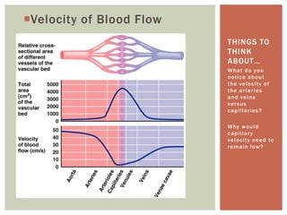
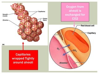
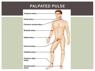
1. It describes the three main types of blood vessels - arteries, veins, and capillaries - and notes their distinguishing characteristics like direction of blood flow and velocity.
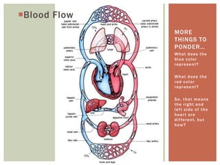
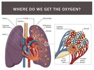
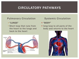
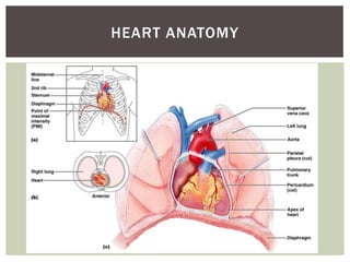
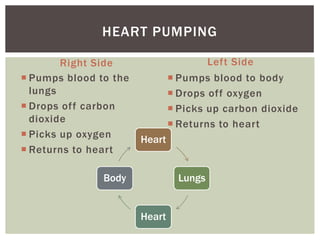
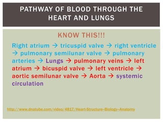
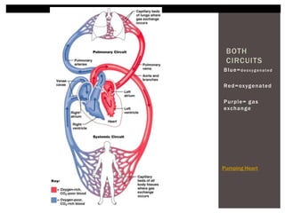
2. It explains the pulmonary and systemic circulations, noting that pulmonary circulation occurs between the heart and lungs while systemic circulation transports blood to the entire body.
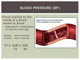
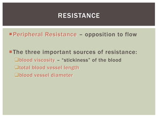
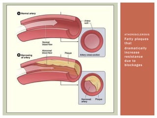
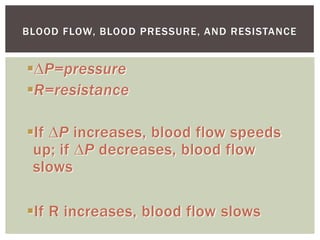
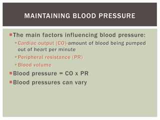
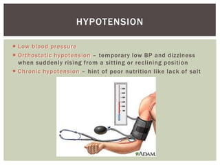
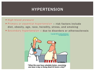
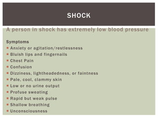
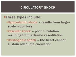
3. Key components of the circulatory system are discussed like blood pressure, resistance, and shock states that can occur if blood pressure drops drastically.