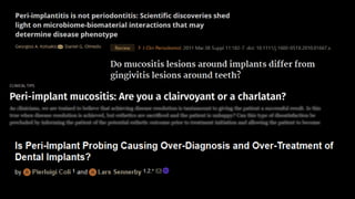
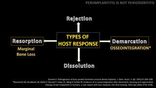
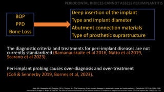
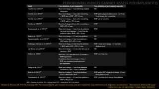
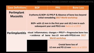
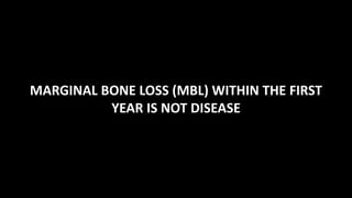
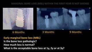
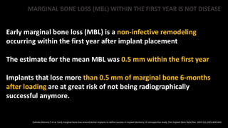
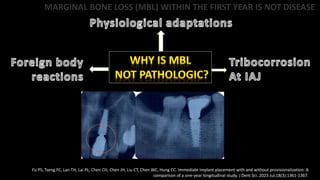
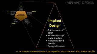
PERIIMPLANTITIS
Clarity on the Concepts
PERIIMPLANTITIS IS NOT PERIODONTITIS
*Consensus report of 7th European Workshop on Periodontology, 2011
It was always
assumed that’s that
the common
etiology for peri-
implant disease is
the presence of a
bacterial biofilm
and excessive
occlusal load*
PERIIMPLANTITIS IS NOT PERIODONTITIS
Donath K. Pathogenesis of bony pocket formation around dental implants. J. Dent. Assoc. S. Afr. 1992;47:204–208.
*Bosshardt DD, Brodbeck UR, Rathe F, Stumpf T, Imber JC, Weigl P, Schlee M. Evidence of re-osseointegration after electrolytic cleaning and regenerative
therapy of peri-implantitis in humans: a case report with four implants. Clin Oral Investig. 2022 Apr;26(4):3735-3746.
PERIIMPLANTITIS IS NOT PERIODONTITIS
Marginal
Bone Loss
OSSEOINTEGRATION*
Kotsakis GA, Olmedo DG. Peri-implantitis is not periodontitis: Scientific discoveries shed light on microbiome-biomaterial interactions that may determine disease phenotype. Periodontol 2000. 2021
Jun;86(1):231-240. doi: 10.1111/prd.12372. Epub 2021 Mar 10. PMID: 33690947.
Antibiotic Resistance
Titanium Dissolution Products
PERIIMPLANTITIS IS NOT PERIODONTITIS
Mombelli A, Hashim D, Cionca N. What is the impact of titanium particles and biocorrosion on implant survival and complications? A critical review. Clin Oral
Implants Res. 2018 Oct;29 Suppl 18:37-53. doi: 10.1111/clr.13305. PMID: 30306693.
PERIIMPLANTITIS IS NOT PERIODONTITIS
1.52 µm-94.00 µm
Gianfreda F, Punzo A, Pistilli V, Bollero P, Cervino G, D'Amico C, Cairo F, Cicciù M. Electrolytic Cleaning and Regenerative Therapy of Peri-implantitis in the
Esthetic Area: A Case Report. Eur J Dent. 2022 Oct;16(4):950-956. doi: 10.1055/s-0042-1750773. Epub 2022 Jul 4. PMID: 35785819; PMCID: PMC9683897
PERIIMPLANT MUCOSITIS IS NOT GINGIVITIS
• More PMNs
• Less plasma cells
and lymphocytes
• Peri-implant lesion
was less
immunologically
regulated
Peri-implant mucositis has to be diagnosed properly
Peri-implant soft tissues develop a stronger inflammatory
response
Inflammation was reversible only at the biomarker level
3-weeks was not sufficient for inflammation reversal
PERIIMPLANT MUCOSITIS IS NOT GINGIVITIS
Non-surgical therapy has not been shown to be effective for the treatment
of peri-implant mucositis
Peri-implant mucositis appears to be reversible
Peri-implant mucositis may be present for extensive periods of time without
progression to peri-Implantitis
Optimal biofilm control may take longer than 3 weeks
PERIIMPLANT MUCOSITIS IS NOT A DISEASE
Inflammatory connective tissue (ICT) infiltrate of the peri-
implant mucosa in the microgap*
This ICT zone indicates a response of the immune system to
microorganisms and in generally considered as protective**
If the microgap is located more coronally, ie, further from the
alveolar crest, then we should expect less bone loss because of
the greater distance between the bacteria and the alveolar
Crest***
*Ericsson I, Persson LG, Berglundh T, Mariniello CP, Lindhe J, Klinge B. Different types of inflammatory reactions in periimplant soft tissues. J Clin Periodontol 1995;22:255–261.
**Piattelli A, Vrespa G, Petrone G, Iezzi G, Annibali S, Scarano A. Role of the Microgap between implant and abutment: A retrospective histologic evaluation in monkeys. J Periodontol 2003;74:346–352.
***Luongo R, Traini T, Guidone PC, Bianco G, Cocchetto R, Celletti R. Hard and soft tissue responses to the platform-switching technique. Int J Periodontics Restorative Dent. 2008 Dec;28(6):551-7. PMID:
19146050.
PERIIMPLANT MUCOSITIS IS NOT A DISEASE
The ICT is never in contact with the bone but was
separated from it by an approximately 1-mm-wide layer of
healthy connective tissue*
This should be considered as a sort of barrier or protective
seal capable of preserving bone complementing the
biologic width
IF IT IS PROTECTIVE, WHY IS IT A DISEASE?
*Luongo R, Traini T, Guidone PC, Bianco G, Cocchetto R, Celletti R. Hard and soft tissue responses to the platform-switching technique. Int J Periodontics Restorative Dent. 2008 Dec;28(6):551-7.
**Berglundh T, Lindhe J. Dimension of the periimplant mucosa. Biologic width revisited. J Clin Periodontol 1996;23:971–973
PERIODONTAL INDICES CANNOT ASSESS
PERIIMPLANTITIS
PERIODONTAL INDICES CANNOT ASSESS PERIIMPLANTITIS
BOP
PPD
Bone Loss
Deep insertion of the implant
Type and implant diameter
Abutment connection materials
Type of prosthetic suprastructure
Atieh MA, Alsabeeha NH, Faggion CM Jr, Duncan WJ. The frequency of peri-implant diseases: a systematic review and meta-analysis. J Periodontol. 2013;84:1586–1598.
Hammerle CH, Bragger U, Burgin W, Lang NP. The effect of subcrestal placement of the polished surface of ITI implants on marginal soft and hard tissues. Clin Oral Implants Res. 1996;7:111–119.
The diagnostic criteria and treatments for peri-implant diseases are not
currently standardized (Ramanauskaite et al 2016, Natto et al 2019,
Scarano et al 2023).
Peri-implant probing causes over-diagnosis and over-treatment
(Coli & Sennerby 2019, Bornes et al, 2023).
PERIODONTAL INDICES CANNOT ASSESS PERIIMPLANTITIS
Renvert S, Persson GR, Pirih FQ, Camargo PM. Peri-implant health, peri-implant mucositis, and peri-implantitis: Case definitions and diagnostic considerations. J Periodontol. 2018 Jun;89 Suppl
1:S304-S312. doi: 10.1002/JPER.17-0588. PMID: 29926953.
PERIODONTAL INDICES CANNOT ASSESS PERIIMPLANTITIS
Case Definitions BOP PPD Bone Loss
Periimplant
Mucositis
Erythema & BOP+ & PPD↗ & Absence of bone loss beyond
initial remodeling 2017 World workshop
BOP+ with ≤2 mm in the first year and ≤0.2 mm in each
subsequent year and PD≥5 mm Natto et al 2019
Periimplantitis visual inflammatory changes + PPD↗ + Progressive bone loss
+ evidence of bone loss ≥3 mm with PPD≥6 mm 2017
World workshop
Crestal bone loss of
≥2 mm and PD ≥3 mm Natto et al 2019
MARGINAL BONE LOSS (MBL) WITHIN THE FIRST
YEAR IS NOT DISEASE
6 Months 3 Months 9 Months
MARGINAL BONE LOSS (MBL) WITHIN THE FIRST YEAR IS NOT DISEASE
Early marginal bone loss (MBL)
Is the bone loss pathologic?
How much loss is normal?
What is the acceptable bone loss at 1y, 2y or at 3y?
MARGINAL BONE LOSS (MBL) WITHIN THE FIRST YEAR IS NOT DISEASE
Early marginal bone loss (MBL) is a non-infective remodeling
occurring within the first year after implant placement
The estimate for the mean MBL was 0.5 mm within the first year
Implants that lose more than 0.5 mm of marginal bone 6-months
after loading are at great risk of not being radiographically
successful anymore.
Galindo-Moreno P et al. Early marginal bone loss around dental implants to define success in implant dentistry: A retrospective study. Clin Implant Dent Relat Res. 2022 Oct;24(5):630-642.
MARGINAL BONE LOSS (MBL) WITHIN THE FIRST YEAR IS NOT DISEASE
Fu PS, Tseng FC, Lan TH, Lai PL, Chen CH, Chen JH, Liu CT, Chen WC, Hung CC. Immediate implant placement with and without provisionalization: A
comparison of a one-year longitudinal study. J Dent Sci. 2023 Jul;18(3):1361-1367.
IMPLANT DESIGN DOES NOT CORRELATE WITH
INCIDENCE OF PERIIMPLANTITIS
Fu JH, Wang HL. Breaking the wave of peri-implantitis. Periodontol 2000. 2020 Oct;84(1):145-160.
IMPLANT DESIGN
1.5 Yrs 3 months
Zirconia Implants
IMPLANT DESIGN
THANK YOU

PeriImplantitis Concepts.pptx

  • 1.
  • 4.
  • 5.
    *Consensus report of7th European Workshop on Periodontology, 2011 It was always assumed that’s that the common etiology for peri- implant disease is the presence of a bacterial biofilm and excessive occlusal load* PERIIMPLANTITIS IS NOT PERIODONTITIS
  • 6.
    Donath K. Pathogenesisof bony pocket formation around dental implants. J. Dent. Assoc. S. Afr. 1992;47:204–208. *Bosshardt DD, Brodbeck UR, Rathe F, Stumpf T, Imber JC, Weigl P, Schlee M. Evidence of re-osseointegration after electrolytic cleaning and regenerative therapy of peri-implantitis in humans: a case report with four implants. Clin Oral Investig. 2022 Apr;26(4):3735-3746. PERIIMPLANTITIS IS NOT PERIODONTITIS Marginal Bone Loss OSSEOINTEGRATION*
  • 7.
    Kotsakis GA, OlmedoDG. Peri-implantitis is not periodontitis: Scientific discoveries shed light on microbiome-biomaterial interactions that may determine disease phenotype. Periodontol 2000. 2021 Jun;86(1):231-240. doi: 10.1111/prd.12372. Epub 2021 Mar 10. PMID: 33690947. Antibiotic Resistance Titanium Dissolution Products
  • 8.
    PERIIMPLANTITIS IS NOTPERIODONTITIS Mombelli A, Hashim D, Cionca N. What is the impact of titanium particles and biocorrosion on implant survival and complications? A critical review. Clin Oral Implants Res. 2018 Oct;29 Suppl 18:37-53. doi: 10.1111/clr.13305. PMID: 30306693.
  • 9.
    PERIIMPLANTITIS IS NOTPERIODONTITIS 1.52 µm-94.00 µm Gianfreda F, Punzo A, Pistilli V, Bollero P, Cervino G, D'Amico C, Cairo F, Cicciù M. Electrolytic Cleaning and Regenerative Therapy of Peri-implantitis in the Esthetic Area: A Case Report. Eur J Dent. 2022 Oct;16(4):950-956. doi: 10.1055/s-0042-1750773. Epub 2022 Jul 4. PMID: 35785819; PMCID: PMC9683897
  • 10.
  • 11.
    • More PMNs •Less plasma cells and lymphocytes • Peri-implant lesion was less immunologically regulated Peri-implant mucositis has to be diagnosed properly Peri-implant soft tissues develop a stronger inflammatory response Inflammation was reversible only at the biomarker level 3-weeks was not sufficient for inflammation reversal PERIIMPLANT MUCOSITIS IS NOT GINGIVITIS
  • 12.
    PERIIMPLANT MUCOSITIS ISNOT GINGIVITIS Non-surgical therapy has not been shown to be effective for the treatment of peri-implant mucositis Peri-implant mucositis appears to be reversible Peri-implant mucositis may be present for extensive periods of time without progression to peri-Implantitis Optimal biofilm control may take longer than 3 weeks
  • 13.
  • 14.
    Inflammatory connective tissue(ICT) infiltrate of the peri- implant mucosa in the microgap* This ICT zone indicates a response of the immune system to microorganisms and in generally considered as protective** If the microgap is located more coronally, ie, further from the alveolar crest, then we should expect less bone loss because of the greater distance between the bacteria and the alveolar Crest*** *Ericsson I, Persson LG, Berglundh T, Mariniello CP, Lindhe J, Klinge B. Different types of inflammatory reactions in periimplant soft tissues. J Clin Periodontol 1995;22:255–261. **Piattelli A, Vrespa G, Petrone G, Iezzi G, Annibali S, Scarano A. Role of the Microgap between implant and abutment: A retrospective histologic evaluation in monkeys. J Periodontol 2003;74:346–352. ***Luongo R, Traini T, Guidone PC, Bianco G, Cocchetto R, Celletti R. Hard and soft tissue responses to the platform-switching technique. Int J Periodontics Restorative Dent. 2008 Dec;28(6):551-7. PMID: 19146050. PERIIMPLANT MUCOSITIS IS NOT A DISEASE
  • 15.
    PERIIMPLANT MUCOSITIS ISNOT A DISEASE The ICT is never in contact with the bone but was separated from it by an approximately 1-mm-wide layer of healthy connective tissue* This should be considered as a sort of barrier or protective seal capable of preserving bone complementing the biologic width IF IT IS PROTECTIVE, WHY IS IT A DISEASE? *Luongo R, Traini T, Guidone PC, Bianco G, Cocchetto R, Celletti R. Hard and soft tissue responses to the platform-switching technique. Int J Periodontics Restorative Dent. 2008 Dec;28(6):551-7. **Berglundh T, Lindhe J. Dimension of the periimplant mucosa. Biologic width revisited. J Clin Periodontol 1996;23:971–973
  • 16.
    PERIODONTAL INDICES CANNOTASSESS PERIIMPLANTITIS
  • 17.
    PERIODONTAL INDICES CANNOTASSESS PERIIMPLANTITIS BOP PPD Bone Loss Deep insertion of the implant Type and implant diameter Abutment connection materials Type of prosthetic suprastructure Atieh MA, Alsabeeha NH, Faggion CM Jr, Duncan WJ. The frequency of peri-implant diseases: a systematic review and meta-analysis. J Periodontol. 2013;84:1586–1598. Hammerle CH, Bragger U, Burgin W, Lang NP. The effect of subcrestal placement of the polished surface of ITI implants on marginal soft and hard tissues. Clin Oral Implants Res. 1996;7:111–119. The diagnostic criteria and treatments for peri-implant diseases are not currently standardized (Ramanauskaite et al 2016, Natto et al 2019, Scarano et al 2023). Peri-implant probing causes over-diagnosis and over-treatment (Coli & Sennerby 2019, Bornes et al, 2023).
  • 18.
    PERIODONTAL INDICES CANNOTASSESS PERIIMPLANTITIS Renvert S, Persson GR, Pirih FQ, Camargo PM. Peri-implant health, peri-implant mucositis, and peri-implantitis: Case definitions and diagnostic considerations. J Periodontol. 2018 Jun;89 Suppl 1:S304-S312. doi: 10.1002/JPER.17-0588. PMID: 29926953.
  • 19.
    PERIODONTAL INDICES CANNOTASSESS PERIIMPLANTITIS Case Definitions BOP PPD Bone Loss Periimplant Mucositis Erythema & BOP+ & PPD↗ & Absence of bone loss beyond initial remodeling 2017 World workshop BOP+ with ≤2 mm in the first year and ≤0.2 mm in each subsequent year and PD≥5 mm Natto et al 2019 Periimplantitis visual inflammatory changes + PPD↗ + Progressive bone loss + evidence of bone loss ≥3 mm with PPD≥6 mm 2017 World workshop Crestal bone loss of ≥2 mm and PD ≥3 mm Natto et al 2019
  • 20.
    MARGINAL BONE LOSS(MBL) WITHIN THE FIRST YEAR IS NOT DISEASE
  • 21.
    6 Months 3Months 9 Months MARGINAL BONE LOSS (MBL) WITHIN THE FIRST YEAR IS NOT DISEASE Early marginal bone loss (MBL) Is the bone loss pathologic? How much loss is normal? What is the acceptable bone loss at 1y, 2y or at 3y?
  • 22.
    MARGINAL BONE LOSS(MBL) WITHIN THE FIRST YEAR IS NOT DISEASE Early marginal bone loss (MBL) is a non-infective remodeling occurring within the first year after implant placement The estimate for the mean MBL was 0.5 mm within the first year Implants that lose more than 0.5 mm of marginal bone 6-months after loading are at great risk of not being radiographically successful anymore. Galindo-Moreno P et al. Early marginal bone loss around dental implants to define success in implant dentistry: A retrospective study. Clin Implant Dent Relat Res. 2022 Oct;24(5):630-642.
  • 23.
    MARGINAL BONE LOSS(MBL) WITHIN THE FIRST YEAR IS NOT DISEASE Fu PS, Tseng FC, Lan TH, Lai PL, Chen CH, Chen JH, Liu CT, Chen WC, Hung CC. Immediate implant placement with and without provisionalization: A comparison of a one-year longitudinal study. J Dent Sci. 2023 Jul;18(3):1361-1367.
  • 24.
    IMPLANT DESIGN DOESNOT CORRELATE WITH INCIDENCE OF PERIIMPLANTITIS
  • 25.
    Fu JH, WangHL. Breaking the wave of peri-implantitis. Periodontol 2000. 2020 Oct;84(1):145-160. IMPLANT DESIGN
  • 26.
    1.5 Yrs 3months Zirconia Implants IMPLANT DESIGN
  • 28.

Editor's Notes

  • #4 a person falsely claiming to have a special knowledge or skil a person who claims to have a supernatural ability to perceive events in the future or beyond normal sensory contact.
  • #6 Risk factors for periimplant diseases are very similar to periodontitis risk factors Common etiology for peri-implant disease is the presence of a bacterial biofilm and excessive occlusal load*
  • #9 Most dental implants have a 2-stage design in which (1) the implant is placed and the surrounding tissues are allowed to heal, and then (2) the abutment is placed and the restoration is completed. To prevent the unsightly metal ring that occurs with supragingival placement of the IAJ, most implants involve subgingival placement. The implant and the abutment cannot be accurately matched because of the precision limit during production . This subgingival placement makes the IAJ inaccessible for routine hygiene care and raises legitimate concerns about bacterial colonization within the IAJ micro-gap. In vitro studies show that biofilm in sliding contact surfaces could act like a lubricant, resulting in a friction behavior and reducing the mechanical integrity of the joint vs COLD WELDING Around implant-abutment connections, a lower pH value of 3–4 can induce corrosion at the contacting surfaces. Galvanic corrosion is the most common type of corrosion in dental implants. The study of the simultaneous degradation by wear and corrosion that occurs at the sliding contacts is known as tribocorrosion 
  • #10 The internal cavity of implant is similar to a reservoir (Nayak et al., 2014; Orsini et al., 2000; Proff et al., 2006). When the abutment is removed and replaced, bacteria can enter the implant internal cavity, where they reside and proliferate. The bacteria with their toxic by-products and small nutritious molecules can freely penetrate into the implant internal cavity or reverse through the IAI microgap. The destruction of the IAI micromotion is mainly displayed in two aspects. First, micromotion interferes the attachment of soft tissue around the implant neck and disrupts the stability of soft tissue that has completed integration (Passos, Gressler May, Faria, Ozcan, & Bottino, 2013). Second, micromotion causes a micropumping effect (Ericsson, et al., 1995), which intensifies the leakage of bacteria and their toxic by-products and accelerates the blood, saliva, and proteoglycans (including the extracellular matrix and mucus layer) into the internal cavity of implant (Baixe, Tenenbaum, & Etienne, 2016). Mechanical damages of microgap and micromotion include fretting wear, adhesive wear, and screw loosening (Jorn, Kohorst, Besdo, Borchers, & Stiesch, 2016; Sakamoto et al., 2016). Fretting wear refers to microfracture and chipping between the IAI, whereas adhesive wear is defined as the plastic deformation in the IAI (
  • #12 The host response to biofilms does not differ substantially at teeth or implants. The most obvious sign clinically is the development of an inflammatory lesion as a result of the bacterial challenge. Gingivitis at teeth or peri-implant mucositis at implants are precursors for more detrimental lesions, and hence have to be diagnosed properly and prevented by applying anti-infective therapy. Non-surgical interventions are usually sufficient for the treatment of both gingivitis and mucositis.