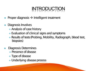
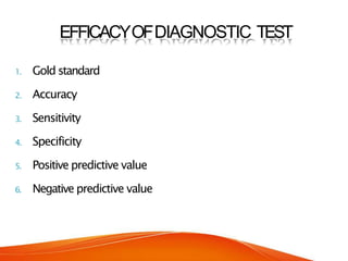
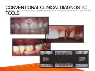
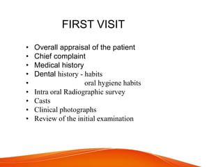
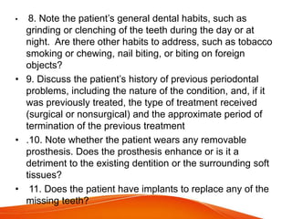
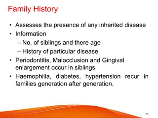
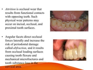
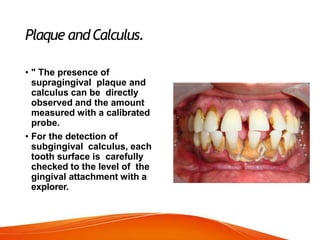
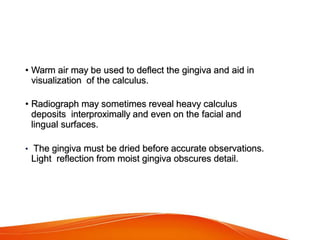
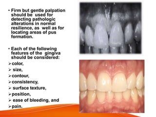
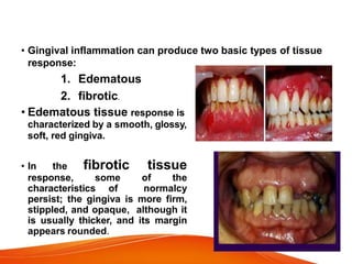
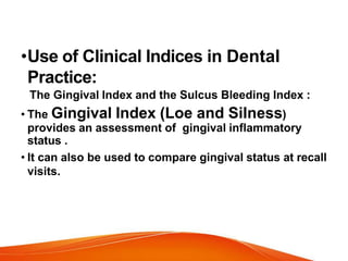
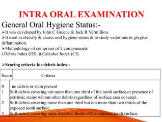
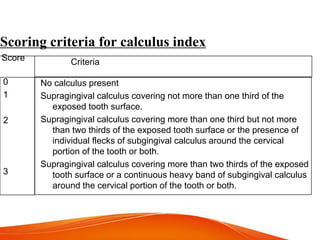
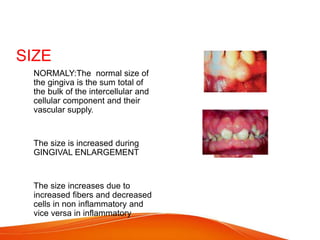
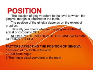
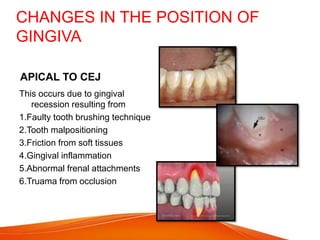
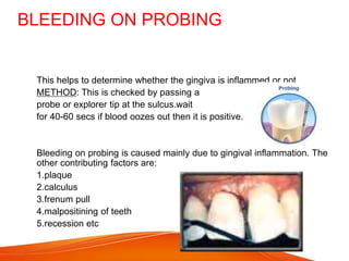
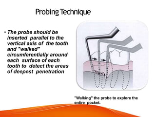
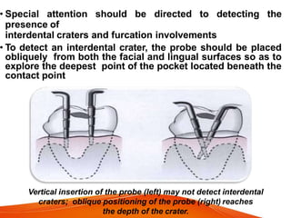
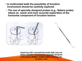
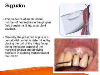
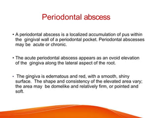
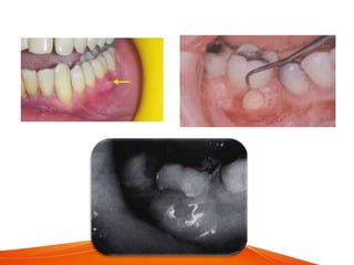
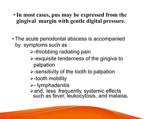
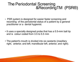
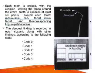
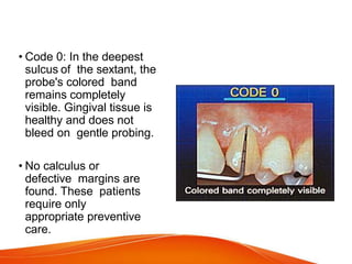
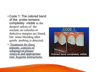
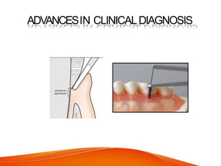
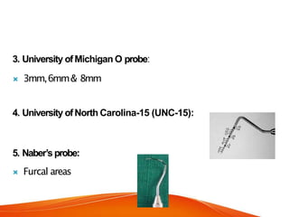
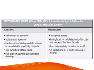
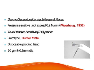
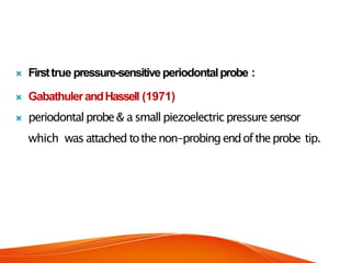
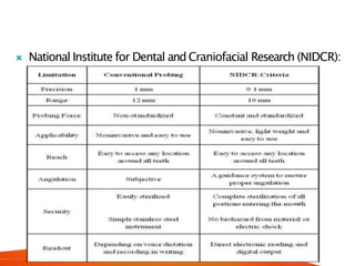
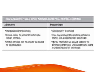
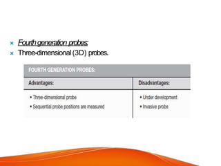
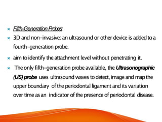
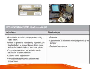
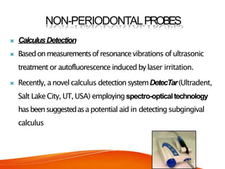
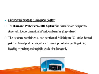
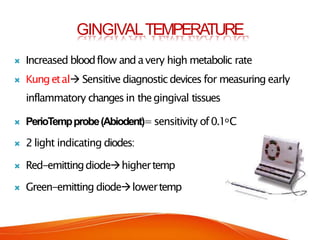
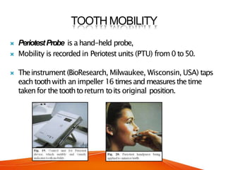
SlideShare a Scribd company logo
CLINICALDIAGNOSIS
Guided by:
Dr. Rupal Mehta
Prof. & HOD of
Periodontics
Presented by :
Rutu Dabhi
(Part II PG)
CONTENTS
Introduction
Efficacy ofdiagnostic test
Firstvisit
Second visit
Laboratoryaidstoclinicaldaignosis
Periodontalscreeningandrecordingsystem
Limitations ofconventional methods
Advancesin Clinical diagnosis
Conclusion
References
INTRODUCTION
 Proper diagnosis Intelligent treatment
 Diagnosis Involves
 Analysis ofcasehistory
 Evaluation ofclinical signsandsymptoms
 Resultsoftests(Probing, Mobility, Radiograph, blood test,
biopsies)
 Diagnosis Determines
 Presenceofdisease
 Typeofdisease
 Underlying diseaseprocess
 A“Diagnostic”refers totools,procedures or technologiesthat are
usedin determination of diagnosis
 usedto:
a) predisposingrisk factors
b) identify early disease
c) specifictypeof disease
Kornman,2005
EFFICACYOFDIAGNOSTIC TEST
1. Gold standard
2. Accuracy
3. Sensitivity
4. Specificity
5. Positive predictive value
6. Negative predictive value
CONVENTIONALCLINICALDIAGNOSTIC
TOOLS
FIRST VISIT
• Overall appraisal of the patient
• Chief complaint
• Medical history
• Dental history - habits
• oral hygiene habits
• Intra oral Radiographic survey
• Casts
• Clinical photographs
• Review of the initial examination
SECOND VISIT
• Oral examination
• Examination of teeth and implants
• Examination of periodontium
• A Periodontal daignosis should determine
1. Presence of disease 2. Identify the type 3.
Extent 4.Distribution 5. Severity 6.Understanding
of cause
CHIEF COMPLAINT
The chief complaint is established by asking the patient to describe the
problem for which he or she is seeking help or treatment.
 It is recorded in patients own words as much as possible no
documentary or technical language should be used.
 It is recorded in chronological order of their appearance and in the
order of their severity
 The chief complaint aids in the diagnosis and treatment planning
and should be given the 1st priority.
Common chief complaint are: pain, bleeding, loose teeth, halitosis,
swelling, bad taste
HISTORY OF CHIEF COMPLAINT
Initially the patient may not volunteer the detailed history of the
problem, so the examiner has to elicit of out the information by the
possible questionnaire about the symptoms. The patient’s response to
these questions is termed history of present illness.
 The questions can be asked in the manner.
1.When did the problem start?
2.What did you do first?
3.Do you have any symptoms related to this?
4.What makes the problem worse/better?
5.Have any tests being performed to diagnose the complaint?
6.Have you ever consulted any examiner for this problem?
7.What have you done to treat this problem?
Detailed history of particular symptom
• Anatomical location
• Origin & mode of onset
• Intensity of pain
• Nature of pain
• Progression of pain
• Duration of pain
• Movement of pain
• Effect of functional activity
Medical History
• 1. If the patient is under the care of a physician, the nature and
duration of the problem and its therapy should be discussed. The
name, address, and telephone number of the physician should be
recorded.
• 2. Details regarding hospitalizations and operations, including
the diagnosis, the type of operation, and any untoward events
(e.g., anesthetic, hemorrhagic, or infectious complications) should
be provided.
• 3. A list of all medications being taken .Especially dosage and
duration of therapy with anticoagulants and corticosteroids.
Patients who are taking the family of drugs called
bisphosphonates which are prescribed for patients with
osteoporosis.
• 4. All medical problems (e.g., cardiovascular, hematologic,
endocrine), including infectious diseases, sexually transmitted
diseases, and high-risk behavior for human immunodeficiency
virus infection, should be listed.
• 5. Any possibility of occupational disease should be noted.
• 6. Abnormal bleeding tendencies, such as nosebleeds,
prolonged bleeding from minor cuts, spontaneous ecchymoses,
a tendency toward excessive bruising, and excessive menstrual
bleeding, should be cited. These symptoms should be correlated
with the medications that the patient is taking.
• 7. The patient’s allergy history should be taken, including that
related to hay fever, asthma, sensitivity to foods, sensitivity to
drugs (e.g., aspirin, codeine, barbiturates, sulfonamides,
antibiotics, procaine, laxatives), and sensitivity to dental
materials (e.g., eugenol, acrylic resins).
• 8. Information is needed regarding the onset of puberty and for
females, menopause, menstrual disorders, hysterectomy,
pregnancies, and miscarriages.
• 9. A family medical history should be taken, including that of
bleeding disorders and diabetes.
• ORAL CONTRACEPTIVES: they cause an exaggerated response to local
irritants occurring in gingiva ranging from mild edema and erythema to severe
inflammation with hemorrhagic or hyperplastic gingival tissues.
• DRUG INDUCED GINGIVAL ENLARGEMENT: with drugs like
cyclosporins ,anti- convulsants, calcium channel blockers etc.,. It should be
treated by substituting the causative drug. For eg: replace phenytoin with
carbamazepane and valproic acid ;nifedipin with diltiazem or verapamil.
Dilantin induced gingival enlargement
4. Medical problems: It includes the following
(A) Cardiovascular system.
• Hypertension
• Congestive heart failure
• Ischaemic heart disease
• Infective endocarditis
(B) Endocrine system.
• Diabetes
• Thyroid disorders
• Adrenal insufficiency
(C) Hematological disorder.
• Leukemia
• Hemophilia
(D) Hepatitis.
(E) Radiation therapy.
(F) Prosthetic joint replacement.
(G) Renal disease
Dental History
• The dental history should include reference to the
following:
• 1. Visits to the dentist should be listed, including their
frequency, , the nature of the treatment, and oral prophylaxis
or cleaning by a dentist or hygienist, including the
frequency and date of most recent cleaning.
• 2. The patient’s oral hygiene regimen should be described,
including toothbrushing frequency, time of day, method,
type of toothbrush and dentifrice, and interval at which
brushes are replaced. Other methods for mouth care, such as
mouthwashes, interdental brushes, other devices, water
irrigation, and dental floss, should also be listed.
Past Dental History
• Attitude towards dentistry
• Awareness towards oral health
• History of bad experience
• Previous Treatment
18
Past Dental History
 Frequency of dental visits
 Date of most recent visit
 Nature of treatment and oral prophylaxis or cleaning by a
dentist or hygienist including frequency and date of most
recent cleaning.
 Any orthodontic treatment including duration and
approximate date of termination.
 Pain in teeth or gums – nature, duration, relieving and
provoking factors.
 Any history of previous periodontal problems – nature,
treatment received.
19
• 3. Any orthodontic treatment, including its duration and the
approximate date of termination, should be noted.
• 4. If the patient is experiencing pain in the teeth or in the gingiva,
the manner in which the pain is provoked, its nature and duration,
and the manner in which it is relieved should be described.
• 5. Note the presence of any gingival bleeding, including
when it first occurred; whether it occurs spontaneously, on
brushing or eating, at night, or with regular periodicity;
whether it is associated with the menstrual period or other
specific factors; and the duration of the bleeding and the
manner in which it is stopped.
• 6. A bad taste in the mouth and areas of food impaction
should be mentioned. Assess whether the patient’s teeth feel
“loose” or insecure, if he or she has any difficulty chewing,
and whether there is any tooth mobility.
• 8. Note the patient’s general dental habits, such as
grinding or clenching of the teeth during the day or at
night. Are there other habits to address, such as tobacco
smoking or chewing, nail biting, or biting on foreign
objects?
• 9. Discuss the patient’s history of previous periodontal
problems, including the nature of the condition, and, if it
was previously treated, the type of treatment received
(surgical or nonsurgical) and the approximate period of
termination of the previous treatment
• .10. Note whether the patient wears any removable
prosthesis. Does the prosthesis enhance or is it a
detriment to the existing dentition or the surrounding soft
tissues?
• 11. Does the patient have implants to replace any of the
missing teeth?
Gingival Diseases
• Chronic marginal gingivitis
• Acute necrotizing ulcerative gingivitis
• Acute herpetic gingivostomatitis
• Allergic gingivitis
• Gingivitis associated with skin diseases
• Gingivitis associated with endocrine–metabolic
disturbances
• Gingivitis associated with hematologic–immunologic
disturbances
• Gingival enlargement associated with medications
• Gingival tumors
Various features of types periodontitis
Family History
• Assesses the presence of any inherited disease
• Information
– No. of siblings and there age
– History of particular disease
• Periodontitis, Malocclusion and Gingival
enlargement occur in siblings
• Haemophilia, diabetes, hypertension recur in
families generation after generation.
24
Patient Cleaning Habits
• Brushing
– Regularity, Frequency, Method
• Tooth Pastes
– Fluoridated/Non-fluoridated
• Brush
– Type, Frequency of change
• Tongue cleaning
• Mouth care
– Mouthwashes, finger massage, inter-dental stimulation, water
irrigation, dental floss
25
OTHER HABITS :-
I. Adverse habits:-
i. Smoking
ii. Alcohol
iii. Tobacco
iv. Betel nut
• For any of the above habits patient is to be asked the following set of
questions:-
a. The frequency?
b. The duration?
c. The amount?
EXTRA ORAL EXAMINATIONS
• SYMMETRY OF FACE
• LYMPH NODE
• LIPS
• T.M.JOINT
Effects of Smoking on Prevalence & Severity of
Periodontal Disease.
Periodontal Disease
Gingivitis
Periodontitis
Impact of Smoking
Gingival inflammation & bleeding on probing
Prevalence & severity of periodontal destruction
Pocket depth, attachment loss & bone loss
Rate of periodontal destruction
Prevalence of severe periodontitis
Tooth loss
Prevalence with increased number of cigarettes smoked per day
Prevalence & severity with smoking cessation
Examination of thePeriodontium
• Should be systematic: molar region in either maxilla or mandible
and proceeding around the arch.
• Charts to record periodontal and associated findings provide a
guide for a thorough examination and record of the patient's
condition. They are used for evaluating response to treatment and
for comparision at recall visits.
Intra oral radiographic survey
Clinical photographs and casts
• Color photographs are useful for recording the
appearance of the tissue before and after
treatment.
• Position of gingival margins
• Proximal contact relationship
• Food impaction areas
• View of lingual cuspal relationship
• Visual aid in discussion and comparision
• Position of the implant placement if required
Review of initial examination
• If no emergency care is required, the patient is
dismissed and instructed about when to report for
the second visit.
Second visit
• ORAL EXAMINATION
• Oral Hygiene
• Extent of accumulated food debris, plaque, and tooth
surface stains .
• Oral Malodor
• Examination of the Oral Cavity Includes the lips, the
floor of the mouth, the tongue, the palate, and the
oropharyngeal region as well as the quality and
quantity of saliva.
• Examination of the Lymph Nodes. Because
periodontal, periapical, and other oral diseases may
result in lymph node changes, the diagnostician should
routinely examine and evaluate the lymph nodes of the
head and neck.
• Examination of the Teeth and Implants The teeth are
examined for caries, poor restorations, developmental
defects, anomalies of tooth form, wasting,
hypersensitivity, and proximal contact relationships. The
stability, position, and number of implants and their
relationship to the adjacent natural dentition are also
examined.
Wasting Disease of the Teeth.
• Erosion, which is a sharply defined
wedge-shaped depression in the
cervical area of the facial tooth
surface. The long axis of the eroded
area is perpendicular to the vertical
axis of the tooth. The surfaces are
smooth, hard, and polished.
• Abrasion refers to the loss of tooth
substance that is induced by
mechanical wear other than that of
mastication. Saucer-shaped or
wedge-shaped indentations with a
smooth, shiny surface. Starts on the
exposed cementum surfaces rather
than on the enamel.
• Attrition is occlusal wear that
results from functional contacts
with opposing teeth. Such
physical wear patterns may
occur on incisal, occlusal, and
proximal tooth surfaces.
• Angular facets direct occlusal
forces laterally and increase the
risk of periodontal damage
called abfraction, and it results
from occlusal loading surfaces
causing tooth flexure and
mechanical microfractures and
tooth substance loss in the
cervical area.
Plaque andCalculus.
• " The presence of
supragingival plaque and
calculus can be directly
observed and the amount
measured with a calibrated
probe.
• For the detection of
subgingival calculus, each
tooth surface is carefully
checked to the level of the
gingival attachment with a
explorer.
• Warm air may be used to deflect the gingiva and aid in
visualization of the calculus.
• Radiograph may sometimes reveal heavy calculus
deposits interproximally and even on the facial and
lingual surfaces.
• The gingiva must be dried before accurate observations.
Light reflection from moist gingiva obscures detail.
• Firm but gentle palpation
should be used for
detecting pathologic
alterations in normal
resilience, as well as for
locating areas of pus
formation.
• Each of the following
features of the gingiva
should be considered:
color,
 size,
contour,
consistency,
 surface texture,
position,
 ease of bleeding, and
pain.
• Gingival inflammation can produce two basic types of tissue
response:
1. Edematous
2. fibrotic.
• Edematous tissue response is
characterized by a smooth, glossy,
soft, red gingiva.
• In the fibrotic tissue
response, some of the
characteristics of normalcy
persist; the gingiva is more firm,
stippled, and opaque, although it
is usually thicker, and its margin
appears rounded.
•Use of Clinical Indices in Dental
Practice:
The Gingival Index and the Sulcus Bleeding Index :
• The Gingival Index (Loe and Silness)
provides an assessment of gingival inflammatory
status .
• It can also be used to compare gingival status at recall
visits.
INTRA ORAL EXAMINATION
General Oral Hygiene Status:-
It was developed by John C Greene & Jack R Vermillion.
It used to classify & assess oral hygiene status & to study variations in gingival
inflammation.
Methodology:-it comprises of 2 componenets
i.Debris Index (DI) ii.Calculuc Index (CI).
Scoring criteria for debris index:-
Score Criteria
0 no debris or stain present
1 Soft debris covering not more than one third of the tooth surface,or presence of
extrinsic stains wihout other debris regardless of surface area covered.
2 Soft debris covering more than one third but not more than two thirds of the
exposed tooth surface.
3 Soft debris covering more than two thirds of the exposed tooth surface.
Scoring criteria for calculus index
Score Criteria
0
1
2
3
No calculus present
Supragingival calculus covering not more than one third of the
exposed tooth surface.
Supragingival calculus covering more than one third but not more
than two thirds of the exposed tooth surface or the presence of
individual flecks of subgingival calculus around the cervical
portion of the tooth or both.
Supragingival calculus covering more than two thirds of the exposed
tooth surface or a continuous heavy band of subgingival calculus
around the cervical portion of the tooth or both.
Surfaces & teeth to be examined:-
16buccal
11labial
26buccal
36lingual
31labial
46lingual
Exclusions:-natrual teeth with full crown restorations & surfaces reduced in height by
caries or trauma are not scored.
Calculation:- DI/CI score=total score/no. of surfaces examined
INTERPRETATION:-
For DI & CI Score,
Good0.0—0.6
Fair 0.7—1.8
Poor 1.9—3.0
For OHI,
Good0.0—1.2
Fair 1.3—3.0
Poor 3.1—6.0
TheSulcusBleedingIndex (Mulhlemann and
Son).
• It is useful for detecting
early inflammatory
changes and presence
of inflammatory lesions
located at base of the
periodontal pocket, an
area inaccessible to
visual examination.
-COLOUR
-CONSISTENCY
-CONTOUR
-SURFACE TEXTURE
-SIZE AND SHAPE
-POSITION
-BLEEDING ON PROBING
-WIDTH OF ATTACHED GINGIVA
COLOUR
NORMAL : ideally gingiva is ”
CORAL PINK” or “SALMON PINK”
but in clinical practice the normal
colour is described as being pale
pink
Factors that affect the colour of
gingiva :
1.Vascularity
2.Thickness of epithelium
3.Presence of physiological
pigments
4.Degree of keratinization
Sometimes there may be
pigmentations present :
The pigmentations may be due to ,.
•Melanin – this gives brownish or blackish
patchy appearance. It may be localised or
generalized.
•The degree of pigmentation is more in black individuals while it is
decreased or absent in albinos. It is also commonly seen in
1 Addisons disease
2 Puetz jeghers syndrome
3 Albrights syndrome etc.
The pigmentation can also be due to systemically absorbed heavy
metals such as
-Bismuth
-Lead
-Arsenic
-Mercury
-Silver
METHOD TO CHECK THE CONSISTENCY OF GINGIVA
• INSTRUMENT USED: straight probe
• METHOD : The probes surface and not tip is pressed at the
marginal gingiva , if the gingiva is healthy then there will be no
pitting i.e. persistence of a depression.
• In case of a diseased i.e. edematous gingiva the marginal gingiva
appears swollen , shiny and during probing the impression persists
• The gingiva is edematous in the following conditions:
• 1.in acute gingivitis
• 2.scurvy
• 3.chronic gingivitis
• 4.ANUG
CONTOUR
 NORMAL: The normal contour of
the gingiva is scalloped and knife
edged
i.e. the crest of the gingiva is
higher then the cervical margin
The contour depends on;
1.Shape of the teeth
2.The alignment of the teeth in the
arch
3.Location and size of the proximal
contact
4.Dimention of the facial and lingual
gingival embrassure
 DISEASED: The gingiva in
diseased states becomes
accentuated scalloped or flat.
The margins become rounded or
rolled. In cases such as gingival
recession it becomes
accentuated
 In normal physiology when teeth
are lingually placed the gingiva
is horizontal and thickened
SURFACE TEXTURE
• NORMALY: the gingiva is
stippled i.e. it has an orange
peal appearance.
• Stippling is a form of adaptive
specialization or reinforcement
for function
• Stippling is a feature of healthy
gingiva absence indicates
gingival disease
 This stippling is due to
attachment of gingival fibers to
the underlying bone.
 The attached gingiva and the
central portion of interdental
papilla demonstrate stippling.
 Stippling is less prominent in
the lingual areas than facial
surfaces.
 stippling also varies with age.
 stippling is produced by
alternate rounded
protuberances and depressions
in the gingival surface.
 The papillary layer of the
connective tissue project into
these elevations.
• Method: The attached gingiva and the central portion of
interdental papilla are dried using cotton then it is
observed in visible light. The surface may appear
stippled or smooth and shiny.
Diseases causing loss of
stippling are
1.Exudative chronic gingivitis
2. Fibrotic chronic gingivitis
3.Hyperkeratosis
4.Non inflammatory gingival hyperplasia
5.Chronic desquamative gingivitis
SIZE
 NORMALY:The normal size of
the gingiva is the sum total of
the bulk of the intercellular and
cellular component and their
vascular supply.
 The size is increased during
GINGIVAL ENLARGEMENT
 The size increases due to
increased fibers and decreased
cells in non inflammatory and
vice versa in inflammatory
The position of gingiva refers to the level at which the
gingival margin is attached to the tooth.
The position of the gingiva depends on the extent of
eruption.
clinically ;we check whether the gingiva is either at,
apical or coronal to CEJ
NORMALY:THE POSITION OF THE GINGIVA IS 1MM
CORONAL TO CEJ
FACTORS AFFECTING THE POSITION OF GINGIVA:
1.Position of the tooth in the arch
2.Root bone angle
3.The mesio distal curvature of the tooth
CHANGES IN THE POSITION OF
GINGIVA
APICAL TO CEJ
This occurs due to gingival
recession resulting from
1.Faulty tooth brushing technique
2.Tooth malpositioning
3.Friction from soft tissues
4.Gingival inflammation
5.Abnormal frenal attachments
6.Truama from occlusion
BLEEDING ON PROBING
 This helps to determine whether the gingiva is inflammed or not
 METHOD: This is checked by passing a
 probe or explorer tip at the sulcus.wait
 for 40-60 secs if blood oozes out then it is positive.
 Bleeding on probing is caused mainly due to gingival inflammation. The
other contributing factors are:
 1.plaque
 2.calculus
 3.frenum pull
 4.malpositining of teeth
 5.recession etc
• Examination for
periodontal pockets
must include
consideration of the
following:
• presence and
distribution on each
tooth surface,
• pocket depth,
• level of attachment
on the root and
• type of pocket
(suprabony or
intrabony).
Signs AndSymptoms
• a bluish-red marginal gingiva or a bluish-red vertical zone
that extends from the gingival margin to the attached gingiva
• a “rolled” edge separating the gingival margin from the tooth
surface; or an enlarged, edematous gingiva.
• The presence of bleeding, suppuration, and loose, extruded
teeth.
• Localized or sometimes radiating pain or the sensation of
pressure after eating that gradually diminishes.
• A foul taste in localized areas, sensitivity to hot and cold,
and toothache in the absence of caries is also sometimes
present.
A)Extrusion of the maxillary left central incisor and
diastema associated with a periodontal pocket
B)Deep periodontal pocket revealed by probing. The
probe has penetrated to its entire length.
•Guttapercha pointsorcalibratedsilver pointscan
beusedwiththeradiographtoassistindeterminingthe
levelofattachmentofperiodontalpockets
Blunted silver points assist in locating the base of
pockets
PocketProbing
The two different pocket depths
are:
• Biologic or histologic depth
• Clinical or probing depth
A)Biologic or histologic
pocket depth
B)Probing or clinical
pocket depth
• The biologic depth is the
distance between the gingival
margin and the base of the
pocket
• The probing depth is the
distance to which a probe
penetrates into the pocket
Tooth Mobility
• • Normal mobility
• • Grade I: Slightly more
than normal
• • Grade II: Moderately
more than normal
• • Grade III: Severe
mobility faciolingually,
mesiodistally, or both in
combination with vertical
displacement .
Etiology
• Loss of tooth support (bone loss)
• Trauma from occlusion
• Extension of inflammation from the gingiva or
from the periapex into the periodontal ligament
• Periodontal surgery
• Tooth mobility is increased during pregnancy, and
associated with the menstrual cycle or the use of
hormonal contraceptives.
• Pathologic processes of the jaws
• Trauma from occlusion occurs when the occlusal
forces exceed the adaptive capacity of periodontal
tissues and tissue injury results.
• Pathologic tooth migration
• The loss of posterior teeth can lead to the facial
“flaring” of the maxillary anterior dentition.
• Sensitivity to Percussion. Sensitivity to
percussion is a feature of acute inflammation of the
periodontal ligament.
• Dentition With the Jaws Closed Can detect
conditions, such as irregularly aligned teeth,
extruded teeth, improper proximal contacts, and
areas of food impaction, all of which may favor
plaque accumulation.
ProbingTechnique
• The probe should be
inserted parallel to the
vertical axis of the tooth
and "walked"
circumferentially around
each surface of each
tooth to detect the areas
of deepest penetration
"Walking" the probe to explore the
entire pocket.
• Special attention should be directed to detecting the
presence of
interdental craters and furcation involvements
• To detect an interdental crater, the probe should be placed
obliquely from both the facial and lingual surfaces so as to
explore the deepest point of the pocket located beneath the
contact point
Vertical insertion of the probe (left) may not detect interdental
craters; oblique positioning of the probe (right) reaches
the depth of the crater.
• In multirooted teeth the possibility of furcation
involvement should be carefully explored.
• The use of specially designed probes (e.g., Nabers probe)
allows an easier and more accurate exploration of the
horizontal component of furcation lesions
Exploring with a periodontal probe (left) may not
detect furcation involvement; specially designed
instruments (Nabers probe) (right) can enter the
furcation area
LEVELOFATTACHMENTVERSUS POCKETDEPTH
• Pocket depth is the
distance between the base
of the pocket and the
gingival margin
• It may change from time to
time even in untreated
periodontal disease owing to
changes in the position of the
gingival margin, and therefore
it may be unrelated to the
existing attachment of the
tooth.
•The level of
attachment, on the
other hand, is the
distance between
the base of the
pocket and a fixed
point on the crown
such as the cemento
enamel junction.
Changes in the level of
attachment can be due only to
gain or loss of attachment and
afford a better indication of the
degree of periodontal
destruction
DETERMINING THE LEVELOFATTACHMENT
• When the gingival margin is located
on the anatomic crown the level of
attachment is determined by
subtracting from the pocket the
distance between gingival margin to
CEJ , if both are same the loss of
attachment is zero.
• When the gingival margin coincides
with the CEJ the loss of attachment
equals the pocket depth.
• When the gingival margin is located
apical to the CEJ the loss of
attachment is greater than the pocket
depth and so distance between CEJ
and gingival margin should be added
to the pocket depth.
Bleeding onProbing
• The insertion of a probe to the bottom of the pocket
elicits bleeding if the gingiva is inflamed and the pocket
epithelium is atrophic or ulcerated.
• In most cases, bleeding on probing is an earlier sign of
inflammation than gingival colour changes.
• Depending on the severity of inflammation, bleeding can
vary from a tenuous red line along the gingival sulcus to
profuse bleeding.
To test for bleeding after probing, the probe is carefully
introduced to the bottom of the pocket and gently moved
laterally along the pocket wall.
When to probe
• The purpose of the initial probing, together with the
clinical and radiographic examination is done, however,
with the main purpose of determining whether the tooth
can be saved or should be extracted.
• After the patient has performed an adequate plaque
control for some time and calculus has been removed,
the major inflammatory changes disappears, and a more
accurate probing of the pockets can be performed.
• This second probing is for the purpose of accurately
establishing the level of attachment and degree of
involvement of roots and furcations.
Probing aroundimplants
• To prevent scratching of the implant surface, plastic
periodontal probes should be used instead of the usual
steel probes used for the natural dentition.
•Inactive lesions may show little or no bleeding on
probing and minimal amounts of gingival fluid.
• Active lesions bleed more readily on probing and have
large amounts of fluid and exudate
Amount of attached
gingiva
The width of the attached gingiva is the distance between
the muco- gingival junction and the projection on the
external surface of the bottom of the gingival sulcus or the
periodontal pocket.
• The width of the attached gingiva is determined by
subtracting the sulcus or pocket depth from the total width
of the gingiva
• This is done by stretching the lip or cheek to demarcate
the mucogingival line while the pocket is being probed.
• The amount of attached gingiva is generally considered to
be insufficient when stretching of the lip or cheek induces
movement of the free gingival margin.
Alveolar bone loss
• Alveolar bone levels are evaluated by clinical and
radiographic examination.
• Probing is helpful for determining
the height and contour of the facial and lingual
bones obscured on the radiograph by the dense
roots and
the architecture of the inter-dental bone.
• Trans-gingival probing, performed after the area is
anesthetized, is a more accurate method of evaluation
and provides additional information on bone
architecture
Palpation
Palpating the oral mucosa in the lateral and apical
areas of the tooth help locate the origin of radiating
pain that the patient cannot localize.
Infection deep in the periodontal tissues and the early
stages of a periodontal abscess may also be detected
by palpation.
Suppuration
• The presence of an abundant
number of neutrophils in the gingival
fluid transforms it into a purulent
exudate.
• Clinically, the presence of pus in a
periodontal pocket is determined by
placing the ball of the index finger
along the lateral aspect of the
marginal gingiva and applying
pressure in a rolling motion toward
the crown
Periodontal abscess
• A periodontal abscess is a localized accumulation of pus within
the gingival wall of a periodontal pocket. Periodontal abscesses
may be acute or chronic.
• The acute periodontal abscess appears as an ovoid elevation
of the gingiva along the lateral aspect of the root.
• The gingiva is edematous and red, with a smooth, shiny
surface. The shape and consistency of the elevated area vary;
the area may be domelike and relatively firm, or pointed and
soft.
• In most cases, pus may be expressed from the
gingival margin with gentle digital pressure.
• The acute periodontal abscess is accompanied
by symptoms such as :
-throbbing radiating pain
-exquisite tenderness of the gingiva to
palpation
-sensitivity of the tooth to palpation
-tooth mobility
- lymphadenitis
and, less frequently, systemic effects
such as fever, leukocytosis, and malaise.
• An acute periodontal abscess without any notable clinical
lesion or radiographic changes.
• The chronic periodontal abscess usually presents draining
sinus along the lateral aspect of the root.
• There may be a history of intermittent exudation.
• The sinus may be covered by a small, pink, beadlike mass of
granulation tissue.
• The chronic periodontal abscess is usually asymptomatic
• The orifice of the sinus may appear as a difficult-to-detect
pinpoint opening, which, when probed, reveals a sinus
tract deep in the periodontium.
• The sinus may be covered by a small, pink, beadlike mass
of granulation tissue.
• The chronic periodontal abscess is usually asymptomatic.
• The orifice may be patent and
draining, or it may be closed
and appear as a red, nodular
mass.
• Exploration of such masses
with a probe usually reveals a
pinpoint orifice that
communicates with an
underlying sinus.
Sinus
Sinus orifice from a buccal
periodontal abscess
A. Pinpoint orifice in the buccal side
indicative of a sinus from a periodontal
abscess.
B. Probe extends into the abscess deep in
Periodontal abscess VS gingivalabscess
• The principal differences between the periodontal abscess
and the gingival abscess are the location and history
• The gingival abscess is confined to the marginal gingiva,
and it often occurs in previously disease-free areas
• It is usually an acute inflammatory response to forcing of
foreign material into the gingiva.
The periodontal abscess involves the supporting
periodontal structures and generally occurs in the course of
chronic destructive periodontitis.
Periodontal abscess & periapicalabscess
• If the tooth is non-vital, the lesion is most likely
periapical.
• However, a previously non-vital tooth can have a deep
periodontal pocket that can abscess. A deep periodontal
pocket can extend to the apex and cause pulpal
involvement and necrosis.
• An apical abscess may spread along the lateral aspect of
the root to the gingival margin.
• However, when the apex and lateral surface of a root are
involved by a single lesion that can be probed directly
from the gingival margin, the lesion is more likely to
have originated in a periodontal abscess.
The Periodontal Screening
&RecordingTM (PSR®)
• PSR system is designed for easier faster screening and
recording of the periodontal status of a patient by a general
practitioner or a dental hygienist.
• It uses a specially designed probe that has a 0.5-mm ball tip
and is colour coded from 3.5 to 5.5 mm
• The patient's mouth is divided into six sextants (maxillary
right, anterior, and left; mandibular left, anterior, and right).
• Each tooth is probed, with the
clinician walking the probe around
the entire tooth to examine at least
six points around each tooth:
mesio-facial, mid- facial, disto-
facial, and thecorresponding
lingual/palatal areas.
• The deepest finding is recorded in
each sextant, along with other
findings, according to the following
code:
• Code 0,
• Code 1,
• Code 2,
• Code 3,
• Code 4.
• Code 0: In the deepest
sulcus of the sextant, the
probe's colored band
remains completely
visible. Gingival tissue is
healthy and does not
bleed on gentle probing.
• No calculus or
defective margins are
found. These patients
require only
appropriate preventive
care.
• Code 1: The colored band
of the probe remains
completely visible in the
deepest sulcus of the
sextant; no calculus or
defective margins are found,
but some bleeding after
gentle probing is detected.
• Treatment for these
patients consists of
subgingival plaque
removal and appropriate
oral hygiene instructions.
• Code 2: The probe's
colored band is still
completely visible, but
there is bleeding on
probing, and supra-
gingival or sub-gingival
calculus and/or defective
margins are found.
Treatment should include
plaque and calculus
removal, correction of
plaque-retentive margins
of restorations, and oral
hygiene instruction.
• Code 3: The colored band is partially submerged. This
indicates the need for a comprehensive periodontal
examination and charting of the affected sextant to
determine the necessary treatment plan.
• If two or more sextants score Code 3, a comprehensive
full-mouth examination and charting is indicated.
• Code 4: The colored band completely disappears in the
pocket, indicating a depth greater than 5.5 mm. In this case a
comprehensive full-mouth periodontal examination, charting,
and treatment planning are needed.
• Code *: When any of the following abnormalities are seen,
an asterisk (*) is entered, in addition to the code number:
- furcation involvement,
-tooth mobility,
- mucogingival problem,
- gingival recession extending to the colored band
of the probe (3.5 mm or greater).
Bloodtests
• Analyses of blood smears, red and
white blood cell counts, white
blood cell differential counts, and
erythrocyte sedimentation rates
are used to evaluate the presence
of blood dyscrasias and
generalized infections.
• Determination of coagulation
time, bleeding time, clot
retraction time, prothrombin
time, capillary fragility test, and
bone marrow studies may be
required at times.
• They may be useful aids in the
differential diagnosis of certain
types of periodontal diseases.
ADVANCESIN CLINICALDIAGNOSIS
PERIODONTALPROBES
 Orban asthe“eyeoftheoperatorbeneaththegingivalmargin”
 Latin word “Probo”,which means “to test”.
 Gold standard
 Simonton(1925) andBox(1928) were amongthefirst to
advocatetheroutine useofcalibrated probes
 locate calculus, measure gingival recession, width ofattached
gingiva and sizeofintraoral lesions,identify toothand soft-tissue
anomalies,locate and measurefurcation involvements and
determine mucogingival relationships and bleeding tendencies.
TYPESOFPROBE:
 Pihlstrom(1992) classified probesinto three generations.
 In 2000, Wattsextendedthis classification byadding fourth-
and fifth-generation probes.
 First-Generation(Conventional)Probes:
 conventional hand-held instruments.
 Probesdonotcontrol for probing pressureand are notsuitedfor
automatic data collection.
1. Willams’Periodontal probe: 1936, CharlesH.M. Williams
2. CommunityPeriodontalIndex ofTreatmentNeed (CPITN):
 ProfessorGeorgeS.Beagrie&JukkaAinamo 1978
 FDI World Dental Federation/WHOJoint WorkingGroup
 CPITN-E (epidemiologic)3.5mm& 5.5mm
 CPITN-C (clinical) 3.5mm,5.5mm,8.5mm& 11.5mm
 5gmwt, ball tip 0.5 mm
3. University ofMichigan O probe:
 3mm,6mm& 8mm
4. University ofNorth Carolina-15 (UNC-15):
5. Naber’sprobe:
 Furcal areas
 Second-Generation(Constant-Pressure) Probes:
 Pressure sensitive,notexceed0.2N/mm2(Waerhaug, 1952)
 TruePressureSensitive(TPS)probe:
 Prototype ,Hunter 1994
 Disposableprobing head
 20 gm& 0.5mm dia
 Firsttrue pressure-sensitiveperiodontalprobe :
 GabathulerandHassell (1971)
 periodontal probe& asmall piezoelectricpressuresensor
which was attached tothenon-probingendoftheprobe tip.
 In 1977, Armitage:Simplepressure-sensitiveperiodontalprobe
holder :
 Tostandardizetheinsertion pressure.
 In 1978, vander Velden presentedthe"PressureProbe",which
allowed probing force tobe adjusted.
 Cylinder & aPistonconnectedtoavariable air pressure system
 Theelectronicpressure-sensitiveprobe, allowing for control of
insertion pressure,was introduced byPolsonin 1980.
 Polson’s original design was modified: the probe is known as the
Yeapleprobe, which is usedin studiesofdentinal
hypersensitivity (Kleinbergetal., 1994).
 Asimple,constant-force, periodontalprobewas presentedby
Borsboomandco-workers(1981). Their instrument useda
stainless steelspring togenerateconstant force.
 Kalkwarf etal 1986:
 force upto30 gJunctional epithelium
50 gperiodontal osseous defects
 Third-Generation (Automated) Probes:
 Controlled force application, automatedmeasurementand
computerizeddatacapture and storage
 Foster-Miller probe(Foster-Miller Inc, Waltham, MA): prototype.
 Jeffcoatetal. in 1986,
 capable of automated cemento-enamel junction (CEJ) detection
and direct measurement of attachment level with a high level of
repeatability and accuracy.
 National Institute for Dental andCraniofacial Research (NIDCR):
 Gibbsetal. (1988) developedtheFloridaProbe®system(Florida
ProbeCorp, Gainesville, FL):
 constantprobing force, precise electronic measurementto0.1
mmand computerstorageofthedataand sterilization ofall
systemparts entering or closetothe mouth
 CAL-Fixedreference point
occlusal surface ofteeth-diskprobe
prefabricated stent-Stent probe
 FloridaP
ASHAProbe-Modified sleeve,tip edge0.125mm
“catch” of the CEJ
 Bireketal. (1981) andMcCulloch etal. (1981) developedthe
TorontoAutomated probe:
 It usedtheocclusal/incisal surface tomeasurerelative clinical
attachment levels.
 GoodsonandKondon(1988) usedfiber optic technologyin their
controlled-force Accutekprobe.
 The InterProbe™ (The Dental Probe Inc, Glen Allen, VA), also
known as the Perio Probe,is a third-generation probe with a
flexible probetip, Jeffcoat 1991
 Fourthgeneration probes:
 Three-dimensional(3D) probes.
 Fifth-GenerationProbes:
 3D and non-invasive:an ultrasoundor other device is addedtoa
fourth-generation probe.
 aim toidentify theattachment level without penetrating it.
 Theonly fifth-generationprobeavailable, theUltrasonographic
(US)probe uses ultrasound waves todetect,imageand mapthe
upper boundary oftheperiodontal ligament and its variation
over timeasan indicator ofthepresenceofperiodontal disease.
NON-PERIODONTALPROBES
 CalculusDetection
 Basedonmeasurementsofresonancevibrations ofultrasonic
treatment or autofluorescence induced bylaser irritation.
 Recently, anovel calculus detectionsystemDetecTar(Ultradent,
Salt LakeCity, UT, USA) employing spectro-opticaltechnology
hasbeensuggestedasapotential aid in detecting subgingival
calculus
 PeriodontalDiseaseEvaluation System
TheDiamondProbe/Perio2000 System®isadentaldevice designedto
detectsulphideconcentrationsofvariousforms in gingivalsulci
The system combines a conventional Michigan “O”style dental
probewithasulphidesensor,which measures periodontal probingdepth,
bleedingonprobingandsulphidelevels simultaneously
GINGIVALTEMPERA
TURE
 Increased bloodflow andavery high metabolic rate
 Kungetal Sensitive diagnosticdevices for measuring early
inflammatory changesin thegingival tissues
 PerioTempprobe(Abiodent)=sensitivity of0.1oC
 2 light indicating diodes:
 Red-emittingdiodehighertemp
 Green-emitting diodelowertemp
Diseased sites
Posterior teeth
Mandibular sites
 Tempincreaseswith probingdepth =Unknown
 Haffajee et.al., 1992  siteswith higher temperaturehave greater
than twice therisk offuture attachment loss
TOOTH MOBILITY
 PeriotestProbe is ahand-held probe,
 Mobility is recorded in Periotest units (PTU) from0 to50.
 Theinstrument (BioResearch, Milwaukee, Wisconsin, USA) taps
each toothwith an impeller 16 timesand measuresthetime
takenfor thetoothtoreturn toits original position.
CONCLUSION
• After all theseyearsofintensiveresearch,we still lack a
proven clinicaldiagnostictestthathasdemonstratedhigh
predictivevalue for diseaseprogression,hasanimpact
ondiseaseincidence & prevalence,& issimple,safe&
cost-effective…
• Future application ofadvanceddiagnostictechniqueswill
beof value in documentingdiseaseactivity & treatment
options.
Refrences
o CARRANZA’S- CLINICAL PERIODONTOLOGY
-Tenth edition
o RamachandraS
S
,Mehta DS,SandeshN,BaligaV,AmarnathJ.
PeriodontalProbingSystems:AReviewofAvailableEquipment.
Dentistry India 2009; 3(3): 2-10.
Clinical diagnosis seminar.pptx

More Related Content

What's hot

Diagnostic methods for detecting periodontal diseases
Diagnostic methods for detecting periodontal diseasesDiagnostic methods for detecting periodontal diseases
Diagnostic methods for detecting periodontal diseasesUsama Madany
 
Splinting in Periodontics
Splinting in PeriodonticsSplinting in Periodontics
Splinting in Periodontics
Aishwarya Hajare
 
Pi,gi Loe and Sliness : ORIGINAL ARTICLE
Pi,gi Loe and Sliness : ORIGINAL ARTICLEPi,gi Loe and Sliness : ORIGINAL ARTICLE
Pi,gi Loe and Sliness : ORIGINAL ARTICLE
Navneet Randhawa
 
Gingival and Periondantal diseases in children
 Gingival and Periondantal diseases in children Gingival and Periondantal diseases in children
Gingival and Periondantal diseases in children
Dr.Shagun Agarwal
 
local drug delivery in periodontics
local drug delivery in periodonticslocal drug delivery in periodontics
local drug delivery in periodontics
Aishwarya Hajare
 
"INFLUENCE OF SYSTEMIC DISEASES (CONDITIONS) ON PERIODONTIUM" -PART-2
"INFLUENCE OF SYSTEMIC DISEASES (CONDITIONS) ON PERIODONTIUM" -PART-2"INFLUENCE OF SYSTEMIC DISEASES (CONDITIONS) ON PERIODONTIUM" -PART-2
"INFLUENCE OF SYSTEMIC DISEASES (CONDITIONS) ON PERIODONTIUM" -PART-2
Perio Files
 
Periodontal management of pt. with diabetes mellitus, hypertension, iinfectiv...
Periodontal management of pt. with diabetes mellitus, hypertension, iinfectiv...Periodontal management of pt. with diabetes mellitus, hypertension, iinfectiv...
Periodontal management of pt. with diabetes mellitus, hypertension, iinfectiv...
Ujwal Gautam
 
Treatment plan In Periodontics
Treatment plan In PeriodonticsTreatment plan In Periodontics
Treatment plan In Periodontics
Arthiie Thangavelu
 
Dental mobility
Dental mobilityDental mobility
Dental mobility
Sannah Jahangir
 
Desquamative gingivitis
Desquamative gingivitisDesquamative gingivitis
Desquamative gingivitis
Dr. Abhishek Ashok Sharma
 
Gingival disease in childhood
Gingival disease in childhoodGingival disease in childhood
Gingival disease in childhood
Dr. Vartika Srivastava
 
Periodontal Diesase Classification (presentation)
Periodontal Diesase Classification (presentation)Periodontal Diesase Classification (presentation)
Periodontal Diesase Classification (presentation)
Neil Pande
 
Periodontal examination and diagnosis
Periodontal examination and diagnosisPeriodontal examination and diagnosis
Periodontal examination and diagnosis
Nael Almasri
 
Direct and indirect pulp capping
Direct and indirect pulp capping Direct and indirect pulp capping
Direct and indirect pulp capping
Weam Faroun
 
Local Anesthesia - Paedodontics
Local Anesthesia - Paedodontics Local Anesthesia - Paedodontics
Local Anesthesia - Paedodontics
Dr Reem Ayesha
 
periodontal pocket
periodontal pocketperiodontal pocket
periodontal pocket
Dr.Jaffar Raza BDS
 
Plaque Indices commonly used in dentistry
Plaque Indices commonly used in dentistryPlaque Indices commonly used in dentistry
Plaque Indices commonly used in dentistry
Amir Rajaey
 
Gingival enlargement
Gingival enlargementGingival enlargement
Gingival enlargement
Pallavi Saxena
 
Periodontal flap
Periodontal flapPeriodontal flap
Periodontal flap
akshay shete
 
Phase 1 periodontal therapy
Phase 1 periodontal therapyPhase 1 periodontal therapy
Phase 1 periodontal therapy
Dr.Shraddha Kode
 

What's hot (20)

Diagnostic methods for detecting periodontal diseases
Diagnostic methods for detecting periodontal diseasesDiagnostic methods for detecting periodontal diseases
Diagnostic methods for detecting periodontal diseases
 
Splinting in Periodontics
Splinting in PeriodonticsSplinting in Periodontics
Splinting in Periodontics
 
Pi,gi Loe and Sliness : ORIGINAL ARTICLE
Pi,gi Loe and Sliness : ORIGINAL ARTICLEPi,gi Loe and Sliness : ORIGINAL ARTICLE
Pi,gi Loe and Sliness : ORIGINAL ARTICLE
 
Gingival and Periondantal diseases in children
 Gingival and Periondantal diseases in children Gingival and Periondantal diseases in children
Gingival and Periondantal diseases in children
 
local drug delivery in periodontics
local drug delivery in periodonticslocal drug delivery in periodontics
local drug delivery in periodontics
 
"INFLUENCE OF SYSTEMIC DISEASES (CONDITIONS) ON PERIODONTIUM" -PART-2
"INFLUENCE OF SYSTEMIC DISEASES (CONDITIONS) ON PERIODONTIUM" -PART-2"INFLUENCE OF SYSTEMIC DISEASES (CONDITIONS) ON PERIODONTIUM" -PART-2
"INFLUENCE OF SYSTEMIC DISEASES (CONDITIONS) ON PERIODONTIUM" -PART-2
 
Periodontal management of pt. with diabetes mellitus, hypertension, iinfectiv...
Periodontal management of pt. with diabetes mellitus, hypertension, iinfectiv...Periodontal management of pt. with diabetes mellitus, hypertension, iinfectiv...
Periodontal management of pt. with diabetes mellitus, hypertension, iinfectiv...
 
Treatment plan In Periodontics
Treatment plan In PeriodonticsTreatment plan In Periodontics
Treatment plan In Periodontics
 
Dental mobility
Dental mobilityDental mobility
Dental mobility
 
Desquamative gingivitis
Desquamative gingivitisDesquamative gingivitis
Desquamative gingivitis
 
Gingival disease in childhood
Gingival disease in childhoodGingival disease in childhood
Gingival disease in childhood
 
Periodontal Diesase Classification (presentation)
Periodontal Diesase Classification (presentation)Periodontal Diesase Classification (presentation)
Periodontal Diesase Classification (presentation)
 
Periodontal examination and diagnosis
Periodontal examination and diagnosisPeriodontal examination and diagnosis
Periodontal examination and diagnosis
 
Direct and indirect pulp capping
Direct and indirect pulp capping Direct and indirect pulp capping
Direct and indirect pulp capping
 
Local Anesthesia - Paedodontics
Local Anesthesia - Paedodontics Local Anesthesia - Paedodontics
Local Anesthesia - Paedodontics
 
periodontal pocket
periodontal pocketperiodontal pocket
periodontal pocket
 
Plaque Indices commonly used in dentistry
Plaque Indices commonly used in dentistryPlaque Indices commonly used in dentistry
Plaque Indices commonly used in dentistry
 
Gingival enlargement
Gingival enlargementGingival enlargement
Gingival enlargement
 
Periodontal flap
Periodontal flapPeriodontal flap
Periodontal flap
 
Phase 1 periodontal therapy
Phase 1 periodontal therapyPhase 1 periodontal therapy
Phase 1 periodontal therapy
 

Similar to Clinical diagnosis seminar.pptx

CLINICAL DIAGNOSIS M.pptx
CLINICAL DIAGNOSIS M.pptxCLINICAL DIAGNOSIS M.pptx
CLINICAL DIAGNOSIS M.pptx
malti19
 
Periodontology lecture 1 part 1 2003
Periodontology lecture 1  part 1 2003Periodontology lecture 1  part 1 2003
Periodontology lecture 1 part 1 2003
Lama K Banna
 
endo diagnosis ppt friday.pptx [Autosaved].pptx
endo diagnosis ppt friday.pptx [Autosaved].pptxendo diagnosis ppt friday.pptx [Autosaved].pptx
endo diagnosis ppt friday.pptx [Autosaved].pptx
AmanSachdeva32
 
Diagnosis and treatment planing of conservative
Diagnosis and treatment planing of conservativeDiagnosis and treatment planing of conservative
Diagnosis and treatment planing of conservative
Ajeet Kumar
 
Case history
Case historyCase history
1 Examination, evaluation, diagnosis and treatment planning
1 Examination, evaluation, diagnosis and treatment planning1 Examination, evaluation, diagnosis and treatment planning
1 Examination, evaluation, diagnosis and treatment planning
Shruti MISHRA
 
The diagnostic process
The diagnostic processThe diagnostic process
The diagnostic process
GousiaAmin
 
History taking- dr. ritesh
History taking- dr. riteshHistory taking- dr. ritesh
History taking- dr. ritesh
UE
 
Art of Diagnosis part 1.pptx
Art of Diagnosis part 1.pptxArt of Diagnosis part 1.pptx
Art of Diagnosis part 1.pptx
Dr. Haydar Muneer Salih
 
prognosis.pdf
prognosis.pdfprognosis.pdf
Diagnosis and treatment planning in conservative dentistry and endodontics
Diagnosis and treatment planning in conservative dentistry and endodonticsDiagnosis and treatment planning in conservative dentistry and endodontics
Diagnosis and treatment planning in conservative dentistry and endodontics
Indian dental academy
 
4_5785435455835080200_111501.pdf
4_5785435455835080200_111501.pdf4_5785435455835080200_111501.pdf
4_5785435455835080200_111501.pdf
AboSami4
 
Perioperative Nursing Care
Perioperative Nursing CarePerioperative Nursing Care
Perioperative Nursing Care
Prof Vijayraddi
 
medical history seminar 1.pptx
medical history seminar 1.pptxmedical history seminar 1.pptx
medical history seminar 1.pptx
PragyaSaran1
 
Case history
Case historyCase history
Case history
lakshminuvvula
 
Preoperative preparation
Preoperative preparationPreoperative preparation
Preoperative preparation
Dr. Quazi Mehranuddin Ahmed
 
History taking.pptxHistory taking.pptxHistory taking.pptx
History taking.pptxHistory taking.pptxHistory taking.pptxHistory taking.pptxHistory taking.pptxHistory taking.pptx
History taking.pptxHistory taking.pptxHistory taking.pptx
ssuser12303b
 
Case history (1)
Case history (1)Case history (1)
Case history (1)
ShrutiAnanya
 
qaqaqa.pptx
qaqaqa.pptxqaqaqa.pptx
qaqaqa.pptx
ssuserc8c2e2
 

Similar to Clinical diagnosis seminar.pptx (20)

CLINICAL DIAGNOSIS M.pptx
CLINICAL DIAGNOSIS M.pptxCLINICAL DIAGNOSIS M.pptx
CLINICAL DIAGNOSIS M.pptx
 
Periodontology lecture 1 part 1 2003
Periodontology lecture 1  part 1 2003Periodontology lecture 1  part 1 2003
Periodontology lecture 1 part 1 2003
 
endo diagnosis ppt friday.pptx [Autosaved].pptx
endo diagnosis ppt friday.pptx [Autosaved].pptxendo diagnosis ppt friday.pptx [Autosaved].pptx
endo diagnosis ppt friday.pptx [Autosaved].pptx
 
Diagnosis and treatment planing of conservative
Diagnosis and treatment planing of conservativeDiagnosis and treatment planing of conservative
Diagnosis and treatment planing of conservative
 
Case history
Case historyCase history
Case history
 
1 Examination, evaluation, diagnosis and treatment planning
1 Examination, evaluation, diagnosis and treatment planning1 Examination, evaluation, diagnosis and treatment planning
1 Examination, evaluation, diagnosis and treatment planning
 
The diagnostic process
The diagnostic processThe diagnostic process
The diagnostic process
 
History taking- dr. ritesh
History taking- dr. riteshHistory taking- dr. ritesh
History taking- dr. ritesh
 
Art of Diagnosis part 1.pptx
Art of Diagnosis part 1.pptxArt of Diagnosis part 1.pptx
Art of Diagnosis part 1.pptx
 
prognosis.pdf
prognosis.pdfprognosis.pdf
prognosis.pdf
 
Diagnosis and treatment planning in conservative dentistry and endodontics
Diagnosis and treatment planning in conservative dentistry and endodonticsDiagnosis and treatment planning in conservative dentistry and endodontics
Diagnosis and treatment planning in conservative dentistry and endodontics
 
4_5785435455835080200_111501.pdf
4_5785435455835080200_111501.pdf4_5785435455835080200_111501.pdf
4_5785435455835080200_111501.pdf
 
Perioperative Nursing Care
Perioperative Nursing CarePerioperative Nursing Care
Perioperative Nursing Care
 
medical history seminar 1.pptx
medical history seminar 1.pptxmedical history seminar 1.pptx
medical history seminar 1.pptx
 
Case history
Case historyCase history
Case history
 
Preoperative preparation
Preoperative preparationPreoperative preparation
Preoperative preparation
 
History taking.pptxHistory taking.pptxHistory taking.pptx
History taking.pptxHistory taking.pptxHistory taking.pptxHistory taking.pptxHistory taking.pptxHistory taking.pptx
History taking.pptxHistory taking.pptxHistory taking.pptx
 
Case history
Case history Case history
Case history
 
Case history (1)
Case history (1)Case history (1)
Case history (1)
 
qaqaqa.pptx
qaqaqa.pptxqaqaqa.pptx
qaqaqa.pptx
 

More from Rutu Dabhi

Aggressive-final.ppt
Aggressive-final.pptAggressive-final.ppt
Aggressive-final.ppt
Rutu Dabhi
 
CASE_PRESENTATION_04.pptx
CASE_PRESENTATION_04.pptxCASE_PRESENTATION_04.pptx
CASE_PRESENTATION_04.pptx
Rutu Dabhi
 
pemphigus_paper_Autosaved.pptx
pemphigus_paper_Autosaved.pptxpemphigus_paper_Autosaved.pptx
pemphigus_paper_Autosaved.pptx
Rutu Dabhi
 
Topical phenytoin effects on palatal wound healing.pptx
Topical phenytoin effects on palatal wound healing.pptxTopical phenytoin effects on palatal wound healing.pptx
Topical phenytoin effects on palatal wound healing.pptx
Rutu Dabhi
 
(11) PERIODONTAL POCKET lecture.pptx
(11) PERIODONTAL POCKET lecture.pptx(11) PERIODONTAL POCKET lecture.pptx
(11) PERIODONTAL POCKET lecture.pptx
Rutu Dabhi
 
geneticfactorsandperiodontaldisease-170426182246.pptx
geneticfactorsandperiodontaldisease-170426182246.pptxgeneticfactorsandperiodontaldisease-170426182246.pptx
geneticfactorsandperiodontaldisease-170426182246.pptx
Rutu Dabhi
 
osseodensification-221027015308-2fae2d6f.pdf
osseodensification-221027015308-2fae2d6f.pdfosseodensification-221027015308-2fae2d6f.pdf
osseodensification-221027015308-2fae2d6f.pdf
Rutu Dabhi
 
ppp.pptx
ppp.pptxppp.pptx
ppp.pptx
Rutu Dabhi
 
9. TRAUMA FROM OCCLUSION PPT.pptx
9. TRAUMA FROM OCCLUSION PPT.pptx9. TRAUMA FROM OCCLUSION PPT.pptx
9. TRAUMA FROM OCCLUSION PPT.pptx
Rutu Dabhi
 
tepratz-150406235602-conversion-gate01.pptx
tepratz-150406235602-conversion-gate01.pptxtepratz-150406235602-conversion-gate01.pptx
tepratz-150406235602-conversion-gate01.pptx
Rutu Dabhi
 
Presentation1 seminar classification of periodontal diseases.pptx
Presentation1 seminar classification of periodontal diseases.pptxPresentation1 seminar classification of periodontal diseases.pptx
Presentation1 seminar classification of periodontal diseases.pptx
Rutu Dabhi
 
Healing seminar.pptx
Healing seminar.pptxHealing seminar.pptx
Healing seminar.pptx
Rutu Dabhi
 
comparison between sticky bone and concentrated growth factor
comparison between sticky bone and concentrated growth factorcomparison between sticky bone and concentrated growth factor
comparison between sticky bone and concentrated growth factor
Rutu Dabhi
 
Presentation .pptx
Presentation .pptxPresentation .pptx
Presentation .pptx
Rutu Dabhi
 
vdocuments.net_surgical-periodontal-therapy-56ebcbc25be52.ppt
vdocuments.net_surgical-periodontal-therapy-56ebcbc25be52.pptvdocuments.net_surgical-periodontal-therapy-56ebcbc25be52.ppt
vdocuments.net_surgical-periodontal-therapy-56ebcbc25be52.ppt
Rutu Dabhi
 
Presentation1 rutu.pptx
Presentation1 rutu.pptxPresentation1 rutu.pptx
Presentation1 rutu.pptx
Rutu Dabhi
 
female and periodontium ppt
female and periodontium pptfemale and periodontium ppt
female and periodontium ppt
Rutu Dabhi
 

More from Rutu Dabhi (17)

Aggressive-final.ppt
Aggressive-final.pptAggressive-final.ppt
Aggressive-final.ppt
 
CASE_PRESENTATION_04.pptx
CASE_PRESENTATION_04.pptxCASE_PRESENTATION_04.pptx
CASE_PRESENTATION_04.pptx
 
pemphigus_paper_Autosaved.pptx
pemphigus_paper_Autosaved.pptxpemphigus_paper_Autosaved.pptx
pemphigus_paper_Autosaved.pptx
 
Topical phenytoin effects on palatal wound healing.pptx
Topical phenytoin effects on palatal wound healing.pptxTopical phenytoin effects on palatal wound healing.pptx
Topical phenytoin effects on palatal wound healing.pptx
 
(11) PERIODONTAL POCKET lecture.pptx
(11) PERIODONTAL POCKET lecture.pptx(11) PERIODONTAL POCKET lecture.pptx
(11) PERIODONTAL POCKET lecture.pptx
 
geneticfactorsandperiodontaldisease-170426182246.pptx
geneticfactorsandperiodontaldisease-170426182246.pptxgeneticfactorsandperiodontaldisease-170426182246.pptx
geneticfactorsandperiodontaldisease-170426182246.pptx
 
osseodensification-221027015308-2fae2d6f.pdf
osseodensification-221027015308-2fae2d6f.pdfosseodensification-221027015308-2fae2d6f.pdf
osseodensification-221027015308-2fae2d6f.pdf
 
ppp.pptx
ppp.pptxppp.pptx
ppp.pptx
 
9. TRAUMA FROM OCCLUSION PPT.pptx
9. TRAUMA FROM OCCLUSION PPT.pptx9. TRAUMA FROM OCCLUSION PPT.pptx
9. TRAUMA FROM OCCLUSION PPT.pptx
 
tepratz-150406235602-conversion-gate01.pptx
tepratz-150406235602-conversion-gate01.pptxtepratz-150406235602-conversion-gate01.pptx
tepratz-150406235602-conversion-gate01.pptx
 
Presentation1 seminar classification of periodontal diseases.pptx
Presentation1 seminar classification of periodontal diseases.pptxPresentation1 seminar classification of periodontal diseases.pptx
Presentation1 seminar classification of periodontal diseases.pptx
 
Healing seminar.pptx
Healing seminar.pptxHealing seminar.pptx
Healing seminar.pptx
 
comparison between sticky bone and concentrated growth factor
comparison between sticky bone and concentrated growth factorcomparison between sticky bone and concentrated growth factor
comparison between sticky bone and concentrated growth factor
 
Presentation .pptx
Presentation .pptxPresentation .pptx
Presentation .pptx
 
vdocuments.net_surgical-periodontal-therapy-56ebcbc25be52.ppt
vdocuments.net_surgical-periodontal-therapy-56ebcbc25be52.pptvdocuments.net_surgical-periodontal-therapy-56ebcbc25be52.ppt
vdocuments.net_surgical-periodontal-therapy-56ebcbc25be52.ppt
 
Presentation1 rutu.pptx
Presentation1 rutu.pptxPresentation1 rutu.pptx
Presentation1 rutu.pptx
 
female and periodontium ppt
female and periodontium pptfemale and periodontium ppt
female and periodontium ppt
 

Recently uploaded

Home assignment II on Spectroscopy 2024 Answers.pdf
Home assignment II on Spectroscopy 2024 Answers.pdfHome assignment II on Spectroscopy 2024 Answers.pdf
Home assignment II on Spectroscopy 2024 Answers.pdf
Tamralipta Mahavidyalaya
 
TESDA TM1 REVIEWER FOR NATIONAL ASSESSMENT WRITTEN AND ORAL QUESTIONS WITH A...
TESDA TM1 REVIEWER  FOR NATIONAL ASSESSMENT WRITTEN AND ORAL QUESTIONS WITH A...TESDA TM1 REVIEWER  FOR NATIONAL ASSESSMENT WRITTEN AND ORAL QUESTIONS WITH A...
TESDA TM1 REVIEWER FOR NATIONAL ASSESSMENT WRITTEN AND ORAL QUESTIONS WITH A...
EugeneSaldivar
 
Additional Benefits for Employee Website.pdf
Additional Benefits for Employee Website.pdfAdditional Benefits for Employee Website.pdf
Additional Benefits for Employee Website.pdf
joachimlavalley1
 
Synthetic Fiber Construction in lab .pptx
Synthetic Fiber Construction in lab .pptxSynthetic Fiber Construction in lab .pptx
Synthetic Fiber Construction in lab .pptx
Pavel ( NSTU)
 
The Art Pastor's Guide to Sabbath | Steve Thomason
The Art Pastor's Guide to Sabbath | Steve ThomasonThe Art Pastor's Guide to Sabbath | Steve Thomason
The Art Pastor's Guide to Sabbath | Steve Thomason
Steve Thomason
 
The approach at University of Liverpool.pptx
The approach at University of Liverpool.pptxThe approach at University of Liverpool.pptx
The approach at University of Liverpool.pptx
Jisc
 
aaaaaaaaaaaaaaaaaaaaaaaaaaaaaaaaaaaaaaaaaaaaaaaaaaaaaaa
aaaaaaaaaaaaaaaaaaaaaaaaaaaaaaaaaaaaaaaaaaaaaaaaaaaaaaaaaaaaaaaaaaaaaaaaaaaaaaaaaaaaaaaaaaaaaaaaaaaaaaaaaaaaaa
aaaaaaaaaaaaaaaaaaaaaaaaaaaaaaaaaaaaaaaaaaaaaaaaaaaaaaa
siemaillard
 
How to Break the cycle of negative Thoughts
How to Break the cycle of negative ThoughtsHow to Break the cycle of negative Thoughts
How to Break the cycle of negative Thoughts
Col Mukteshwar Prasad
 
Cambridge International AS A Level Biology Coursebook - EBook (MaryFosbery J...
Cambridge International AS  A Level Biology Coursebook - EBook (MaryFosbery J...Cambridge International AS  A Level Biology Coursebook - EBook (MaryFosbery J...
Cambridge International AS A Level Biology Coursebook - EBook (MaryFosbery J...
AzmatAli747758
 
Unit 2- Research Aptitude (UGC NET Paper I).pdf
Unit 2- Research Aptitude (UGC NET Paper I).pdfUnit 2- Research Aptitude (UGC NET Paper I).pdf
Unit 2- Research Aptitude (UGC NET Paper I).pdf
Thiyagu K
 
1.4 modern child centered education - mahatma gandhi-2.pptx
1.4 modern child centered education - mahatma gandhi-2.pptx1.4 modern child centered education - mahatma gandhi-2.pptx
1.4 modern child centered education - mahatma gandhi-2.pptx
JosvitaDsouza2
 
How libraries can support authors with open access requirements for UKRI fund...
How libraries can support authors with open access requirements for UKRI fund...How libraries can support authors with open access requirements for UKRI fund...
How libraries can support authors with open access requirements for UKRI fund...
Jisc
 
Mule 4.6 & Java 17 Upgrade | MuleSoft Mysore Meetup #46
Mule 4.6 & Java 17 Upgrade | MuleSoft Mysore Meetup #46Mule 4.6 & Java 17 Upgrade | MuleSoft Mysore Meetup #46
Mule 4.6 & Java 17 Upgrade | MuleSoft Mysore Meetup #46
MysoreMuleSoftMeetup
 
Palestine last event orientationfvgnh .pptx
Palestine last event orientationfvgnh .pptxPalestine last event orientationfvgnh .pptx
Palestine last event orientationfvgnh .pptx
RaedMohamed3
 
Instructions for Submissions thorugh G- Classroom.pptx
Instructions for Submissions thorugh G- Classroom.pptxInstructions for Submissions thorugh G- Classroom.pptx
Instructions for Submissions thorugh G- Classroom.pptx
Jheel Barad
 
MARUTI SUZUKI- A Successful Joint Venture in India.pptx
MARUTI SUZUKI- A Successful Joint Venture in India.pptxMARUTI SUZUKI- A Successful Joint Venture in India.pptx
MARUTI SUZUKI- A Successful Joint Venture in India.pptx
bennyroshan06
 
Thesis Statement for students diagnonsed withADHD.ppt
Thesis Statement for students diagnonsed withADHD.pptThesis Statement for students diagnonsed withADHD.ppt
Thesis Statement for students diagnonsed withADHD.ppt
EverAndrsGuerraGuerr
 
Supporting (UKRI) OA monographs at Salford.pptx
Supporting (UKRI) OA monographs at Salford.pptxSupporting (UKRI) OA monographs at Salford.pptx
Supporting (UKRI) OA monographs at Salford.pptx
Jisc
 
aaaaaaaaaaaaaaaaaaaaaaaaaaaaaaaaaaaaaaaaa
aaaaaaaaaaaaaaaaaaaaaaaaaaaaaaaaaaaaaaaaaaaaaaaaaaaaaaaaaaaaaaaaaaaaaaaaaaaaaaaaaa
aaaaaaaaaaaaaaaaaaaaaaaaaaaaaaaaaaaaaaaaa
siemaillard
 
Template Jadual Bertugas Kelas (Boleh Edit)
Template Jadual Bertugas Kelas (Boleh Edit)Template Jadual Bertugas Kelas (Boleh Edit)
Template Jadual Bertugas Kelas (Boleh Edit)
rosedainty
 

Recently uploaded (20)

Home assignment II on Spectroscopy 2024 Answers.pdf
Home assignment II on Spectroscopy 2024 Answers.pdfHome assignment II on Spectroscopy 2024 Answers.pdf
Home assignment II on Spectroscopy 2024 Answers.pdf
 
TESDA TM1 REVIEWER FOR NATIONAL ASSESSMENT WRITTEN AND ORAL QUESTIONS WITH A...
TESDA TM1 REVIEWER  FOR NATIONAL ASSESSMENT WRITTEN AND ORAL QUESTIONS WITH A...TESDA TM1 REVIEWER  FOR NATIONAL ASSESSMENT WRITTEN AND ORAL QUESTIONS WITH A...
TESDA TM1 REVIEWER FOR NATIONAL ASSESSMENT WRITTEN AND ORAL QUESTIONS WITH A...
 
Additional Benefits for Employee Website.pdf
Additional Benefits for Employee Website.pdfAdditional Benefits for Employee Website.pdf
Additional Benefits for Employee Website.pdf
 
Synthetic Fiber Construction in lab .pptx
Synthetic Fiber Construction in lab .pptxSynthetic Fiber Construction in lab .pptx
Synthetic Fiber Construction in lab .pptx
 
The Art Pastor's Guide to Sabbath | Steve Thomason
The Art Pastor's Guide to Sabbath | Steve ThomasonThe Art Pastor's Guide to Sabbath | Steve Thomason
The Art Pastor's Guide to Sabbath | Steve Thomason
 
The approach at University of Liverpool.pptx
The approach at University of Liverpool.pptxThe approach at University of Liverpool.pptx
The approach at University of Liverpool.pptx
 
aaaaaaaaaaaaaaaaaaaaaaaaaaaaaaaaaaaaaaaaaaaaaaaaaaaaaaa
aaaaaaaaaaaaaaaaaaaaaaaaaaaaaaaaaaaaaaaaaaaaaaaaaaaaaaaaaaaaaaaaaaaaaaaaaaaaaaaaaaaaaaaaaaaaaaaaaaaaaaaaaaaaaa
aaaaaaaaaaaaaaaaaaaaaaaaaaaaaaaaaaaaaaaaaaaaaaaaaaaaaaa
 
How to Break the cycle of negative Thoughts
How to Break the cycle of negative ThoughtsHow to Break the cycle of negative Thoughts
How to Break the cycle of negative Thoughts
 
Cambridge International AS A Level Biology Coursebook - EBook (MaryFosbery J...
Cambridge International AS  A Level Biology Coursebook - EBook (MaryFosbery J...Cambridge International AS  A Level Biology Coursebook - EBook (MaryFosbery J...
Cambridge International AS A Level Biology Coursebook - EBook (MaryFosbery J...
 
Unit 2- Research Aptitude (UGC NET Paper I).pdf
Unit 2- Research Aptitude (UGC NET Paper I).pdfUnit 2- Research Aptitude (UGC NET Paper I).pdf
Unit 2- Research Aptitude (UGC NET Paper I).pdf
 
1.4 modern child centered education - mahatma gandhi-2.pptx
1.4 modern child centered education - mahatma gandhi-2.pptx1.4 modern child centered education - mahatma gandhi-2.pptx
1.4 modern child centered education - mahatma gandhi-2.pptx
 
How libraries can support authors with open access requirements for UKRI fund...
How libraries can support authors with open access requirements for UKRI fund...How libraries can support authors with open access requirements for UKRI fund...
How libraries can support authors with open access requirements for UKRI fund...
 
Mule 4.6 & Java 17 Upgrade | MuleSoft Mysore Meetup #46
Mule 4.6 & Java 17 Upgrade | MuleSoft Mysore Meetup #46Mule 4.6 & Java 17 Upgrade | MuleSoft Mysore Meetup #46
Mule 4.6 & Java 17 Upgrade | MuleSoft Mysore Meetup #46
 
Palestine last event orientationfvgnh .pptx
Palestine last event orientationfvgnh .pptxPalestine last event orientationfvgnh .pptx
Palestine last event orientationfvgnh .pptx
 
Instructions for Submissions thorugh G- Classroom.pptx
Instructions for Submissions thorugh G- Classroom.pptxInstructions for Submissions thorugh G- Classroom.pptx
Instructions for Submissions thorugh G- Classroom.pptx
 
MARUTI SUZUKI- A Successful Joint Venture in India.pptx
MARUTI SUZUKI- A Successful Joint Venture in India.pptxMARUTI SUZUKI- A Successful Joint Venture in India.pptx
MARUTI SUZUKI- A Successful Joint Venture in India.pptx
 
Thesis Statement for students diagnonsed withADHD.ppt
Thesis Statement for students diagnonsed withADHD.pptThesis Statement for students diagnonsed withADHD.ppt
Thesis Statement for students diagnonsed withADHD.ppt
 
Supporting (UKRI) OA monographs at Salford.pptx
Supporting (UKRI) OA monographs at Salford.pptxSupporting (UKRI) OA monographs at Salford.pptx
Supporting (UKRI) OA monographs at Salford.pptx
 
aaaaaaaaaaaaaaaaaaaaaaaaaaaaaaaaaaaaaaaaa
aaaaaaaaaaaaaaaaaaaaaaaaaaaaaaaaaaaaaaaaaaaaaaaaaaaaaaaaaaaaaaaaaaaaaaaaaaaaaaaaaa
aaaaaaaaaaaaaaaaaaaaaaaaaaaaaaaaaaaaaaaaa
 
Template Jadual Bertugas Kelas (Boleh Edit)
Template Jadual Bertugas Kelas (Boleh Edit)Template Jadual Bertugas Kelas (Boleh Edit)
Template Jadual Bertugas Kelas (Boleh Edit)
 

Clinical diagnosis seminar.pptx

  • 1. CLINICALDIAGNOSIS Guided by: Dr. Rupal Mehta Prof. & HOD of Periodontics Presented by : Rutu Dabhi (Part II PG)
  • 2. CONTENTS Introduction Efficacy ofdiagnostic test Firstvisit Second visit Laboratoryaidstoclinicaldaignosis Periodontalscreeningandrecordingsystem Limitations ofconventional methods Advancesin Clinical diagnosis Conclusion References
  • 3. INTRODUCTION  Proper diagnosis Intelligent treatment  Diagnosis Involves  Analysis ofcasehistory  Evaluation ofclinical signsandsymptoms  Resultsoftests(Probing, Mobility, Radiograph, blood test, biopsies)  Diagnosis Determines  Presenceofdisease  Typeofdisease  Underlying diseaseprocess
  • 4.  A“Diagnostic”refers totools,procedures or technologiesthat are usedin determination of diagnosis  usedto: a) predisposingrisk factors b) identify early disease c) specifictypeof disease Kornman,2005
  • 5. EFFICACYOFDIAGNOSTIC TEST 1. Gold standard 2. Accuracy 3. Sensitivity 4. Specificity 5. Positive predictive value 6. Negative predictive value
  • 7. FIRST VISIT • Overall appraisal of the patient • Chief complaint • Medical history • Dental history - habits • oral hygiene habits • Intra oral Radiographic survey • Casts • Clinical photographs • Review of the initial examination
  • 8. SECOND VISIT • Oral examination • Examination of teeth and implants • Examination of periodontium • A Periodontal daignosis should determine 1. Presence of disease 2. Identify the type 3. Extent 4.Distribution 5. Severity 6.Understanding of cause
  • 9. CHIEF COMPLAINT The chief complaint is established by asking the patient to describe the problem for which he or she is seeking help or treatment.  It is recorded in patients own words as much as possible no documentary or technical language should be used.  It is recorded in chronological order of their appearance and in the order of their severity  The chief complaint aids in the diagnosis and treatment planning and should be given the 1st priority. Common chief complaint are: pain, bleeding, loose teeth, halitosis, swelling, bad taste
  • 10. HISTORY OF CHIEF COMPLAINT Initially the patient may not volunteer the detailed history of the problem, so the examiner has to elicit of out the information by the possible questionnaire about the symptoms. The patient’s response to these questions is termed history of present illness.  The questions can be asked in the manner. 1.When did the problem start? 2.What did you do first? 3.Do you have any symptoms related to this? 4.What makes the problem worse/better? 5.Have any tests being performed to diagnose the complaint? 6.Have you ever consulted any examiner for this problem? 7.What have you done to treat this problem?
  • 11. Detailed history of particular symptom • Anatomical location • Origin & mode of onset • Intensity of pain • Nature of pain • Progression of pain • Duration of pain • Movement of pain • Effect of functional activity
  • 12. Medical History • 1. If the patient is under the care of a physician, the nature and duration of the problem and its therapy should be discussed. The name, address, and telephone number of the physician should be recorded. • 2. Details regarding hospitalizations and operations, including the diagnosis, the type of operation, and any untoward events (e.g., anesthetic, hemorrhagic, or infectious complications) should be provided. • 3. A list of all medications being taken .Especially dosage and duration of therapy with anticoagulants and corticosteroids. Patients who are taking the family of drugs called bisphosphonates which are prescribed for patients with osteoporosis.
  • 13. • 4. All medical problems (e.g., cardiovascular, hematologic, endocrine), including infectious diseases, sexually transmitted diseases, and high-risk behavior for human immunodeficiency virus infection, should be listed. • 5. Any possibility of occupational disease should be noted. • 6. Abnormal bleeding tendencies, such as nosebleeds, prolonged bleeding from minor cuts, spontaneous ecchymoses, a tendency toward excessive bruising, and excessive menstrual bleeding, should be cited. These symptoms should be correlated with the medications that the patient is taking.
  • 14. • 7. The patient’s allergy history should be taken, including that related to hay fever, asthma, sensitivity to foods, sensitivity to drugs (e.g., aspirin, codeine, barbiturates, sulfonamides, antibiotics, procaine, laxatives), and sensitivity to dental materials (e.g., eugenol, acrylic resins). • 8. Information is needed regarding the onset of puberty and for females, menopause, menstrual disorders, hysterectomy, pregnancies, and miscarriages. • 9. A family medical history should be taken, including that of bleeding disorders and diabetes.
  • 15. • ORAL CONTRACEPTIVES: they cause an exaggerated response to local irritants occurring in gingiva ranging from mild edema and erythema to severe inflammation with hemorrhagic or hyperplastic gingival tissues. • DRUG INDUCED GINGIVAL ENLARGEMENT: with drugs like cyclosporins ,anti- convulsants, calcium channel blockers etc.,. It should be treated by substituting the causative drug. For eg: replace phenytoin with carbamazepane and valproic acid ;nifedipin with diltiazem or verapamil. Dilantin induced gingival enlargement
  • 16. 4. Medical problems: It includes the following (A) Cardiovascular system. • Hypertension • Congestive heart failure • Ischaemic heart disease • Infective endocarditis (B) Endocrine system. • Diabetes • Thyroid disorders • Adrenal insufficiency (C) Hematological disorder. • Leukemia • Hemophilia (D) Hepatitis. (E) Radiation therapy. (F) Prosthetic joint replacement. (G) Renal disease
  • 17. Dental History • The dental history should include reference to the following: • 1. Visits to the dentist should be listed, including their frequency, , the nature of the treatment, and oral prophylaxis or cleaning by a dentist or hygienist, including the frequency and date of most recent cleaning. • 2. The patient’s oral hygiene regimen should be described, including toothbrushing frequency, time of day, method, type of toothbrush and dentifrice, and interval at which brushes are replaced. Other methods for mouth care, such as mouthwashes, interdental brushes, other devices, water irrigation, and dental floss, should also be listed.
  • 18. Past Dental History • Attitude towards dentistry • Awareness towards oral health • History of bad experience • Previous Treatment 18
  • 19. Past Dental History  Frequency of dental visits  Date of most recent visit  Nature of treatment and oral prophylaxis or cleaning by a dentist or hygienist including frequency and date of most recent cleaning.  Any orthodontic treatment including duration and approximate date of termination.  Pain in teeth or gums – nature, duration, relieving and provoking factors.  Any history of previous periodontal problems – nature, treatment received. 19
  • 20. • 3. Any orthodontic treatment, including its duration and the approximate date of termination, should be noted. • 4. If the patient is experiencing pain in the teeth or in the gingiva, the manner in which the pain is provoked, its nature and duration, and the manner in which it is relieved should be described. • 5. Note the presence of any gingival bleeding, including when it first occurred; whether it occurs spontaneously, on brushing or eating, at night, or with regular periodicity; whether it is associated with the menstrual period or other specific factors; and the duration of the bleeding and the manner in which it is stopped. • 6. A bad taste in the mouth and areas of food impaction should be mentioned. Assess whether the patient’s teeth feel “loose” or insecure, if he or she has any difficulty chewing, and whether there is any tooth mobility.
  • 21. • 8. Note the patient’s general dental habits, such as grinding or clenching of the teeth during the day or at night. Are there other habits to address, such as tobacco smoking or chewing, nail biting, or biting on foreign objects? • 9. Discuss the patient’s history of previous periodontal problems, including the nature of the condition, and, if it was previously treated, the type of treatment received (surgical or nonsurgical) and the approximate period of termination of the previous treatment • .10. Note whether the patient wears any removable prosthesis. Does the prosthesis enhance or is it a detriment to the existing dentition or the surrounding soft tissues? • 11. Does the patient have implants to replace any of the missing teeth?
  • 22. Gingival Diseases • Chronic marginal gingivitis • Acute necrotizing ulcerative gingivitis • Acute herpetic gingivostomatitis • Allergic gingivitis • Gingivitis associated with skin diseases • Gingivitis associated with endocrine–metabolic disturbances • Gingivitis associated with hematologic–immunologic disturbances • Gingival enlargement associated with medications • Gingival tumors
  • 23. Various features of types periodontitis
  • 24. Family History • Assesses the presence of any inherited disease • Information – No. of siblings and there age – History of particular disease • Periodontitis, Malocclusion and Gingival enlargement occur in siblings • Haemophilia, diabetes, hypertension recur in families generation after generation. 24
  • 25. Patient Cleaning Habits • Brushing – Regularity, Frequency, Method • Tooth Pastes – Fluoridated/Non-fluoridated • Brush – Type, Frequency of change • Tongue cleaning • Mouth care – Mouthwashes, finger massage, inter-dental stimulation, water irrigation, dental floss 25
  • 26. OTHER HABITS :- I. Adverse habits:- i. Smoking ii. Alcohol iii. Tobacco iv. Betel nut • For any of the above habits patient is to be asked the following set of questions:- a. The frequency? b. The duration? c. The amount?
  • 27. EXTRA ORAL EXAMINATIONS • SYMMETRY OF FACE • LYMPH NODE • LIPS • T.M.JOINT
  • 28. Effects of Smoking on Prevalence & Severity of Periodontal Disease. Periodontal Disease Gingivitis Periodontitis Impact of Smoking Gingival inflammation & bleeding on probing Prevalence & severity of periodontal destruction Pocket depth, attachment loss & bone loss Rate of periodontal destruction Prevalence of severe periodontitis Tooth loss Prevalence with increased number of cigarettes smoked per day Prevalence & severity with smoking cessation
  • 29. Examination of thePeriodontium • Should be systematic: molar region in either maxilla or mandible and proceeding around the arch. • Charts to record periodontal and associated findings provide a guide for a thorough examination and record of the patient's condition. They are used for evaluating response to treatment and for comparision at recall visits.
  • 31. Clinical photographs and casts • Color photographs are useful for recording the appearance of the tissue before and after treatment. • Position of gingival margins • Proximal contact relationship • Food impaction areas • View of lingual cuspal relationship • Visual aid in discussion and comparision • Position of the implant placement if required
  • 32. Review of initial examination • If no emergency care is required, the patient is dismissed and instructed about when to report for the second visit.
  • 33. Second visit • ORAL EXAMINATION • Oral Hygiene • Extent of accumulated food debris, plaque, and tooth surface stains . • Oral Malodor • Examination of the Oral Cavity Includes the lips, the floor of the mouth, the tongue, the palate, and the oropharyngeal region as well as the quality and quantity of saliva.
  • 34. • Examination of the Lymph Nodes. Because periodontal, periapical, and other oral diseases may result in lymph node changes, the diagnostician should routinely examine and evaluate the lymph nodes of the head and neck. • Examination of the Teeth and Implants The teeth are examined for caries, poor restorations, developmental defects, anomalies of tooth form, wasting, hypersensitivity, and proximal contact relationships. The stability, position, and number of implants and their relationship to the adjacent natural dentition are also examined.
  • 35. Wasting Disease of the Teeth.
  • 36. Wasting Disease of the Teeth. • Erosion, which is a sharply defined wedge-shaped depression in the cervical area of the facial tooth surface. The long axis of the eroded area is perpendicular to the vertical axis of the tooth. The surfaces are smooth, hard, and polished. • Abrasion refers to the loss of tooth substance that is induced by mechanical wear other than that of mastication. Saucer-shaped or wedge-shaped indentations with a smooth, shiny surface. Starts on the exposed cementum surfaces rather than on the enamel.
  • 37. • Attrition is occlusal wear that results from functional contacts with opposing teeth. Such physical wear patterns may occur on incisal, occlusal, and proximal tooth surfaces. • Angular facets direct occlusal forces laterally and increase the risk of periodontal damage called abfraction, and it results from occlusal loading surfaces causing tooth flexure and mechanical microfractures and tooth substance loss in the cervical area.
  • 38. Plaque andCalculus. • " The presence of supragingival plaque and calculus can be directly observed and the amount measured with a calibrated probe. • For the detection of subgingival calculus, each tooth surface is carefully checked to the level of the gingival attachment with a explorer.
  • 39. • Warm air may be used to deflect the gingiva and aid in visualization of the calculus. • Radiograph may sometimes reveal heavy calculus deposits interproximally and even on the facial and lingual surfaces. • The gingiva must be dried before accurate observations. Light reflection from moist gingiva obscures detail.
  • 40. • Firm but gentle palpation should be used for detecting pathologic alterations in normal resilience, as well as for locating areas of pus formation. • Each of the following features of the gingiva should be considered: color,  size, contour, consistency,  surface texture, position,  ease of bleeding, and pain.
  • 41. • Gingival inflammation can produce two basic types of tissue response: 1. Edematous 2. fibrotic. • Edematous tissue response is characterized by a smooth, glossy, soft, red gingiva. • In the fibrotic tissue response, some of the characteristics of normalcy persist; the gingiva is more firm, stippled, and opaque, although it is usually thicker, and its margin appears rounded.
  • 42. •Use of Clinical Indices in Dental Practice: The Gingival Index and the Sulcus Bleeding Index : • The Gingival Index (Loe and Silness) provides an assessment of gingival inflammatory status . • It can also be used to compare gingival status at recall visits.
  • 43. INTRA ORAL EXAMINATION General Oral Hygiene Status:- It was developed by John C Greene & Jack R Vermillion. It used to classify & assess oral hygiene status & to study variations in gingival inflammation. Methodology:-it comprises of 2 componenets i.Debris Index (DI) ii.Calculuc Index (CI). Scoring criteria for debris index:- Score Criteria 0 no debris or stain present 1 Soft debris covering not more than one third of the tooth surface,or presence of extrinsic stains wihout other debris regardless of surface area covered. 2 Soft debris covering more than one third but not more than two thirds of the exposed tooth surface. 3 Soft debris covering more than two thirds of the exposed tooth surface.
  • 44. Scoring criteria for calculus index Score Criteria 0 1 2 3 No calculus present Supragingival calculus covering not more than one third of the exposed tooth surface. Supragingival calculus covering more than one third but not more than two thirds of the exposed tooth surface or the presence of individual flecks of subgingival calculus around the cervical portion of the tooth or both. Supragingival calculus covering more than two thirds of the exposed tooth surface or a continuous heavy band of subgingival calculus around the cervical portion of the tooth or both.
  • 45. Surfaces & teeth to be examined:- 16buccal 11labial 26buccal 36lingual 31labial 46lingual Exclusions:-natrual teeth with full crown restorations & surfaces reduced in height by caries or trauma are not scored. Calculation:- DI/CI score=total score/no. of surfaces examined INTERPRETATION:- For DI & CI Score, Good0.0—0.6 Fair 0.7—1.8 Poor 1.9—3.0 For OHI, Good0.0—1.2 Fair 1.3—3.0 Poor 3.1—6.0
  • 46. TheSulcusBleedingIndex (Mulhlemann and Son). • It is useful for detecting early inflammatory changes and presence of inflammatory lesions located at base of the periodontal pocket, an area inaccessible to visual examination.
  • 47. -COLOUR -CONSISTENCY -CONTOUR -SURFACE TEXTURE -SIZE AND SHAPE -POSITION -BLEEDING ON PROBING -WIDTH OF ATTACHED GINGIVA
  • 48. COLOUR NORMAL : ideally gingiva is ” CORAL PINK” or “SALMON PINK” but in clinical practice the normal colour is described as being pale pink Factors that affect the colour of gingiva : 1.Vascularity 2.Thickness of epithelium 3.Presence of physiological pigments 4.Degree of keratinization
  • 49. Sometimes there may be pigmentations present : The pigmentations may be due to ,. •Melanin – this gives brownish or blackish patchy appearance. It may be localised or generalized. •The degree of pigmentation is more in black individuals while it is decreased or absent in albinos. It is also commonly seen in 1 Addisons disease 2 Puetz jeghers syndrome 3 Albrights syndrome etc. The pigmentation can also be due to systemically absorbed heavy metals such as -Bismuth -Lead -Arsenic -Mercury -Silver
  • 50. METHOD TO CHECK THE CONSISTENCY OF GINGIVA • INSTRUMENT USED: straight probe • METHOD : The probes surface and not tip is pressed at the marginal gingiva , if the gingiva is healthy then there will be no pitting i.e. persistence of a depression. • In case of a diseased i.e. edematous gingiva the marginal gingiva appears swollen , shiny and during probing the impression persists • The gingiva is edematous in the following conditions: • 1.in acute gingivitis • 2.scurvy • 3.chronic gingivitis • 4.ANUG
  • 51. CONTOUR  NORMAL: The normal contour of the gingiva is scalloped and knife edged i.e. the crest of the gingiva is higher then the cervical margin The contour depends on; 1.Shape of the teeth 2.The alignment of the teeth in the arch 3.Location and size of the proximal contact 4.Dimention of the facial and lingual gingival embrassure  DISEASED: The gingiva in diseased states becomes accentuated scalloped or flat. The margins become rounded or rolled. In cases such as gingival recession it becomes accentuated  In normal physiology when teeth are lingually placed the gingiva is horizontal and thickened
  • 52. SURFACE TEXTURE • NORMALY: the gingiva is stippled i.e. it has an orange peal appearance. • Stippling is a form of adaptive specialization or reinforcement for function • Stippling is a feature of healthy gingiva absence indicates gingival disease  This stippling is due to attachment of gingival fibers to the underlying bone.  The attached gingiva and the central portion of interdental papilla demonstrate stippling.  Stippling is less prominent in the lingual areas than facial surfaces.  stippling also varies with age.  stippling is produced by alternate rounded protuberances and depressions in the gingival surface.  The papillary layer of the connective tissue project into these elevations.
  • 53. • Method: The attached gingiva and the central portion of interdental papilla are dried using cotton then it is observed in visible light. The surface may appear stippled or smooth and shiny. Diseases causing loss of stippling are 1.Exudative chronic gingivitis 2. Fibrotic chronic gingivitis 3.Hyperkeratosis 4.Non inflammatory gingival hyperplasia 5.Chronic desquamative gingivitis
  • 54. SIZE  NORMALY:The normal size of the gingiva is the sum total of the bulk of the intercellular and cellular component and their vascular supply.  The size is increased during GINGIVAL ENLARGEMENT  The size increases due to increased fibers and decreased cells in non inflammatory and vice versa in inflammatory
  • 55. The position of gingiva refers to the level at which the gingival margin is attached to the tooth. The position of the gingiva depends on the extent of eruption. clinically ;we check whether the gingiva is either at, apical or coronal to CEJ NORMALY:THE POSITION OF THE GINGIVA IS 1MM CORONAL TO CEJ FACTORS AFFECTING THE POSITION OF GINGIVA: 1.Position of the tooth in the arch 2.Root bone angle 3.The mesio distal curvature of the tooth
  • 56. CHANGES IN THE POSITION OF GINGIVA APICAL TO CEJ This occurs due to gingival recession resulting from 1.Faulty tooth brushing technique 2.Tooth malpositioning 3.Friction from soft tissues 4.Gingival inflammation 5.Abnormal frenal attachments 6.Truama from occlusion
  • 57. BLEEDING ON PROBING  This helps to determine whether the gingiva is inflammed or not  METHOD: This is checked by passing a  probe or explorer tip at the sulcus.wait  for 40-60 secs if blood oozes out then it is positive.  Bleeding on probing is caused mainly due to gingival inflammation. The other contributing factors are:  1.plaque  2.calculus  3.frenum pull  4.malpositining of teeth  5.recession etc
  • 58. • Examination for periodontal pockets must include consideration of the following: • presence and distribution on each tooth surface, • pocket depth, • level of attachment on the root and • type of pocket (suprabony or intrabony).
  • 59. Signs AndSymptoms • a bluish-red marginal gingiva or a bluish-red vertical zone that extends from the gingival margin to the attached gingiva • a “rolled” edge separating the gingival margin from the tooth surface; or an enlarged, edematous gingiva. • The presence of bleeding, suppuration, and loose, extruded teeth. • Localized or sometimes radiating pain or the sensation of pressure after eating that gradually diminishes. • A foul taste in localized areas, sensitivity to hot and cold, and toothache in the absence of caries is also sometimes present.
  • 60. A)Extrusion of the maxillary left central incisor and diastema associated with a periodontal pocket B)Deep periodontal pocket revealed by probing. The probe has penetrated to its entire length.
  • 62. PocketProbing The two different pocket depths are: • Biologic or histologic depth • Clinical or probing depth A)Biologic or histologic pocket depth B)Probing or clinical pocket depth • The biologic depth is the distance between the gingival margin and the base of the pocket • The probing depth is the distance to which a probe penetrates into the pocket
  • 63. Tooth Mobility • • Normal mobility • • Grade I: Slightly more than normal • • Grade II: Moderately more than normal • • Grade III: Severe mobility faciolingually, mesiodistally, or both in combination with vertical displacement .
  • 64. Etiology • Loss of tooth support (bone loss) • Trauma from occlusion • Extension of inflammation from the gingiva or from the periapex into the periodontal ligament • Periodontal surgery • Tooth mobility is increased during pregnancy, and associated with the menstrual cycle or the use of hormonal contraceptives. • Pathologic processes of the jaws
  • 65. • Trauma from occlusion occurs when the occlusal forces exceed the adaptive capacity of periodontal tissues and tissue injury results. • Pathologic tooth migration • The loss of posterior teeth can lead to the facial “flaring” of the maxillary anterior dentition. • Sensitivity to Percussion. Sensitivity to percussion is a feature of acute inflammation of the periodontal ligament. • Dentition With the Jaws Closed Can detect conditions, such as irregularly aligned teeth, extruded teeth, improper proximal contacts, and areas of food impaction, all of which may favor plaque accumulation.
  • 66. ProbingTechnique • The probe should be inserted parallel to the vertical axis of the tooth and "walked" circumferentially around each surface of each tooth to detect the areas of deepest penetration "Walking" the probe to explore the entire pocket.
  • 67. • Special attention should be directed to detecting the presence of interdental craters and furcation involvements • To detect an interdental crater, the probe should be placed obliquely from both the facial and lingual surfaces so as to explore the deepest point of the pocket located beneath the contact point Vertical insertion of the probe (left) may not detect interdental craters; oblique positioning of the probe (right) reaches the depth of the crater.
  • 68. • In multirooted teeth the possibility of furcation involvement should be carefully explored. • The use of specially designed probes (e.g., Nabers probe) allows an easier and more accurate exploration of the horizontal component of furcation lesions Exploring with a periodontal probe (left) may not detect furcation involvement; specially designed instruments (Nabers probe) (right) can enter the furcation area
  • 69. LEVELOFATTACHMENTVERSUS POCKETDEPTH • Pocket depth is the distance between the base of the pocket and the gingival margin • It may change from time to time even in untreated periodontal disease owing to changes in the position of the gingival margin, and therefore it may be unrelated to the existing attachment of the tooth. •The level of attachment, on the other hand, is the distance between the base of the pocket and a fixed point on the crown such as the cemento enamel junction. Changes in the level of attachment can be due only to gain or loss of attachment and afford a better indication of the degree of periodontal destruction
  • 70. DETERMINING THE LEVELOFATTACHMENT • When the gingival margin is located on the anatomic crown the level of attachment is determined by subtracting from the pocket the distance between gingival margin to CEJ , if both are same the loss of attachment is zero. • When the gingival margin coincides with the CEJ the loss of attachment equals the pocket depth. • When the gingival margin is located apical to the CEJ the loss of attachment is greater than the pocket depth and so distance between CEJ and gingival margin should be added to the pocket depth.
  • 71.
  • 72. Bleeding onProbing • The insertion of a probe to the bottom of the pocket elicits bleeding if the gingiva is inflamed and the pocket epithelium is atrophic or ulcerated. • In most cases, bleeding on probing is an earlier sign of inflammation than gingival colour changes. • Depending on the severity of inflammation, bleeding can vary from a tenuous red line along the gingival sulcus to profuse bleeding. To test for bleeding after probing, the probe is carefully introduced to the bottom of the pocket and gently moved laterally along the pocket wall.
  • 73. When to probe • The purpose of the initial probing, together with the clinical and radiographic examination is done, however, with the main purpose of determining whether the tooth can be saved or should be extracted. • After the patient has performed an adequate plaque control for some time and calculus has been removed, the major inflammatory changes disappears, and a more accurate probing of the pockets can be performed. • This second probing is for the purpose of accurately establishing the level of attachment and degree of involvement of roots and furcations.
  • 74. Probing aroundimplants • To prevent scratching of the implant surface, plastic periodontal probes should be used instead of the usual steel probes used for the natural dentition. •Inactive lesions may show little or no bleeding on probing and minimal amounts of gingival fluid. • Active lesions bleed more readily on probing and have large amounts of fluid and exudate
  • 75. Amount of attached gingiva The width of the attached gingiva is the distance between the muco- gingival junction and the projection on the external surface of the bottom of the gingival sulcus or the periodontal pocket. • The width of the attached gingiva is determined by subtracting the sulcus or pocket depth from the total width of the gingiva • This is done by stretching the lip or cheek to demarcate the mucogingival line while the pocket is being probed. • The amount of attached gingiva is generally considered to be insufficient when stretching of the lip or cheek induces movement of the free gingival margin.
  • 76. Alveolar bone loss • Alveolar bone levels are evaluated by clinical and radiographic examination. • Probing is helpful for determining the height and contour of the facial and lingual bones obscured on the radiograph by the dense roots and the architecture of the inter-dental bone. • Trans-gingival probing, performed after the area is anesthetized, is a more accurate method of evaluation and provides additional information on bone architecture
  • 77. Palpation Palpating the oral mucosa in the lateral and apical areas of the tooth help locate the origin of radiating pain that the patient cannot localize. Infection deep in the periodontal tissues and the early stages of a periodontal abscess may also be detected by palpation.
  • 78. Suppuration • The presence of an abundant number of neutrophils in the gingival fluid transforms it into a purulent exudate. • Clinically, the presence of pus in a periodontal pocket is determined by placing the ball of the index finger along the lateral aspect of the marginal gingiva and applying pressure in a rolling motion toward the crown
  • 79. Periodontal abscess • A periodontal abscess is a localized accumulation of pus within the gingival wall of a periodontal pocket. Periodontal abscesses may be acute or chronic. • The acute periodontal abscess appears as an ovoid elevation of the gingiva along the lateral aspect of the root. • The gingiva is edematous and red, with a smooth, shiny surface. The shape and consistency of the elevated area vary; the area may be domelike and relatively firm, or pointed and soft.
  • 80.
  • 81. • In most cases, pus may be expressed from the gingival margin with gentle digital pressure. • The acute periodontal abscess is accompanied by symptoms such as : -throbbing radiating pain -exquisite tenderness of the gingiva to palpation -sensitivity of the tooth to palpation -tooth mobility - lymphadenitis and, less frequently, systemic effects such as fever, leukocytosis, and malaise.
  • 82. • An acute periodontal abscess without any notable clinical lesion or radiographic changes. • The chronic periodontal abscess usually presents draining sinus along the lateral aspect of the root. • There may be a history of intermittent exudation. • The sinus may be covered by a small, pink, beadlike mass of granulation tissue. • The chronic periodontal abscess is usually asymptomatic
  • 83. • The orifice of the sinus may appear as a difficult-to-detect pinpoint opening, which, when probed, reveals a sinus tract deep in the periodontium. • The sinus may be covered by a small, pink, beadlike mass of granulation tissue. • The chronic periodontal abscess is usually asymptomatic.
  • 84.
  • 85. • The orifice may be patent and draining, or it may be closed and appear as a red, nodular mass. • Exploration of such masses with a probe usually reveals a pinpoint orifice that communicates with an underlying sinus.
  • 86. Sinus Sinus orifice from a buccal periodontal abscess A. Pinpoint orifice in the buccal side indicative of a sinus from a periodontal abscess. B. Probe extends into the abscess deep in
  • 87. Periodontal abscess VS gingivalabscess • The principal differences between the periodontal abscess and the gingival abscess are the location and history • The gingival abscess is confined to the marginal gingiva, and it often occurs in previously disease-free areas • It is usually an acute inflammatory response to forcing of foreign material into the gingiva. The periodontal abscess involves the supporting periodontal structures and generally occurs in the course of chronic destructive periodontitis.
  • 88.
  • 89. Periodontal abscess & periapicalabscess • If the tooth is non-vital, the lesion is most likely periapical. • However, a previously non-vital tooth can have a deep periodontal pocket that can abscess. A deep periodontal pocket can extend to the apex and cause pulpal involvement and necrosis. • An apical abscess may spread along the lateral aspect of the root to the gingival margin. • However, when the apex and lateral surface of a root are involved by a single lesion that can be probed directly from the gingival margin, the lesion is more likely to have originated in a periodontal abscess.
  • 90. The Periodontal Screening &RecordingTM (PSR®) • PSR system is designed for easier faster screening and recording of the periodontal status of a patient by a general practitioner or a dental hygienist. • It uses a specially designed probe that has a 0.5-mm ball tip and is colour coded from 3.5 to 5.5 mm • The patient's mouth is divided into six sextants (maxillary right, anterior, and left; mandibular left, anterior, and right).
  • 91. • Each tooth is probed, with the clinician walking the probe around the entire tooth to examine at least six points around each tooth: mesio-facial, mid- facial, disto- facial, and thecorresponding lingual/palatal areas. • The deepest finding is recorded in each sextant, along with other findings, according to the following code: • Code 0, • Code 1, • Code 2, • Code 3, • Code 4.
  • 92. • Code 0: In the deepest sulcus of the sextant, the probe's colored band remains completely visible. Gingival tissue is healthy and does not bleed on gentle probing. • No calculus or defective margins are found. These patients require only appropriate preventive care.
  • 93. • Code 1: The colored band of the probe remains completely visible in the deepest sulcus of the sextant; no calculus or defective margins are found, but some bleeding after gentle probing is detected. • Treatment for these patients consists of subgingival plaque removal and appropriate oral hygiene instructions.
  • 94. • Code 2: The probe's colored band is still completely visible, but there is bleeding on probing, and supra- gingival or sub-gingival calculus and/or defective margins are found. Treatment should include plaque and calculus removal, correction of plaque-retentive margins of restorations, and oral hygiene instruction.
  • 95. • Code 3: The colored band is partially submerged. This indicates the need for a comprehensive periodontal examination and charting of the affected sextant to determine the necessary treatment plan. • If two or more sextants score Code 3, a comprehensive full-mouth examination and charting is indicated. • Code 4: The colored band completely disappears in the pocket, indicating a depth greater than 5.5 mm. In this case a comprehensive full-mouth periodontal examination, charting, and treatment planning are needed. • Code *: When any of the following abnormalities are seen, an asterisk (*) is entered, in addition to the code number: - furcation involvement, -tooth mobility, - mucogingival problem, - gingival recession extending to the colored band of the probe (3.5 mm or greater).
  • 96. Bloodtests • Analyses of blood smears, red and white blood cell counts, white blood cell differential counts, and erythrocyte sedimentation rates are used to evaluate the presence of blood dyscrasias and generalized infections. • Determination of coagulation time, bleeding time, clot retraction time, prothrombin time, capillary fragility test, and bone marrow studies may be required at times. • They may be useful aids in the differential diagnosis of certain types of periodontal diseases.
  • 98. PERIODONTALPROBES  Orban asthe“eyeoftheoperatorbeneaththegingivalmargin”  Latin word “Probo”,which means “to test”.  Gold standard  Simonton(1925) andBox(1928) were amongthefirst to advocatetheroutine useofcalibrated probes  locate calculus, measure gingival recession, width ofattached gingiva and sizeofintraoral lesions,identify toothand soft-tissue anomalies,locate and measurefurcation involvements and determine mucogingival relationships and bleeding tendencies.
  • 99. TYPESOFPROBE:  Pihlstrom(1992) classified probesinto three generations.  In 2000, Wattsextendedthis classification byadding fourth- and fifth-generation probes.
  • 100.  First-Generation(Conventional)Probes:  conventional hand-held instruments.  Probesdonotcontrol for probing pressureand are notsuitedfor automatic data collection. 1. Willams’Periodontal probe: 1936, CharlesH.M. Williams
  • 101. 2. CommunityPeriodontalIndex ofTreatmentNeed (CPITN):  ProfessorGeorgeS.Beagrie&JukkaAinamo 1978  FDI World Dental Federation/WHOJoint WorkingGroup  CPITN-E (epidemiologic)3.5mm& 5.5mm  CPITN-C (clinical) 3.5mm,5.5mm,8.5mm& 11.5mm  5gmwt, ball tip 0.5 mm
  • 102. 3. University ofMichigan O probe:  3mm,6mm& 8mm 4. University ofNorth Carolina-15 (UNC-15): 5. Naber’sprobe:  Furcal areas
  • 103.
  • 104.  Second-Generation(Constant-Pressure) Probes:  Pressure sensitive,notexceed0.2N/mm2(Waerhaug, 1952)  TruePressureSensitive(TPS)probe:  Prototype ,Hunter 1994  Disposableprobing head  20 gm& 0.5mm dia
  • 105.  Firsttrue pressure-sensitiveperiodontalprobe :  GabathulerandHassell (1971)  periodontal probe& asmall piezoelectricpressuresensor which was attached tothenon-probingendoftheprobe tip.
  • 106.  In 1977, Armitage:Simplepressure-sensitiveperiodontalprobe holder :  Tostandardizetheinsertion pressure.  In 1978, vander Velden presentedthe"PressureProbe",which allowed probing force tobe adjusted.  Cylinder & aPistonconnectedtoavariable air pressure system
  • 107.  Theelectronicpressure-sensitiveprobe, allowing for control of insertion pressure,was introduced byPolsonin 1980.  Polson’s original design was modified: the probe is known as the Yeapleprobe, which is usedin studiesofdentinal hypersensitivity (Kleinbergetal., 1994).  Asimple,constant-force, periodontalprobewas presentedby Borsboomandco-workers(1981). Their instrument useda stainless steelspring togenerateconstant force.
  • 108.  Kalkwarf etal 1986:  force upto30 gJunctional epithelium 50 gperiodontal osseous defects
  • 109.  Third-Generation (Automated) Probes:  Controlled force application, automatedmeasurementand computerizeddatacapture and storage  Foster-Miller probe(Foster-Miller Inc, Waltham, MA): prototype.  Jeffcoatetal. in 1986,  capable of automated cemento-enamel junction (CEJ) detection and direct measurement of attachment level with a high level of repeatability and accuracy.
  • 110.  National Institute for Dental andCraniofacial Research (NIDCR):
  • 111.  Gibbsetal. (1988) developedtheFloridaProbe®system(Florida ProbeCorp, Gainesville, FL):  constantprobing force, precise electronic measurementto0.1 mmand computerstorageofthedataand sterilization ofall systemparts entering or closetothe mouth
  • 112.  CAL-Fixedreference point occlusal surface ofteeth-diskprobe prefabricated stent-Stent probe  FloridaP ASHAProbe-Modified sleeve,tip edge0.125mm “catch” of the CEJ
  • 113.  Bireketal. (1981) andMcCulloch etal. (1981) developedthe TorontoAutomated probe:  It usedtheocclusal/incisal surface tomeasurerelative clinical attachment levels.  GoodsonandKondon(1988) usedfiber optic technologyin their controlled-force Accutekprobe.  The InterProbe™ (The Dental Probe Inc, Glen Allen, VA), also known as the Perio Probe,is a third-generation probe with a flexible probetip, Jeffcoat 1991
  • 114.
  • 115.  Fourthgeneration probes:  Three-dimensional(3D) probes.
  • 116.  Fifth-GenerationProbes:  3D and non-invasive:an ultrasoundor other device is addedtoa fourth-generation probe.  aim toidentify theattachment level without penetrating it.  Theonly fifth-generationprobeavailable, theUltrasonographic (US)probe uses ultrasound waves todetect,imageand mapthe upper boundary oftheperiodontal ligament and its variation over timeasan indicator ofthepresenceofperiodontal disease.
  • 117.
  • 118. NON-PERIODONTALPROBES  CalculusDetection  Basedonmeasurementsofresonancevibrations ofultrasonic treatment or autofluorescence induced bylaser irritation.  Recently, anovel calculus detectionsystemDetecTar(Ultradent, Salt LakeCity, UT, USA) employing spectro-opticaltechnology hasbeensuggestedasapotential aid in detecting subgingival calculus
  • 119.  PeriodontalDiseaseEvaluation System TheDiamondProbe/Perio2000 System®isadentaldevice designedto detectsulphideconcentrationsofvariousforms in gingivalsulci The system combines a conventional Michigan “O”style dental probewithasulphidesensor,which measures periodontal probingdepth, bleedingonprobingandsulphidelevels simultaneously
  • 120. GINGIVALTEMPERA TURE  Increased bloodflow andavery high metabolic rate  Kungetal Sensitive diagnosticdevices for measuring early inflammatory changesin thegingival tissues  PerioTempprobe(Abiodent)=sensitivity of0.1oC  2 light indicating diodes:  Red-emittingdiodehighertemp  Green-emitting diodelowertemp
  • 121. Diseased sites Posterior teeth Mandibular sites  Tempincreaseswith probingdepth =Unknown  Haffajee et.al., 1992  siteswith higher temperaturehave greater than twice therisk offuture attachment loss
  • 122. TOOTH MOBILITY  PeriotestProbe is ahand-held probe,  Mobility is recorded in Periotest units (PTU) from0 to50.  Theinstrument (BioResearch, Milwaukee, Wisconsin, USA) taps each toothwith an impeller 16 timesand measuresthetime takenfor thetoothtoreturn toits original position.
  • 123. CONCLUSION • After all theseyearsofintensiveresearch,we still lack a proven clinicaldiagnostictestthathasdemonstratedhigh predictivevalue for diseaseprogression,hasanimpact ondiseaseincidence & prevalence,& issimple,safe& cost-effective… • Future application ofadvanceddiagnostictechniqueswill beof value in documentingdiseaseactivity & treatment options.
  • 124. Refrences o CARRANZA’S- CLINICAL PERIODONTOLOGY -Tenth edition o RamachandraS S ,Mehta DS,SandeshN,BaligaV,AmarnathJ. PeriodontalProbingSystems:AReviewofAvailableEquipment. Dentistry India 2009; 3(3): 2-10.

Editor's Notes

  1. The radiographic survey should consist of a minimum of 14 intraoral films and 4 posterior bite-wing films . They are helpful for the detection of developmental anomalies, pathologic lesions of the teeth and jaws, and fractures as well as for the dental screening examinations of large groups. They provide an informative overall radiographic picture of the distribution and severity of bone destruction with periodontal disease.