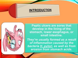
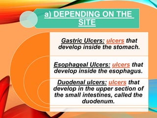
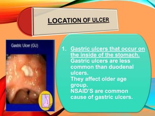
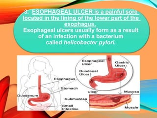
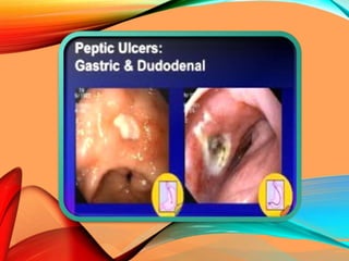
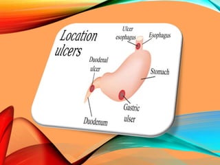
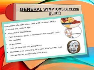
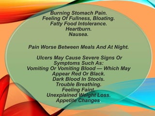
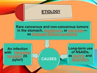
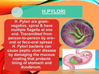
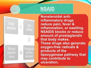
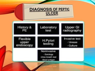
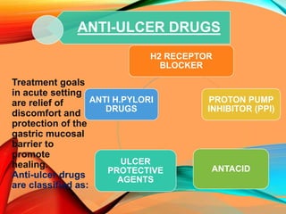
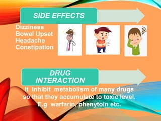
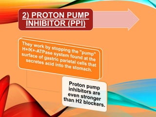
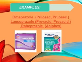
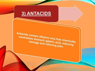
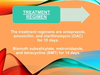
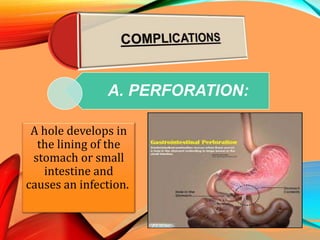
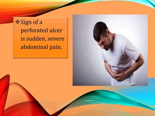
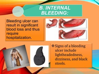
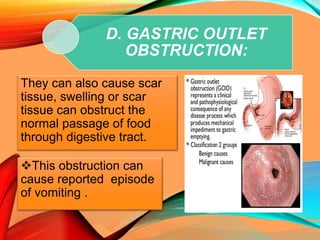
INTRODUCTION
PATHOGENESIS
GENERAL SYMPTOMS
CAUSES
LABORATORY TESTS
RISK FACTORS
TREATMENT
COMPLICATIONS
PREVENTION
Peptic ulcers are sores that
develop in the lining of the
stomach, lower esophagus, or
small intestine.
They’re usually formed as a result
of inflammation caused by the
bacteria H. pylori, as well as from
erosion from stomach acids.
A) Depending On The Site
B) Depending On The
Duration
a) DEPENDING ON THE
SITE
Gastric Ulcers: ulcers that
develop inside the stomach.
Esophageal Ulcers: ulcers that
develop inside the esophagus.
Duodenal ulcers: ulcers that
develop in the upper section of
the small intestines, called the
duodenum.
1. Gastric ulcers that occur on
the inside of the stomach.
Gastric ulcers are less
common than duodenal
ulcers.
They affect older age
group.
NSAID’S are common
cause of gastric ulcers.
2. Duodenal ulcers that occur
on the inside of the upper
portion of small intestine
(duodenum).
All duodenal ulcers occur in
duodenal bulb. This part of
duodenum is in the direct
path of the acid contents of
stomach.
They tend to occur in
younger patients and more
common in men than in
women.
3. ESOPHAGEAL ULCER is a painful sore
located in the lining of the lower part of the
esophagus.
Esophageal ulcers usually form as a result
of an infection with a bacterium
called helicobacter pylori.
Although the prevalence of peptic
ulcer is decreasing in many
western countries it still affects
approximately 10% of all adults at
some time in their lives. The male
to female ratio for duodenal ulcers
varies from 5:1 to 2:1 while for
gastric ulcers is 2:1 or less.
ACUTE PEPTIC ULCER
CHRONIC PEPTIC ULCER
b) DEPENDING ON THE
DURATION:
DIFFERENCE BETWEEN ACUTE
AND CHRONIC PUD:
Burning Stomach Pain.
Feeling Of Fullness, Bloating.
Fatty Food Intolerance.
Heartburn.
Nausea.
Pain Worse Between Meals And At Night.
Ulcers May Cause Severe Signs Or
Symptoms Such As:
Vomiting Or Vomiting Blood — Which May
Appear Red Or Black.
Dark Blood In Stools.
Trouble Breathing.
Feeling Faint.
Unexplained Weight Loss.
Appetite Changes.
CAUSES
Rare cancerous and non-cancerous tumors
in the stomach, duodenum, or pancreas—
as Zollinger-Ellison syndrome
An infection
with Helicobac
ter pylori (H.
pylori)
Long-term use
of NSAIDs,
as aspirin and
ibuprofen.
H.PYLORI
H. Pylori are gram-
negative, spiral & have
multiple flagella at one
end. Transmitted from
person-to-person by oro–
oral or feco-oral spread .
H. Pylori bacteria can
cause peptic ulcer disease
by damaging the mucous
coating that protects
lining of stomach and
duodenum.
NSAID
Nonsteroidal anti-
inflammatory drugs
reduce pain, fever &
inflammation, or swelling.
NSAIDS blocks or reduce
amount of prostaglandin
that body makes.
These drugs also generate
oxygen-free radicals &
products of the
lipoxygenase pathway that
may contribute to
ulceration.
ZOLLINGER ELLISON
SYNDROME
Zollinger-Ellison
syndrome is a rare
disorder.
The tumors release large
amounts of gastrin, a
hormone that causes
your stomach to produce
large amounts of acid.
The extra acid causes
peptic ulcers to form in
your duodenum and in
the upper intestine.
To help diagnose a peptic
ulcer, your doctor will ask you
questions about your medical
history, symptoms, and
medicines you take.
Be sure to mention medicines
that you take without a
prescription, especially
NSAIDS such as
ASPIRIN (bayer aspirin)
IBUPROFEN (motrin, advil)
MEDICAL HISTORY
If you have a (H. Pylori) infection,
your doctor will order these
tests:
1. BLOOD TEST:
A blood test involves drawing a
sample of your blood at lab.
A health care professional tests
the blood sample to see if the
results fall within the normal
range for different disorders or
infections.
2. UREA BREATHE TEST:
For a urea breath test, you will
drink a special liquid that
contains urea,.
If H. pylori are present, the
bacteria will change this waste
product into carbon dioxide—a
harmless gas.
If your breath sample has
higher levels of carbon dioxide
than normal, you have H.
pylori in your stomach or small
intestine.
3. STOOL TEST:
Doctors use a stool test to study a
sample of your stool.
A doctor will give you a container for
catching and storing your stool at home.
You return the sample to the doctor or a
commercial facility, who then sends it to
a lab for analysis. Stool tests can show
the presence of H. Pylori.
4. UPPER GI ENDOSCOPY:
More reliable method and visual diagnosis.
An intravenous (IV) needle will be placed in
your arm to provide a sedative. Sedatives
help you stay relaxed and comfortable
during the procedure.
The doctor may perform a biopsy with the
endoscope by taking a small piece of tissue
from the lining of your esophagus.
5. UPPER GI SERIES:
During the procedure, you’ll stand or sit
in front of an x-ray machine and drink
barium, a chalky liquid. Barium coats
your esophagus, stomach, and small
intestine so your doctor can see the
shapes of these organs more clearly on
x-rays.
6. H.PYLORI TESTING:
Invasive tests using an
endoscopy procedure are less
frequently performed than
non-invasive tests as they
require a tissue biopsy
collection. Tests include:
Histology – examination of
tissue under a microscope.
Rapid urease testing – detects
urease, an enzyme produced
by H. Pylori which lead to
change a ph.
Culture – growing H.
Pylori in/on a nutrient
solution.
1) H2 RECEPTOR
BLOCKERS
Treatment goals
in acute setting
are relief of
discomfort and
protection of the
gastric mucosal
barrier to
promote
healing.
Anti-ulcer drugs
are classified as:
ANTI-ULCER DRUGS
H2 RECEPTOR
BLOCKER
PROTON PUMP
INHIBITOR (PPI)
ANTACID
ULCER
PROTECTIVE
AGENTS
ANTI H.PYLORI
DRUGS
Dizziness
Bowel Upset
Headache
Constipation
SIDE EFFECTS
DRUG
INTERACTION
It Inhibit metabolism of many drugs
so that they accumulate to toxic level.
E.g warfarin, phenytoin etc.
Cimetidine (tagamet)
Ranitidine (zantac)
Famotidine (pepcid)
EXAMPLES
2) PROTON PUMP
INHIBITOR (PPI)
Omeprazole (Prilosec, Prilosec )
Lansoprazole (Prevacid, Prevacid )
Rabeprazole (Aciphex)
EXAMPLES:
3) ANTACIDS
Constipation (caused by
aluminum-containing
antacids).
Diarrhea (caused by
magnesium-containing
antacids).
SIDE EFFECTS
DRUG
INTERACTION
Certain medications may cause complications
when taken with antacids or acid reducers,
particularly those that require an acidic
environment in the stomach for absorption
like aspirin.
Alka-seltzer
Milk Of Magnesia
Amphojel
EXAMPLES:
4) ULCER
PROTECTIVE
DRUGS
.
DRUG
INTERACTION:
Activated by acid,Don't take with H2 blockers or
antacids, which reduce the acidic environment
required for activation of these drugs.
Sucralfate (Carafate)
Misoprostol (Cytotec)
EXAMPLES:
5) ANTI H. PYLORI
DRUGS
The treatment regimens are omeprazole,
amoxicillin, and clarithromycin (OAC)
for 10 days.
Bismuth subsalicylate, metronidazole,
and tetracycline (BMT) for 14 days.
TREATMENT
REGIMEN
A. PERFORATION:
A hole develops in
the lining of the
stomach or small
intestine and
causes an infection.
Sign of a
perforated ulcer
is sudden, severe
abdominal pain.
B. INTERNAL
BLEEDING:
Bleeding ulcer can
result in significant
blood loss and thus
require
hospitalization.
Signs of a bleeding
ulcer include
lightheadedness,
dizziness, and black
stools.
C. SCAR TISSUE:
This is thick tissue that
develops after an injury.
This tissue makes it
difficult for food to pass
through your digestive
tract.
Signs of scar tissue
include vomiting
and weight loss.
D. GASTRIC OUTLET
OBSTRUCTION:
They can also cause scar
tissue, swelling or scar
tissue can obstruct the
normal passage of food
through digestive tract.
This obstruction can
cause reported episode
of vomiting .
AVOID
TOBACCO
PRODUCTS
AVOID
ALCOHOL
USE
CAUTION
WITH
ASPIRIN
AND/OR
NSAIDS
WASH
HANDS
REGULARL
Y
To reduce risk of
developing a peptic ulcer,
Peptic Ulcer

Peptic Ulcer

  • 2.
  • 4.
    Peptic ulcers aresores that develop in the lining of the stomach, lower esophagus, or small intestine. They’re usually formed as a result of inflammation caused by the bacteria H. pylori, as well as from erosion from stomach acids.
  • 5.
    A) Depending OnThe Site B) Depending On The Duration
  • 6.
    a) DEPENDING ONTHE SITE Gastric Ulcers: ulcers that develop inside the stomach. Esophageal Ulcers: ulcers that develop inside the esophagus. Duodenal ulcers: ulcers that develop in the upper section of the small intestines, called the duodenum.
  • 7.
    1. Gastric ulcersthat occur on the inside of the stomach. Gastric ulcers are less common than duodenal ulcers. They affect older age group. NSAID’S are common cause of gastric ulcers.
  • 8.
    2. Duodenal ulcersthat occur on the inside of the upper portion of small intestine (duodenum). All duodenal ulcers occur in duodenal bulb. This part of duodenum is in the direct path of the acid contents of stomach. They tend to occur in younger patients and more common in men than in women.
  • 9.
    3. ESOPHAGEAL ULCERis a painful sore located in the lining of the lower part of the esophagus. Esophageal ulcers usually form as a result of an infection with a bacterium called helicobacter pylori.
  • 12.
    Although the prevalenceof peptic ulcer is decreasing in many western countries it still affects approximately 10% of all adults at some time in their lives. The male to female ratio for duodenal ulcers varies from 5:1 to 2:1 while for gastric ulcers is 2:1 or less.
  • 13.
    ACUTE PEPTIC ULCER CHRONICPEPTIC ULCER b) DEPENDING ON THE DURATION:
  • 14.
  • 17.
    Burning Stomach Pain. FeelingOf Fullness, Bloating. Fatty Food Intolerance. Heartburn. Nausea. Pain Worse Between Meals And At Night. Ulcers May Cause Severe Signs Or Symptoms Such As: Vomiting Or Vomiting Blood — Which May Appear Red Or Black. Dark Blood In Stools. Trouble Breathing. Feeling Faint. Unexplained Weight Loss. Appetite Changes.
  • 18.
    CAUSES Rare cancerous andnon-cancerous tumors in the stomach, duodenum, or pancreas— as Zollinger-Ellison syndrome An infection with Helicobac ter pylori (H. pylori) Long-term use of NSAIDs, as aspirin and ibuprofen.
  • 19.
    H.PYLORI H. Pylori aregram- negative, spiral & have multiple flagella at one end. Transmitted from person-to-person by oro– oral or feco-oral spread . H. Pylori bacteria can cause peptic ulcer disease by damaging the mucous coating that protects lining of stomach and duodenum.
  • 20.
    NSAID Nonsteroidal anti- inflammatory drugs reducepain, fever & inflammation, or swelling. NSAIDS blocks or reduce amount of prostaglandin that body makes. These drugs also generate oxygen-free radicals & products of the lipoxygenase pathway that may contribute to ulceration.
  • 21.
    ZOLLINGER ELLISON SYNDROME Zollinger-Ellison syndrome isa rare disorder. The tumors release large amounts of gastrin, a hormone that causes your stomach to produce large amounts of acid. The extra acid causes peptic ulcers to form in your duodenum and in the upper intestine.
  • 23.
    To help diagnosea peptic ulcer, your doctor will ask you questions about your medical history, symptoms, and medicines you take. Be sure to mention medicines that you take without a prescription, especially NSAIDS such as ASPIRIN (bayer aspirin) IBUPROFEN (motrin, advil) MEDICAL HISTORY
  • 24.
    If you havea (H. Pylori) infection, your doctor will order these tests: 1. BLOOD TEST: A blood test involves drawing a sample of your blood at lab. A health care professional tests the blood sample to see if the results fall within the normal range for different disorders or infections.
  • 25.
    2. UREA BREATHETEST: For a urea breath test, you will drink a special liquid that contains urea,. If H. pylori are present, the bacteria will change this waste product into carbon dioxide—a harmless gas. If your breath sample has higher levels of carbon dioxide than normal, you have H. pylori in your stomach or small intestine.
  • 26.
    3. STOOL TEST: Doctorsuse a stool test to study a sample of your stool. A doctor will give you a container for catching and storing your stool at home. You return the sample to the doctor or a commercial facility, who then sends it to a lab for analysis. Stool tests can show the presence of H. Pylori.
  • 27.
    4. UPPER GIENDOSCOPY: More reliable method and visual diagnosis. An intravenous (IV) needle will be placed in your arm to provide a sedative. Sedatives help you stay relaxed and comfortable during the procedure. The doctor may perform a biopsy with the endoscope by taking a small piece of tissue from the lining of your esophagus.
  • 28.
    5. UPPER GISERIES: During the procedure, you’ll stand or sit in front of an x-ray machine and drink barium, a chalky liquid. Barium coats your esophagus, stomach, and small intestine so your doctor can see the shapes of these organs more clearly on x-rays.
  • 29.
    6. H.PYLORI TESTING: Invasivetests using an endoscopy procedure are less frequently performed than non-invasive tests as they require a tissue biopsy collection. Tests include: Histology – examination of tissue under a microscope. Rapid urease testing – detects urease, an enzyme produced by H. Pylori which lead to change a ph. Culture – growing H. Pylori in/on a nutrient solution.
  • 31.
  • 33.
    Treatment goals in acutesetting are relief of discomfort and protection of the gastric mucosal barrier to promote healing. Anti-ulcer drugs are classified as: ANTI-ULCER DRUGS H2 RECEPTOR BLOCKER PROTON PUMP INHIBITOR (PPI) ANTACID ULCER PROTECTIVE AGENTS ANTI H.PYLORI DRUGS
  • 34.
    Dizziness Bowel Upset Headache Constipation SIDE EFFECTS DRUG INTERACTION ItInhibit metabolism of many drugs so that they accumulate to toxic level. E.g warfarin, phenytoin etc.
  • 35.
  • 36.
  • 37.
    Omeprazole (Prilosec, Prilosec) Lansoprazole (Prevacid, Prevacid ) Rabeprazole (Aciphex) EXAMPLES:
  • 38.
  • 39.
    Constipation (caused by aluminum-containing antacids). Diarrhea(caused by magnesium-containing antacids). SIDE EFFECTS DRUG INTERACTION Certain medications may cause complications when taken with antacids or acid reducers, particularly those that require an acidic environment in the stomach for absorption like aspirin.
  • 40.
  • 41.
  • 42.
    . DRUG INTERACTION: Activated by acid,Don'ttake with H2 blockers or antacids, which reduce the acidic environment required for activation of these drugs.
  • 43.
  • 44.
    5) ANTI H.PYLORI DRUGS
  • 45.
    The treatment regimensare omeprazole, amoxicillin, and clarithromycin (OAC) for 10 days. Bismuth subsalicylate, metronidazole, and tetracycline (BMT) for 14 days. TREATMENT REGIMEN
  • 46.
    A. PERFORATION: A holedevelops in the lining of the stomach or small intestine and causes an infection.
  • 47.
    Sign of a perforatedulcer is sudden, severe abdominal pain.
  • 48.
    B. INTERNAL BLEEDING: Bleeding ulcercan result in significant blood loss and thus require hospitalization. Signs of a bleeding ulcer include lightheadedness, dizziness, and black stools.
  • 49.
    C. SCAR TISSUE: Thisis thick tissue that develops after an injury. This tissue makes it difficult for food to pass through your digestive tract. Signs of scar tissue include vomiting and weight loss.
  • 50.
    D. GASTRIC OUTLET OBSTRUCTION: Theycan also cause scar tissue, swelling or scar tissue can obstruct the normal passage of food through digestive tract. This obstruction can cause reported episode of vomiting .
  • 51.