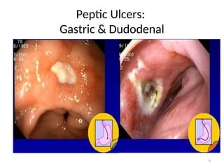
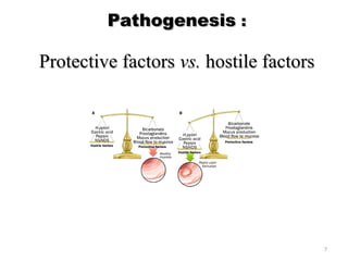
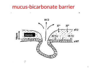
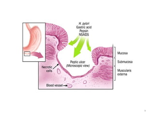
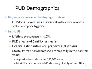
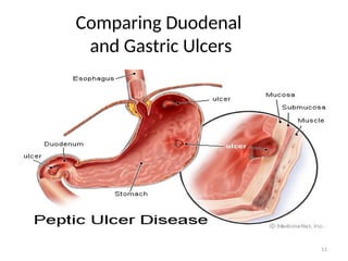
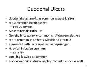
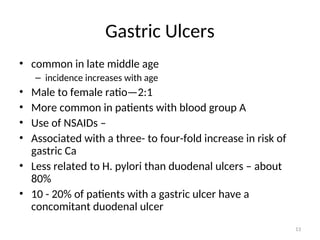
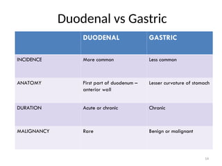
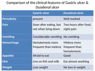
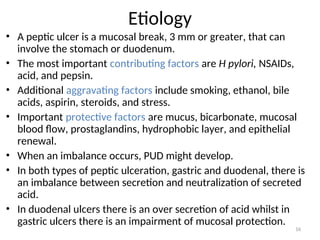
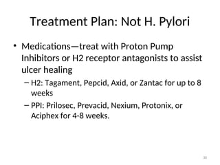
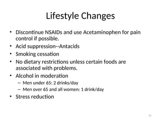
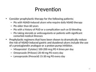
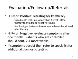
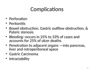
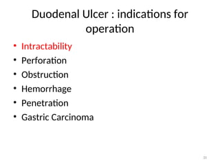
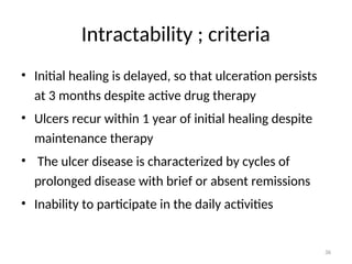
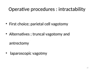
Peptic Ulcer Disease
Dr Tassew Tadesse(MD)
General Surgeon
Y12HMC,Department of surgery
1
Peptic Ulcer Disease (PUD)
Objectives:
Objectives:
1.Define the following terms: peptic ulcer, gastric ulcer, duodenal ulcer
2.Discuss the different etiologic factors of PUD
3.List the role of H.pylori as the main cause of PUD.
4.Discuss the different diagnostic methods of PUD
5.Discuss the complications of PUD
6.Discuss the treatment of PUD
2
Normal Esophagus & Stomach
3
Regulation of gastric acid secretion
Proglumide
4
Definition
Definition
• Peptic ulcer
 refers to erosion of the mucosa lining any portion of the G.I. tract.
 It is defined as : A circumscribed ulceration of the gastrointestinal mucosa
occurring in areas exposed to acid and pepsin and most often caused by
Helicobacter pylori infection.
 gastric ulcer : the ulcer that occurs in the stomach lining ,some of them
may be malignant
 duodenal ulcer : most often seen in first portion of duodenum (>95%)
5
Peptic Ulcers:
Gastric & Dudodenal
6
Pathogenesis
Pathogenesis :
:
Protective factors
Protective factors vs.
vs. hostile factors
hostile factors
7
mucus-bicarbonate barrier
mucus
8
9
PUD Demographics
• Higher prevalence in developing countries
– H. Pylori is sometimes associated with socioeconomic
status and poor hygiene
• In the US:
– Lifetime prevalence is ~10%.
– PUD affects ~4.5 million annually.
– Hospitalization rate is ~30 pts per 100,000 cases.
– Mortality rate has decreased dramatically in the past 20
years
• approximately 1 death per 100,000 cases.
• Mortality rate decreased d/t discovery of H. Pylori and PPI’s.
10
Comparing Duodenal
and Gastric Ulcers
11
Duodenal Ulcers
• duodenal sites are 4x as common as gastric sites
• most common in middle age
– peak 30-50 years
• Male to female ratio—4:1
• Genetic link: 3x more common in 1st
degree relatives
• more common in patients with blood group O
• associated with increased serum pepsinogen
• H. pylori infection common
– up to 95%
• smoking is twice as common
• Socioeconomic status may play into risk factors as well.
12
Gastric Ulcers
• common in late middle age
– incidence increases with age
• Male to female ratio—2:1
• More common in patients with blood group A
• Use of NSAIDs –
• Associated with a three- to four-fold increase in risk of
gastric Ca
• Less related to H. pylori than duodenal ulcers – about
80%
• 10 - 20% of patients with a gastric ulcer have a
concomitant duodenal ulcer
13
Duodenal vs Gastric
DUODENAL GASTRIC
INCIDENCE More common Less common
ANATOMY First part of duodenum –
anterior wall
Lesser curvature of stomach
DURATION Acute or chronic Chronic
MALIGNANCY Rare Benign or malignant
14
Comparison of the clinical features of Gastric ulcer &
Duodenal ulcer
Gastric ulcer Duodenal ulcer
Periodicity present Well marked
Pain Soon after eating, but
not when lying down
Two hours after food,
night pain
Vomiting Considerable vomiting No vomiting
Hemorrhage Hematemesis more
frequent than melena
Melena more
frequent than
hematemesis
Appetite Afraid to eat Good
Diet Live on fish and milk Eat almost anything
Weight Loss weight No loss in weight 15
Etiology
• A peptic ulcer is a mucosal break, 3 mm or greater, that can
involve the stomach or duodenum.
• The most important contributing factors are H pylori, NSAIDs,
acid, and pepsin.
• Additional aggravating factors include smoking, ethanol, bile
acids, aspirin, steroids, and stress.
• Important protective factors are mucus, bicarbonate, mucosal
blood flow, prostaglandins, hydrophobic layer, and epithelial
renewal.
• When an imbalance occurs, PUD might develop.
• In both types of peptic ulceration, gastric and duodenal, there is
an imbalance between secretion and neutralization of secreted
acid.
• In duodenal ulcers there is an over secretion of acid whilst in
gastric ulcers there is an impairment of mucosal protection.
16
• The causes of peptic ulcer disease include the following:
 Infection with the bacteria Helicobacter pylori occurs in 80 to 95% of
patients with peptic ulcer disease. H. pylori infection impairs the protective
mechanisms of the G.I. tract against low pH and digestive enzymes and leads
to ulceration of the mucosa.
 Stress — Emotional, trauma, surgical.
 Injury or death of mucus-producing cells.
 Excess acid production in the stomach. The hormone gastrin stimulates the
production of acid in the stomach; therefore, any factors that increase
gastrin production will in turn increase the production of stomach acid.
 Drugs: Chronic use of aspirins and NSAIDs, or Corticosteroids
17
ETIOLOGIC FACTORS OF PUD
18
Risk factors
• HELICOBACTER PYLORI
• Non Steroidal Anti-inflammatory Drugs
• Steroid therapy
• Smoking
• Excess alcohol intake
• Genetic factors
• Zollinger Ellison syndrome – rare syndrome caused by
gastrin-secreting tumour
• Blood group O and A
• Hyperparathyroidism
19
Helicobacter pylori:
 Most common infection in the world (20%)
Most common infection in the world (20%)
 10% of men, 4% women develop PUD
10% of men, 4% women develop PUD
 Positive in 70-100% of PUD patients.
Positive in 70-100% of PUD patients.
 H. pylori related disorders:
H. pylori related disorders:
 Chronic gastritis – 90%
Chronic gastritis – 90%
 Peptic ulcer disease – 95-100%
Peptic ulcer disease – 95-100%
 Gastric carcinoma – 70%
Gastric carcinoma – 70%
 Gastric lymphoma
Gastric lymphoma
 Reflux Esophagitis.
Reflux Esophagitis.
 Non ulcer dyspepsia
Non ulcer dyspepsia
No acid
No ulcer
OLD TESTAMENT
No HP No ulcer
NEW TESTAMENT
20
H.PYLORI
 Gram negative, Spiral bacilli
 Spirochetes
 Do not invade cells – only mucous
 Breakdown urea - ammonia
 Break down mucosal defense
 Chronic Superficial inflammation
 Stimulates Gastrin secretion
21
22
Actions of H. Pylori
23
Subjective Data
• Pain—”gnawing”, “aching”, or “burning”
– Duodenal ulcers: occurs 1-3 hours after a meal and may awaken
patient from sleep. Pain is relieved by food, antacids, or
vomiting. Pain radiates to the back.
– Gastric ulcers: food may exacerbate the pain while vomiting
relieves it.
• Nausea, vomiting, belching, dyspepsia, bloating, chest
discomfort, anorexia, hematemesis, &/or melena may
also occur.
– nausea, vomiting, & weight loss more common with Gastric
ulcers
24
Objective Data
• Epigastric tenderness
• Guaic-positive stool resulting from occult blood loss
• Succussion splash resulting from scaring or edema due to
partial or complete gastric outlet obstruction
– A succussion splash describes the sound obtained by shaking an
individual who has free fluid and air or gas in a hollow organ or
body cavity.
– Usually elicited to confirm intestinal or pyloric obstruction.
– Done by gently shaking the abdomen by holding either side of
the pelvis. A positive test occurs when a splashing noise is
heard, either with or without a stethoscope. It is not valid if the
pt has eaten or drunk fluid within the last three hours.
25
Differential Diagnosis
• Neoplasm of the stomach
• Pancreatitis
• Pancreatic cancer
• Diverticulitis
• Nonulcer dyspepsia (also called functional dyspepsia)
• Cholecystitis
• Gastritis
• GERD
• MI—not to be missed if having chest pain
• Pneumonia
26
Diagnostic Plan
• Stool for fecal occult blood
• Labs: CBC (R/O bleeding), liver function test, amylase, and
lipase.
• H. Pylori can be diagnosed by urea breath test, blood test,
stool antigen assays, & rapid urease test on a biopsy
sample.
• Upper GI Endoscopy: Any pt >50 yrs with new onset of
symptoms or those with alarm markings including anemia,
weight loss, or GI bleeding.
– Preferred diagnostic test b/c its highly sensitive for dx of ulcers and
allows for biopsy to rule out malignancy and rapid urease tests for
testing for H. Pylori.
27
• Rapid urease tests are considered the endoscopic
diagnostic test of choice.
• The presence of H pylori in gastric mucosal biopsy
specimens is detected by testing for the bacterial product
urease.
• Three kits (CLOtest, Hpfast, Pyloritek) are commercially
available, each containing a combination of a urea
substrate and a pH sensitive indicator.
• One or more gastric biopsy specimens are placed in the
rapid urease test kit. If H pylori are present, bacterial
urease converts urea to ammonia, which changes pH and
produces a color change. 28
Treatment Plan: H. Pylori
• Medications: Triple therapy for 14 days is considered the treatment of
choice.
– Proton Pump Inhibitor + clarithromycin and amoxicillin
• Omeprazole (Prilosec): 20 mg PO bid for 14 d or
Lansoprazole (Prevacid): 30 mg PO bid for 14 d or
Rabeprazole (Aciphex): 20 mg PO bid for 14 d or
Esomeprazole (Nexium): 40 mg PO qd for 14 d plus
Clarithromycin (Biaxin): 500 mg PO bid for 14 and
Amoxicillin (Amoxil): 1 g PO bid for 14 d
• Can substitute Flagyl 500 mg PO bid for 14 d if allergic to PCN
– In the setting of an active ulcer, continue qd proton pump inhibitor
therapy for additional 2 weeks.
• Goal: complete elimination of H. Pylori. Once achieved reinfection
rates are low. Compliance!
• Want a 50-60% improvement in ulcer.
29
Treatment Plan: Not H. Pylori
• Medications—treat with Proton Pump
Inhibitors or H2 receptor antagonists to assist
ulcer healing
– H2: Tagament, Pepcid, Axid, or Zantac for up to 8
weeks
– PPI: Prilosec, Prevacid, Nexium, Protonix, or
Aciphex for 4-8 weeks.
30
Lifestyle Changes
• Discontinue NSAIDs and use Acetaminophen for pain
control if possible.
• Acid suppression--Antacids
• Smoking cessation
• No dietary restrictions unless certain foods are
associated with problems.
• Alcohol in moderation
– Men under 65: 2 drinks/day
– Men over 65 and all women: 1 drink/day
• Stress reduction
31
Prevention
• Consider prophylactic therapy for the following patients:
– Pts with NSAID-induced ulcers who require daily NSAID therapy
– Pts older than 60 years
– Pts with a history of PUD or a complication such as GI bleeding
– Pts taking steroids or anticoagulants or patients with significant
comorbid medical illnesses
• Prophylactic regimens that have been shown to dramatically reduce
the risk of NSAID-induced gastric and duodenal ulcers include the use
of a prostaglandin analogue or a proton pump inhibitor.
– Misoprostol (Cytotec) 100-200 mcg PO 4 times per day
– Omeprazole (Prilosec) 20-40 mg PO every day
– Lansoprazole (Prevacid) 15-30 mg PO every day
32
Evaluation/Follow-up/Referrals
• H. Pylori Positive: retesting for tx efficacy
• Urea breath test—no sooner than 4 weeks after
therapy to avoid false negative results
• Stool antigen test—an 8 week interval must be allowed
after therapy.
• H. Pylori Negative: evaluate symptoms after
one month. Patients who are controlled
should cont. 2-4 more weeks.
• If symptoms persist then refer to specialist for
additional diagnostic testing.
33
Complications
• Perforation
• Peritonitis
• Bowel obstruction, Gastric outflow obstruction, &
Pyloric stenosis
• Bleeding--occurs in 25% to 33% of cases and
accounts for 25% of ulcer deaths.
• Penetration to adjacent organs —into pancreas,
liver and retroperitoneal space
• Gastric Carcinoma
• Intractability
34
Duodenal Ulcer : indications for
operation
• Intractability
• Perforation
• Obstruction
• Hemorrhage
• Penetration
• Gastric Carcinoma
35
Intractability ; criteria
• Initial healing is delayed, so that ulceration persists
at 3 months despite active drug therapy
• Ulcers recur within 1 year of initial healing despite
maintenance therapy
• The ulcer disease is characterized by cycles of
prolonged disease with brief or absent remissions
• Inability to participate in the daily activities
36
Operative procedures : intractability
• First choice; parietal cell vagotomy
• Alternatives ; truncal vagotomy and
antrectomy
• laparoscopic vagotmy
37
Duodenal Ulcer : indications for
operation
• Intractability
• Perforation
• Obstruction
• Hemorrhage
• Penetration
• Gastric Ca
38
Clinical features ; perforated duodenal
ulcer
• Symptoms ; sudden onset of severe epigastric
pain spreading throughout the abdomen,
variable degree of shock
• Shoulder pain
• Signs ; abdominal tenderness, guarding and
rigidity
• Plain X-ray ; peritoneal free air
39
The natural history of PUD perforation
has three phases
• Chemical pertonitis/contamination
Sudden onset of severe upper abdominal pain because of spillage
of the gastrodoudenal content(free from bacteria) into the
peritoneal cavity – diffuse or localized. There can be spillage down
the right paracolic gutter mimicking acute appendicitis. This takes
from 6 to 12 hrs.
• Intermediate stage
After 6 to 12 hrs, the patient will get spontaneous relief from the
pain b/c of the dilution of the gastrodoudenal content by the
peritoneal exudates.
• Intra-abdominal infection(peritonitis)
Usually after 12 to 24 hrs - Intra abdominal infections supervene.
The patient will have sign and symptoms of full blown peritonitis.
40
Diagnosis
• History and physical examination.
• Erect plain chest X – ray
• Sudden onset peritonitis + free gas = perforated viscus
• Sudden onset of peritonitis + no free gas + normal
serum amylase = perforated viscus
• When there no free air:- contrast study and CT
scan are valuable.
41
42
Differential diagnosis ;
perforated duodenal ulcer
• Acute cholecystitis
• Acute pancreatitis
• Intestinal obstruction
• Acute appendicitis
• Perforation of other G-I tract(other than PUD)
• Mesenteric thrombosis
43
Operative procedures : perforation
• Simple closure with omental patch
• Definitive surgery
parietal cell vagotomy and omental patch
truncal vagotomy and pyloroplasty
truncal vagotomy and antrectomy
• Eradication treatment for H.Pylori
44
45
Duodenal Ulcer : indications for
operation
• Intractability
• Perforation
• Obstruction
• Hemorrhage
• Penetration
• Gastric Ca
46
Causes of obstruction in duodenal
ulcer
• Inflammation and edema
• Fibrosis
– Tea-pot deformity
– Hour glass contractures
– Pyloric stenosis-GOO
47
Sign/symptoms
• Projectile vomiting
• Signs of dehydration and malnutrition
• Visible peristalsis
• Succession splash
48
49
50
Operative procedures : obstruction
• Truncal vagotomy and antrectomy
• Truncal vagotomy and gastrojejunostomy
• Parietal cell vagotomy with dilatation
51
52
Duodenal Ulcer : indications for
operation
• Intractability
• Perforation
• Obstruction
• Hemorrhage
• Penetration
• Gastric Ca
53
Indications for operative intervention ;
duodenal
• Most common complication(15%)
• Massive hemorrhage leading to shock
• Prolonged blood loss requiring continuing
transfusion
• Recurrent bleeding during medical therapy or
after endoscopic therapy
• Recurrent bleeding requiring hospitalization
54
Operative procedures : hemorrhage
• Truncal vagotomy and pyloroplasty with
suture ligation of bleeding vessel
• Truncal vagotomy and antrectomy including
the ulcer
• suture ligation of bleeding vessel
55
Duodenal Ulcer : indications for
operation
• Intractability
• Perforation
• Obstruction
• Hemorrhage
• Penetration
• Gastric Ca
56
57
• Complications after
gastrgejunostomy(stomach resection)
Early – dehiscence, stenosis of anastomosis, bleeding,
pancreatitis, obstructive icterus, affection of neighbour
tissues
Late - days, weeks
- early dumping syndrome
- late dumping syndrome
- ulcer in anastomosis
58
59

7.Peptic Ulcer Disease with complication 1presentation.ppt

  • 1.
    Peptic Ulcer Disease DrTassew Tadesse(MD) General Surgeon Y12HMC,Department of surgery 1
  • 2.
    Peptic Ulcer Disease(PUD) Peptic Ulcer Disease (PUD) Objectives: Objectives: 1.Define the following terms: peptic ulcer, gastric ulcer, duodenal ulcer 2.Discuss the different etiologic factors of PUD 3.List the role of H.pylori as the main cause of PUD. 4.Discuss the different diagnostic methods of PUD 5.Discuss the complications of PUD 6.Discuss the treatment of PUD 2
  • 3.
  • 4.
    Regulation of gastricacid secretion Proglumide 4
  • 5.
    Definition Definition • Peptic ulcer refers to erosion of the mucosa lining any portion of the G.I. tract.  It is defined as : A circumscribed ulceration of the gastrointestinal mucosa occurring in areas exposed to acid and pepsin and most often caused by Helicobacter pylori infection.  gastric ulcer : the ulcer that occurs in the stomach lining ,some of them may be malignant  duodenal ulcer : most often seen in first portion of duodenum (>95%) 5
  • 6.
  • 7.
    Pathogenesis Pathogenesis : : Protective factors Protectivefactors vs. vs. hostile factors hostile factors 7
  • 8.
  • 9.
  • 10.
    PUD Demographics • Higherprevalence in developing countries – H. Pylori is sometimes associated with socioeconomic status and poor hygiene • In the US: – Lifetime prevalence is ~10%. – PUD affects ~4.5 million annually. – Hospitalization rate is ~30 pts per 100,000 cases. – Mortality rate has decreased dramatically in the past 20 years • approximately 1 death per 100,000 cases. • Mortality rate decreased d/t discovery of H. Pylori and PPI’s. 10
  • 11.
  • 12.
    Duodenal Ulcers • duodenalsites are 4x as common as gastric sites • most common in middle age – peak 30-50 years • Male to female ratio—4:1 • Genetic link: 3x more common in 1st degree relatives • more common in patients with blood group O • associated with increased serum pepsinogen • H. pylori infection common – up to 95% • smoking is twice as common • Socioeconomic status may play into risk factors as well. 12
  • 13.
    Gastric Ulcers • commonin late middle age – incidence increases with age • Male to female ratio—2:1 • More common in patients with blood group A • Use of NSAIDs – • Associated with a three- to four-fold increase in risk of gastric Ca • Less related to H. pylori than duodenal ulcers – about 80% • 10 - 20% of patients with a gastric ulcer have a concomitant duodenal ulcer 13
  • 14.
    Duodenal vs Gastric DUODENALGASTRIC INCIDENCE More common Less common ANATOMY First part of duodenum – anterior wall Lesser curvature of stomach DURATION Acute or chronic Chronic MALIGNANCY Rare Benign or malignant 14
  • 15.
    Comparison of theclinical features of Gastric ulcer & Duodenal ulcer Gastric ulcer Duodenal ulcer Periodicity present Well marked Pain Soon after eating, but not when lying down Two hours after food, night pain Vomiting Considerable vomiting No vomiting Hemorrhage Hematemesis more frequent than melena Melena more frequent than hematemesis Appetite Afraid to eat Good Diet Live on fish and milk Eat almost anything Weight Loss weight No loss in weight 15
  • 16.
    Etiology • A pepticulcer is a mucosal break, 3 mm or greater, that can involve the stomach or duodenum. • The most important contributing factors are H pylori, NSAIDs, acid, and pepsin. • Additional aggravating factors include smoking, ethanol, bile acids, aspirin, steroids, and stress. • Important protective factors are mucus, bicarbonate, mucosal blood flow, prostaglandins, hydrophobic layer, and epithelial renewal. • When an imbalance occurs, PUD might develop. • In both types of peptic ulceration, gastric and duodenal, there is an imbalance between secretion and neutralization of secreted acid. • In duodenal ulcers there is an over secretion of acid whilst in gastric ulcers there is an impairment of mucosal protection. 16
  • 17.
    • The causesof peptic ulcer disease include the following:  Infection with the bacteria Helicobacter pylori occurs in 80 to 95% of patients with peptic ulcer disease. H. pylori infection impairs the protective mechanisms of the G.I. tract against low pH and digestive enzymes and leads to ulceration of the mucosa.  Stress — Emotional, trauma, surgical.  Injury or death of mucus-producing cells.  Excess acid production in the stomach. The hormone gastrin stimulates the production of acid in the stomach; therefore, any factors that increase gastrin production will in turn increase the production of stomach acid.  Drugs: Chronic use of aspirins and NSAIDs, or Corticosteroids 17
  • 18.
  • 19.
    Risk factors • HELICOBACTERPYLORI • Non Steroidal Anti-inflammatory Drugs • Steroid therapy • Smoking • Excess alcohol intake • Genetic factors • Zollinger Ellison syndrome – rare syndrome caused by gastrin-secreting tumour • Blood group O and A • Hyperparathyroidism 19
  • 20.
    Helicobacter pylori:  Mostcommon infection in the world (20%) Most common infection in the world (20%)  10% of men, 4% women develop PUD 10% of men, 4% women develop PUD  Positive in 70-100% of PUD patients. Positive in 70-100% of PUD patients.  H. pylori related disorders: H. pylori related disorders:  Chronic gastritis – 90% Chronic gastritis – 90%  Peptic ulcer disease – 95-100% Peptic ulcer disease – 95-100%  Gastric carcinoma – 70% Gastric carcinoma – 70%  Gastric lymphoma Gastric lymphoma  Reflux Esophagitis. Reflux Esophagitis.  Non ulcer dyspepsia Non ulcer dyspepsia No acid No ulcer OLD TESTAMENT No HP No ulcer NEW TESTAMENT 20
  • 21.
    H.PYLORI  Gram negative,Spiral bacilli  Spirochetes  Do not invade cells – only mucous  Breakdown urea - ammonia  Break down mucosal defense  Chronic Superficial inflammation  Stimulates Gastrin secretion 21
  • 22.
  • 23.
    Actions of H.Pylori 23
  • 24.
    Subjective Data • Pain—”gnawing”,“aching”, or “burning” – Duodenal ulcers: occurs 1-3 hours after a meal and may awaken patient from sleep. Pain is relieved by food, antacids, or vomiting. Pain radiates to the back. – Gastric ulcers: food may exacerbate the pain while vomiting relieves it. • Nausea, vomiting, belching, dyspepsia, bloating, chest discomfort, anorexia, hematemesis, &/or melena may also occur. – nausea, vomiting, & weight loss more common with Gastric ulcers 24
  • 25.
    Objective Data • Epigastrictenderness • Guaic-positive stool resulting from occult blood loss • Succussion splash resulting from scaring or edema due to partial or complete gastric outlet obstruction – A succussion splash describes the sound obtained by shaking an individual who has free fluid and air or gas in a hollow organ or body cavity. – Usually elicited to confirm intestinal or pyloric obstruction. – Done by gently shaking the abdomen by holding either side of the pelvis. A positive test occurs when a splashing noise is heard, either with or without a stethoscope. It is not valid if the pt has eaten or drunk fluid within the last three hours. 25
  • 26.
    Differential Diagnosis • Neoplasmof the stomach • Pancreatitis • Pancreatic cancer • Diverticulitis • Nonulcer dyspepsia (also called functional dyspepsia) • Cholecystitis • Gastritis • GERD • MI—not to be missed if having chest pain • Pneumonia 26
  • 27.
    Diagnostic Plan • Stoolfor fecal occult blood • Labs: CBC (R/O bleeding), liver function test, amylase, and lipase. • H. Pylori can be diagnosed by urea breath test, blood test, stool antigen assays, & rapid urease test on a biopsy sample. • Upper GI Endoscopy: Any pt >50 yrs with new onset of symptoms or those with alarm markings including anemia, weight loss, or GI bleeding. – Preferred diagnostic test b/c its highly sensitive for dx of ulcers and allows for biopsy to rule out malignancy and rapid urease tests for testing for H. Pylori. 27
  • 28.
    • Rapid ureasetests are considered the endoscopic diagnostic test of choice. • The presence of H pylori in gastric mucosal biopsy specimens is detected by testing for the bacterial product urease. • Three kits (CLOtest, Hpfast, Pyloritek) are commercially available, each containing a combination of a urea substrate and a pH sensitive indicator. • One or more gastric biopsy specimens are placed in the rapid urease test kit. If H pylori are present, bacterial urease converts urea to ammonia, which changes pH and produces a color change. 28
  • 29.
    Treatment Plan: H.Pylori • Medications: Triple therapy for 14 days is considered the treatment of choice. – Proton Pump Inhibitor + clarithromycin and amoxicillin • Omeprazole (Prilosec): 20 mg PO bid for 14 d or Lansoprazole (Prevacid): 30 mg PO bid for 14 d or Rabeprazole (Aciphex): 20 mg PO bid for 14 d or Esomeprazole (Nexium): 40 mg PO qd for 14 d plus Clarithromycin (Biaxin): 500 mg PO bid for 14 and Amoxicillin (Amoxil): 1 g PO bid for 14 d • Can substitute Flagyl 500 mg PO bid for 14 d if allergic to PCN – In the setting of an active ulcer, continue qd proton pump inhibitor therapy for additional 2 weeks. • Goal: complete elimination of H. Pylori. Once achieved reinfection rates are low. Compliance! • Want a 50-60% improvement in ulcer. 29
  • 30.
    Treatment Plan: NotH. Pylori • Medications—treat with Proton Pump Inhibitors or H2 receptor antagonists to assist ulcer healing – H2: Tagament, Pepcid, Axid, or Zantac for up to 8 weeks – PPI: Prilosec, Prevacid, Nexium, Protonix, or Aciphex for 4-8 weeks. 30
  • 31.
    Lifestyle Changes • DiscontinueNSAIDs and use Acetaminophen for pain control if possible. • Acid suppression--Antacids • Smoking cessation • No dietary restrictions unless certain foods are associated with problems. • Alcohol in moderation – Men under 65: 2 drinks/day – Men over 65 and all women: 1 drink/day • Stress reduction 31
  • 32.
    Prevention • Consider prophylactictherapy for the following patients: – Pts with NSAID-induced ulcers who require daily NSAID therapy – Pts older than 60 years – Pts with a history of PUD or a complication such as GI bleeding – Pts taking steroids or anticoagulants or patients with significant comorbid medical illnesses • Prophylactic regimens that have been shown to dramatically reduce the risk of NSAID-induced gastric and duodenal ulcers include the use of a prostaglandin analogue or a proton pump inhibitor. – Misoprostol (Cytotec) 100-200 mcg PO 4 times per day – Omeprazole (Prilosec) 20-40 mg PO every day – Lansoprazole (Prevacid) 15-30 mg PO every day 32
  • 33.
    Evaluation/Follow-up/Referrals • H. PyloriPositive: retesting for tx efficacy • Urea breath test—no sooner than 4 weeks after therapy to avoid false negative results • Stool antigen test—an 8 week interval must be allowed after therapy. • H. Pylori Negative: evaluate symptoms after one month. Patients who are controlled should cont. 2-4 more weeks. • If symptoms persist then refer to specialist for additional diagnostic testing. 33
  • 34.
    Complications • Perforation • Peritonitis •Bowel obstruction, Gastric outflow obstruction, & Pyloric stenosis • Bleeding--occurs in 25% to 33% of cases and accounts for 25% of ulcer deaths. • Penetration to adjacent organs —into pancreas, liver and retroperitoneal space • Gastric Carcinoma • Intractability 34
  • 35.
    Duodenal Ulcer :indications for operation • Intractability • Perforation • Obstruction • Hemorrhage • Penetration • Gastric Carcinoma 35
  • 36.
    Intractability ; criteria •Initial healing is delayed, so that ulceration persists at 3 months despite active drug therapy • Ulcers recur within 1 year of initial healing despite maintenance therapy • The ulcer disease is characterized by cycles of prolonged disease with brief or absent remissions • Inability to participate in the daily activities 36
  • 37.
    Operative procedures :intractability • First choice; parietal cell vagotomy • Alternatives ; truncal vagotomy and antrectomy • laparoscopic vagotmy 37
  • 38.
    Duodenal Ulcer :indications for operation • Intractability • Perforation • Obstruction • Hemorrhage • Penetration • Gastric Ca 38
  • 39.
    Clinical features ;perforated duodenal ulcer • Symptoms ; sudden onset of severe epigastric pain spreading throughout the abdomen, variable degree of shock • Shoulder pain • Signs ; abdominal tenderness, guarding and rigidity • Plain X-ray ; peritoneal free air 39
  • 40.
    The natural historyof PUD perforation has three phases • Chemical pertonitis/contamination Sudden onset of severe upper abdominal pain because of spillage of the gastrodoudenal content(free from bacteria) into the peritoneal cavity – diffuse or localized. There can be spillage down the right paracolic gutter mimicking acute appendicitis. This takes from 6 to 12 hrs. • Intermediate stage After 6 to 12 hrs, the patient will get spontaneous relief from the pain b/c of the dilution of the gastrodoudenal content by the peritoneal exudates. • Intra-abdominal infection(peritonitis) Usually after 12 to 24 hrs - Intra abdominal infections supervene. The patient will have sign and symptoms of full blown peritonitis. 40
  • 41.
    Diagnosis • History andphysical examination. • Erect plain chest X – ray • Sudden onset peritonitis + free gas = perforated viscus • Sudden onset of peritonitis + no free gas + normal serum amylase = perforated viscus • When there no free air:- contrast study and CT scan are valuable. 41
  • 42.
  • 43.
    Differential diagnosis ; perforatedduodenal ulcer • Acute cholecystitis • Acute pancreatitis • Intestinal obstruction • Acute appendicitis • Perforation of other G-I tract(other than PUD) • Mesenteric thrombosis 43
  • 44.
    Operative procedures :perforation • Simple closure with omental patch • Definitive surgery parietal cell vagotomy and omental patch truncal vagotomy and pyloroplasty truncal vagotomy and antrectomy • Eradication treatment for H.Pylori 44
  • 45.
  • 46.
    Duodenal Ulcer :indications for operation • Intractability • Perforation • Obstruction • Hemorrhage • Penetration • Gastric Ca 46
  • 47.
    Causes of obstructionin duodenal ulcer • Inflammation and edema • Fibrosis – Tea-pot deformity – Hour glass contractures – Pyloric stenosis-GOO 47
  • 48.
    Sign/symptoms • Projectile vomiting •Signs of dehydration and malnutrition • Visible peristalsis • Succession splash 48
  • 49.
  • 50.
  • 51.
    Operative procedures :obstruction • Truncal vagotomy and antrectomy • Truncal vagotomy and gastrojejunostomy • Parietal cell vagotomy with dilatation 51
  • 52.
  • 53.
    Duodenal Ulcer :indications for operation • Intractability • Perforation • Obstruction • Hemorrhage • Penetration • Gastric Ca 53
  • 54.
    Indications for operativeintervention ; duodenal • Most common complication(15%) • Massive hemorrhage leading to shock • Prolonged blood loss requiring continuing transfusion • Recurrent bleeding during medical therapy or after endoscopic therapy • Recurrent bleeding requiring hospitalization 54
  • 55.
    Operative procedures :hemorrhage • Truncal vagotomy and pyloroplasty with suture ligation of bleeding vessel • Truncal vagotomy and antrectomy including the ulcer • suture ligation of bleeding vessel 55
  • 56.
    Duodenal Ulcer :indications for operation • Intractability • Perforation • Obstruction • Hemorrhage • Penetration • Gastric Ca 56
  • 57.
  • 58.
    • Complications after gastrgejunostomy(stomachresection) Early – dehiscence, stenosis of anastomosis, bleeding, pancreatitis, obstructive icterus, affection of neighbour tissues Late - days, weeks - early dumping syndrome - late dumping syndrome - ulcer in anastomosis 58
  • 59.