
More Related Content
What's hot
What's hot (20)
Similar to Peptic ulcer
Similar to Peptic ulcer (20)
Recently uploaded
Recently uploaded (20)
Peptic ulcer
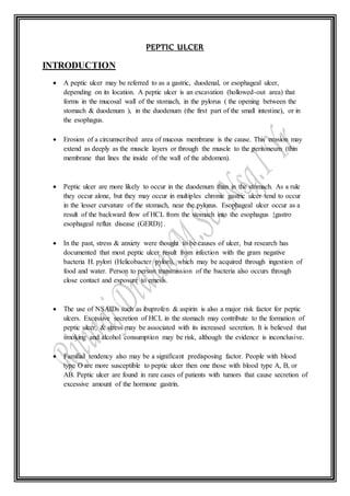
- 1. PEPTIC ULCER INTRODUCTION A peptic ulcer may be referred to as a gastric, duodenal, or esophageal ulcer, depending on its location. A peptic ulcer is an excavation (hollowed-out area) that forms in the mucosal wall of the stomach, in the pylorus ( the opening between the stomach & duodenum ), in the duodenum (the first part of the small intestine), or in the esophagus. Erosion of a circumscribed area of mucous membrane is the cause. This erosion may extend as deeply as the muscle layers or through the muscle to the peritoneum (thin membrane that lines the inside of the wall of the abdomen). Peptic ulcer are more likely to occur in the duodenum than in the stomach. As a rule they occur alone, but they may occur in multiples chronic gastric ulcer tend to occur in the lesser curvature of the stomach, near the pylorus. Esophageal ulcer occur as a result of the backward flow of HCL from the stomach into the esophagus {gastro esophageal reflux disease (GERD)}. In the past, stress & anxiety were thought to be causes of ulcer, but research has documented that most peptic ulcer result from infection with the gram negative bacteria H. pylori (Helicobacter pylori), which may be acquired through ingestion of food and water. Person to person transmission of the bacteria also occurs through close contact and exposure to emesis. The use of NSAIDs such as ibuprofen & aspirin is also a major risk factor for peptic ulcers. Excessive secretion of HCL in the stomach may contribute to the formation of peptic ulcer, & stress may be associated with its increased secretion. It is believed that smoking and alcohol consumption may be risk, although the evidence is inconclusive. Familial tendency also may be a significant predisposing factor. People with blood type O are more susceptible to peptic ulcer then one those with blood type A, B, or AB. Peptic ulcer are found in rare cases of patients with tumors that cause secretion of excessive amount of the hormone gastrin.
- 2. DEFINITION 1. According to Merriam Webster “ An ulcer in the wall of the stomach or duodenum resulting from the digestive action of the gastric juice on the mucous membrane when the latter is rendered susceptible to its action ( as from infection with the bacterium Helicobacter pylori or the chronic use of NSAIDS).” 2. “ peptic ulcer disease is characterized by ulcerations in the esophagus, stomach (gastric), or mucosal layer of either the stomach or duodenum.”small intestine (duodenal) ; approximately 80% are duodenal.” 3. “ peptic ulcer is defined as an ulcer develops when there is erosion of a portion of the 4. “ peptic ulcer disease is caused by erosion of lining cells of the stomach or duodenum; factors such as H. pylori infection and NSAIDs disrupt the normal mucosal defense, making the mucosa more susceptible to the effects of gastric acids.” 5. “An erosion of the mucous membrane of the lower esophagus, stomach or duodenum, causes in part by the corrosive action of the gastric juice.”
- 3. ANATOMY AND PHYSIOLOGY STOMACH The stomach is a muscular, J-shaped organ in the upper part of the abdomen. It is part of the digestive system, which extends from the mouth to the anus. The stomach receives food from the esophagus. As food reaches the end of the esophagus, it enters the stomach through a muscular valve called the lower esophagus sphincter. The stomach secret acid and enzyme that digest food. Ridge of muscles tissue called rogue line the stomach. The stomach muscles contract periodically, churning food to enhance digestion.
- 4. The pyloric sphincter is a muscular valve that opens to allow food to pass from the stomach to the small intestine. REGIONS OF THE STOMACH The stomach is divided into 5 regions: CARDIA :- The cardia is the first part of the stomach below the esophagus. It contains the cardiac sphincter, which is a thin ring of muscle that helps to prevent stomach contents from going back up into the esophagus. FUNDUS :- The fundus is the rounded area that lies to the left of the cardia and below the diaphragm. BODY :- The body is the largest and main part of the stomach. This is where food is mixed and starts to break down. ANTRUM :- The antrum is the lower part of the stomach. The antrum holds the broken-down food until it is ready to be released into the small intestine. It is sometimes called the pyloric antrum. PYLORUS :- The pylorus is the part of the stomach that connects to the small intestine. This region includes the pyloric sphincter, which is a thick ring of muscle that acts as a valve to control the emptying of stomach contents (chyme) into the duodenum (first part of the small intestine). The pyloric sphincter also prevents the contents of the duodenum from going back into the stomach. LAYERS OF THE STOMACHWALL (The stomach is made up of several layers of tissue) The mucosa (mucous membrane) is the inner lining of the stomach. When the stomach is empty the mucosa has a ridged appearance. These ridges (rugae) flatten out as the stomach fills with food. The next layer that covers the mucosa is the submucosa. It is made up of connective tissue that contains larger blood and lymph vessels, nerve cells and fibres. The muscularis propria (or muscularis externa) is the next layer that covers the submucosa. It is the main muscle of the stomach and is made up of 3 layers of muscle. The serosa is the fibrous membrane that covers the outside of the stomach. The serosa of the stomach is also called the visceral peritoneum.
- 5. FUNCTION The stomach has 3 main functions: temporary storage for food, which passes from the esophagus to the stomach where it is held for 2 hours or longer mixing and breakdown of food by contraction and relaxation of the muscle layers in the stomach digestion of food ETIOLOGICAL FACTOR 1. Helicobacterpylori :- A major causative factor (60% -75% of duodenum ulcer ) is chronic inflammation due to helicobacter pylori that colonizes the antral mucosa. The immune system is unable to clear the infection, despite the appearance of antibodies. Thus, the bacterium can cause a chronic active gastritis (type B gastritis). Gastrin stimulate the production of gastric acid by parietal cells. In h. pylori colonization responses to increased gastrin , the increase in acid contribute to the erosion of the mucosa and therefore ulcer formation. 2. NSAIDs :- Another major cause is the use of NSAIDs, such as ibuprofen and aspirin. The gastric mucosa protects itself from gastric acid with a layer of mucus, the secretion of which is stimulated by certain prostaglandins. NSAIDs block the function of cyclooxygenase 1 (COX-1), which is essential for the production of these prostaglandins. COX-2 selective anti-inflammatories (such as celecoxib or the since withdrawn rofecoxib) preferentially inhibit COX-2, which is less essential in the gastric mucosa, and roughly halve the risk of NSAID-related gastric ulceration. 3. Smoking :- The literature reveals a strong positive correlation between cigarette smoking and the incidence of ulcer disease, mortality, complications, recurrences and delay in healing rates. Smokers are about two times more likely to develop ulcer disease than nonsmokers. Cigarette smoking and H. pylori are co-factors for the formation of peptic ulcer disease.
- 6. Cigarette smoking may increase susceptibility, diminish the gastric mucosal defensive factors, or may provide a more favorable milieu for H. pylori infection. 4. Alcohol :- Alcohol can irritate and erode the mucous lining of your stomach, and it increases the amount of stomach acid secretion. 5. Stress :- Acute stress results in increases in pulse rate, blood pressure and anxiety, but only in those patients with duodenal ulcers did acute stress actually result in significant increases in basal acid secretion. Stress due to serious health problems such as those requiring treatment in an intensive care unit is well described as a cause of peptic ulcers, which are termed stress ulcers. While chronic life stress was once believed to be the main cause of ulcers. 6. Eat spicy foods :- Alone, these factors do not cause ulcers, but they can make them worse and more difficult to heal. 7. Others :- a. Gastrinomas (Zollinger – Ellison syndrome) , rare gastrin- secreting tumors, also cause multiple and difficult to heal ulcers. b. Having radiation treatment. c. Stomach cancer.
- 7. PATHOPHYSIOLOGY Multiple factors such as smoking, age, acquisition of infection like H.pylori etc. These factors are mainly reacts in the gastroduodenal mucosa Inability to withstand the digestive action of gastric acid (HCL) & Pepsin Increased concentration or activity of acid-pepsin and decreased resistance of the mucosa Erosion / damage of the mucosa Inability to secret enough mucous to act as a barrier against HCL Hyper secretion of gastric juice can lead gastric ulcer or duodenal ulcer and also lead to gastrinomas (islet cell tumor) in the pancreas Lead to Epigastric pain
- 8. SIGN & SYMPTOMS Symptoms of an ulcer may last for a few days, weeks, or months and may disappear only to reappear, often without an identifiable cause. Many people with ulcers have no symptoms and perforation or hemorrhage may occur in 20% to 30% of patients who had no preceding manifestations. Abdominal Pain or Burning sensation:- abdominal pain classically epigastric strongly correlated to mealtimes. In case of duodenal ulcers the pain appears about 3 hours after taking a meal. In addition, approximately 50% to 80% of patients with duodenal ulcers awake with pain during the night, whereas 30% to 40% of patients with gastric ulcers voice this type of complaint. Pyrosis (heartburn) :- pyrosis is a burning sensation in the stomach and esophagus that moves up to the mouth. It is often accompanied by sour eructation (burping), which is common when patient’s stomach is empty. Nausea and vomiting :- Although vomiting is rare in uncomplicated peptic ulcer, it may be a symptom of a complication of an ulcer. It results from gastric outlet obstruction, caused by either muscular spasm of the pylorus or mechanical obstruction from scarring or acute swelling of the inflamed mucous membrane adjacent to the ulcer. Vomiting may or may not be preceded by nausea usually, it follows a bout of severe pain and bloating, which is relieved by vomiting. Emesis may contain undigested food eaten many hours earlier. Hematemesis (vomiting of blood) :- this can occur due to bleeding directly from a gastric ulcer or from damage to the esophagus from severe or continuing vomiting. Malena :- Malena refers to the dark black, tarry feces that are associated with upper gastrointestinal bleeding. The black color and characteristic strong order are caused due to presence of oxidizing iron from hemoglobin being altered by digestive enzymes and intestinal bacteria. Constipationor Diarrhea :- constipation or diarrhea may occur, probably as a result of diet and medications. Rarely, an ulcer can lead to a gastric or duodenal perforation which leads to acute peritonitis, extreme stabbing pain and requires immediate surgery.
- 9. Others :- Bloating and abdominal fullness Loss of appetite and weight loss Trouble breathing Fatigue Discomfort DIAGNOSTIC EVALUATION 1. History taking :- obtaining a medical history, especially for peptic ulcer disease, H. pylori infection, ingestion of non- steroidal anti-inflammatory drugs or smoking is essential in making the correct diagnosis. Gastric & duodenal ulcer usually cannot be differentiated based on history alone, although some findings may be suggestive. 2. Physicalexamination :- In PUD patient appears in severe stress due to duodenal pain. Common physical examination findings of peptic ulcer disease include :- a. Appearance of the patient :- patients usually appear in severe distress due to severe duodenal pain. b. Vital sign :- patient has normal vital sign in initial stages of peptic ulcer. :- In peptic ulcer perforation vital sign may include – - Tachycardia with regular pulse - Weak pulse - Low blood pressure - High grade fever is present at later stage of peptic ulcer perforation. c. Skin :- pallor is present in patients presenting with hematemesis & melena. d. Lungs :- Normal bilateral vesicular breath sounds. e. Heart :- S1 & S2 normal, no murmur/ rubs/ gallops.
- 10. f. Abdomen :- abdominal tenderness at the epigastrium :- perforated peptic ulcer presents with:- - Decreased bowel sounds - Abdominal rigidity - Abdominal distension - 3. Upper endoscopy :- this is a test to examine the lining of the esophagus, stomach and first part of the small intestine. - An endoscope (a thin , flexible , lighted tube with camera) is passed through the mouth & pharynx & into the esophagus. The forward – viewing scope transmits an image of the esophagus, stomach and duodenum to a monitor visible to the physician. Air may be introduced into the stomach, expanding the folds of tissue and enhancing examination of the stomach. - In addition to identifying the ulcer, its location & size, Esophagogastrduodenoscopy (EGDS) also provide an opportunity to biopsy lesions. - Endoscopic biopsy also appears the best and most accurate diagnostic method for H. pylori. Histological examination with standard hematoxylin and eosin staining provides an excellent means of diagnosis. 4. Barium test :- if the patient don’t have difficulty swallowing and have a low risk of stomach cancer, doctor may recommend an upper gastrointestinal test instead. For this procedure, patient will drink a thick liquid called barium. Then a technician will take an X-ray of patient’s stomach, esophagus and small intestine. The liquid will make it possible for doctor to view and test the ulcer. 5. Hemoglobin blood test to check for anemia . 6. Stool occult blood test to check for blood in stool.
- 11. MANAGEMENT MedicalManagement - Once the diagnosis is established , the patient is informed that the condition can be managed. - Treatment will depend on the underlying cause of the ulcer. If the test show that patient have an H. pylori infection, doctor will prescribe a combination of medication. The medication include antibiotics to help kill infections and proton pump inhibitors (PPI) to help reduce stomach acid. - If doctor determines that patient don’t have an H. pylori infection, they may recommend a prescription or over the counter PPI (such as prolosec or prevacid) for up to eight weeks to reduce stomach acid & help to ulcer heal. Pharmacologicalmanagement Currently, the most commonly used therapy for peptic ulcer is a combination of antibiotics, proton pump inhibitors and bismuth salts that suppress or eradicate H. pylori. Drug regimens for peptic ulcer disease ___ Indication Drug Regimen_______________________ 1. Ulcer healing a. H2 Receptor antagonist:- = Ranitidine 150 mg/bid or 300 mg at bedtime. = famotidine 20mg/ bid or 40mg at bedtime = Nizatidine 150mg/ bid or 300mg at bedtime b. Proton pump inhibitors:- = Omeprazole 20mg daily = pantoprazole 40mg daily = lansoprazole 30 mg daily = Esomeprazole 40mg daily 2. Initial H. pylori therapy a. First line : (antibiotic + PPI) = clarithromycin 500mg/ bid + Amoxicillin 1000mg / bid or metronidazole 500mg/ bid for 10-14 days b. Second line :- = Bismuth subsalicylate 2 tabs qid+
- 12. tetracycline 250mg qid + metronidazole 250mg qid for 14 days 3. Prophylactic therapy for NSAID ulcers = peptic ulcer healing doses of proton pump Inhibitors A . misoprostol 200mg /bid B . metronidazole 250mcg/ bid C . omeprazole 20mg/ daily D . pantoprazole 40mg/ daily Smoking Cessation:- Smoking decreases the secretion of bicarbonate from the pancreas into the duodenum, resulting in increased acidity of the duodenum. Research indicates that continued smoking my significantly inhibit ulcer repair. Therefore, the patient is encourage to stop smoking. Dietary Modification:- The intent of dietary modification for the patients with peptic ulcer is to avoid over secretion of acid and hyper motility in the GI tract. These can be minimized by avoiding extremes of temperature in food and beverages and over stimulation from the consumption of alcohol, coffee and other caffeinated beverages. In addition, an effort is made to neutralize acid by eating three regular meals a day. The patient eats foods that are tolerated and avoids those that produce pain. SurgicalManagement The introduction of antibiotics to eradicate H. pylori and of H2 blockers as treatment for ulcer greatly reduced the need for surgical intervention. However surgery is usually recommended for patient with intractable ulcers, life threatening hemorrhage, perforation or obstruction and for those with ZES (Zollinger Ellison Syndrome) that is unresponsive to medications. Surgical procedure include Vagotomy, with or without pyloroplasty and antrectomy. 1) Vagotomy :- Severing of vagus nerve. Decrease gastric acid by diminishing cholinergic stimulation to the parietal cells, making them less responsive to gastrin. May be performed via
- 13. open surgical approach, laparoscopy or thoracoscopy. May be performed to reduce gastric acid secretion. VAGOTOMY a. Truncal Vagotomy :- severs the right & left vagus nerves as they enter the stomach at the distal part of the esophagus, most commonly used to decrease acid secretion. b. Selective Vagotomy :- severs vagal innervations to the stomach but maintains innervations to the rest of the abdominal organs. c. Proximal gastric Vagotomy without drainage :- Denervates acid secretion parietal cells but preserves vagal innervations to the gastric antrum and pylorus. 2)Pyloroplasty :- Transecting nerves that stimulate acid secretion and opening the pylorus or longitudinal incision is made into the pylorus and transversely sutured closed to enlarged. The outlet and relax the muscle; usually accompanies truncal and selective vagotomies.
- 15. 3)Antrectomy :- antrectomy is removal of the pyloric (antrum) portion of the stomach with anastomosis (surgical connection) to either the duodenum (gastroduodenostomy or Billroth I) or Jejunum (Gastrojejunostomy or Billroth II). a. Gastroduodenostomy :- removal of the lower portion of the antrum of the stomach (which connect the cells that secret gastrin ) as well as a small portion of the duodenum & pylorus. The remaining segment is anastomosed to the duodenum, may be performed in conjuction with a truncal vagotomy. GASTRODUODENOSTOMY
- 16. b. Gastrojejunostomy :- Removal of lower portion of stomach with anastomosis to jejunum. Dotted lines show portion removal (antrectomy). A duodenal stump remain and is over sewn. GASTROJEJUNOSTOMY
- 17. NURSING MANAGEMENT Assessment The nurse asks the patient to describe the pain, its pattern and whether or not it occurs predictably (e.g., after meals during the night), and strategies used to relieve it (e.g., food antacids). If the patient reports a recent history of Vomiting, the nurse determines how often emesis has occurred and notes important characteristics of the vomits: Is it bright red, does it resemble coffee grounds, or is there Undigested food from previous meals? Has the patient noted any bloody or tarry stools? The nurse also asks the patient to list his or her usual food intake for a 72-hour period. Lifestyle and other habits are a concern as well. For example, does he or she smoke cigarettes? If yes, how many? Does the patient ingest alcohol? If yes, how much and how often? Are NSAIDs used? Is there a family history of ulcer disease? The nurse assesses the patient's vital signs and reports tachycardia and hypotension, which may indicate anemia from GI bleeding. The stool is tested for occult blood, and a physical examination, including palpation of the abdomen for localized tenderness, is performed. Nursing Diagnoses Based on the assessment data, nursing diagnoses may include the following: o Acute pain related to the effect of gastric acid secretion on damaged tissue o Anxiety related to an acute illness o Imbalanced nutrition: less than body requirements related to changes in diet. o Deficient Fluid Volume related to hemorrhage. o Deficient Knowledge related to physical, dietary, and pharmacologic treatment of disease. Planning and Goals The goals for the patient may include relief of pain, reduced anxiety, maintenance of nutritional requirements, maintenance of body fluid volume. knowledge about the management and prevention of ulcer recurrence, and absence of complications.
- 18. Nursing Interventions Relieving Pain- The Pain relief can be achieved with prescribed medications patient should avoid aspirin and other NSAIDs and alcohol. In addition, meals should be eaten at regularly paced intervals in a relaxed setting. Medications prescribed to treat the peptic ulcer should provide relief of ulcer-associated pain. Some patients benefit from learning relaxation techniques to help manage stress and pain. Reducing Anxiety The nurse assesses the patient's level of anxiety. Explaining diagnostic tests and administering medications as scheduled help reduce anxiety. The nurse interacts with the patient in a relaxed manner; helps identify stressors; and explains various coping techniques and relaxation methods, such as biofeedback, hypnosis, or behavior modification. The patient's family is also encouraged to participate in care and to provide emotional support. Maintaining Optimal Nutritional Status The nurse assesses the patient for malnutrition and weight loss. After recovery from an acute phase of peptic ulcer disease, the patient is advised about the importance of adhering to the medication regimen and dietary restrictions. Avoiding Fluid Volume Deficit o Monitor intake and output continuously to determine fluid volume status. o Monitor stools for blood and emesis. o Monitor hemoglobin and hematocrit and electrolytes. o Administer prescribed I.V. fluids and blood replacement as prescribed. o Insert NG tube as prescribed, and monitor the tube drainage for signs of visible and occult blood. o Administer medications through the NG tube to neutralize acidity, as prescribed. o Prepare the patient for saline lavage, as ordered. o Observe the patient for an increase in pulse and a decrease in BP (signs of shock).
- 19. o Prepare the patient for diagnostic procedure or surgery to determine or stop the source of bleeding. Educating the Patient About the Treatment Regimen o Explain all tests and procedures to increase knowledge and cooperation; minimize anxiety. o Review the health care provider's recommendations for diet, activity, medication, and treatment. o Allow time for questions, and clarify any misunderstandings. o Give the patient a chart listing medications, dosages, times of administration, and desired effects to promote compliance. COMPLICATIONS Internal bleeding :- Bleeding can occur as slow blood loss that leads to anemia or as severe blood loss that may require hospitalization or a blood transfusion. Severe blood loss may cause black or bloody vomit or black or bloody stools. Obstruction :- Peptic ulcers can lead to swelling, inflammation or scarring that may block passage of food through the digestive tract. A blockage may make you become full easily, vomit and lose weight. Perforation:- A hole develops in the lining of the stomach or small intestine and causes an infection. A sign of a perforated ulcer is sudden, severe abdominal pain.
- 20. HEALTH EDUCATION 1. Advice the patient to take proper rest and sleep at least for 8 to 10 hours. 2. Advice the patient to avoid taking spicy food, fast food, coffee & tea to prevent from recurrence of ulcer. 3. Advice the patient to avoid bad habits (if they does) like cigarette smoking and drinking alcohol which may helps to prevent from recurrence of peptic ulcer. 4. Encourage the patient for doing daily active and passive exercise to improve health status. 5. Encourage the patient for maintaining daily personal hygiene to prevent from possible infection. 6. Encourage the patient for avoid taking stress and tension related to disease. 7. Advice the patient for follow-up care.
- 21. BIBLIOGRAPHY Canadian cancer society. Anatomy & physiology of the stomach. Available from, URL : https://www.cancer.ca/en/cancer-information/cancer-type/stomach/stomach- cancer/the-stomach/?region-com. Chan FKL, Lau JYW. Peptic ulcer disease. In: Feldman M, Friedman LS, Brandt LJ, eds. Sleisenger and Fordtran's Gastrointestinal and Liver Disease. 10th ed. Philadelphia, PA: Elsevier Saunders; 2016:chap 53. Gibson C. Michael, Kaur Manpreet. Peptic ulcer physical examination. Available from, URL: https://www.wikidoc.org/index.php/peptic_ulcer_physical_examination. Hinkle Janice L., Cheever Kerry H. Medical surgical nursing. 13th ed. New Delhi : Wolters Kluwer; 2017. P. 1265-1270. Kuipers EJ, Blaser MJ. Acid peptic disease. In: Goldman L, Schafer AI, eds. Goldman's Cecil Medicine. 25th ed. Philadelphia, PA: Elsevier Saunders; 2016:chap 139. Laine L, Jensen DM. Management of patients with ulcer bleeding. Am J Gastroenterol. 2012;107(3):345-360. PMID: 22310222 www.ncbi.nlm.nih.gov/pubmed/22310222. Mayoclinic. Peptic ulcer. Available from, URL: https://www.mayoclinic.org/diseases- conditions/peptic-ulcer/symptoms-causes/syc-20354223. Morgan DR, Crowe SE. Helicobacter pylori infection. In: Feldman M, Friedman LS, Brandt LJ, eds. Sleisenger and Fordtran's Gastrointestinal and Liver Disease. 10th ed. Philadelphia, PA: Elsevier Saunders; 2016:chap 51. Najm, WI. Peptic ulcer disease. Available from, URL: https://www.en.m.wikipedia.org/wiki/peptic_ulcer_disease. Rogers, Higuera Valencia. Peptic ulcer. Available from, URL: https://www.googleweblight.com/i?u=https://www.healthline.com/health/peptic- ulcer&grqid=sZeiftDs&hl=en-IN.