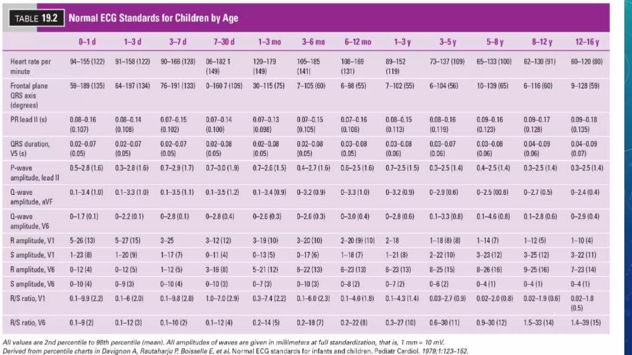
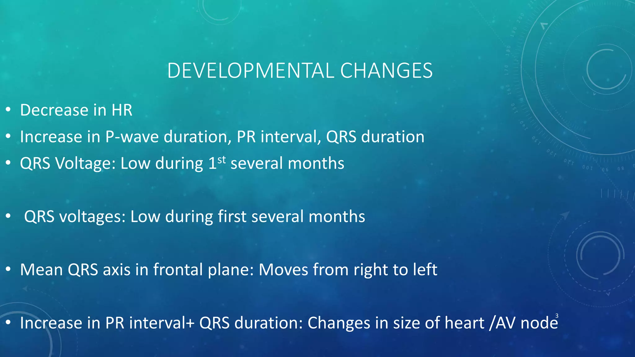
ECG IN CHILDREN:
HOW IS IT DIFFERENT FROM ADULTS?
MURTAZA KAMAL
MURTAZA.VMMC@GMAIL.COM
11/SEPT/2018
1
2
DEVELOPMENTAL CHANGES
• Decrease in HR
• Increase in P-wave duration, PR interval, QRS duration
• QRS Voltage: Low during 1st several months
• QRS voltages: Low during first several months
• Mean QRS axis in frontal plane: Moves from right to left
• Increase in PR interval+ QRS duration: Changes in size of heart /AV node
3
VENTRICULAR DOMINANCE
• Newborn: QRS potentials result from RVRV dominance
• Transition of ECG from RV dominance at birth to pattern of LV: Lags behind
hemodynamic changes
• Loss of RV dominance:
• Starts at 1 month of age
• LV dominance is well established by 1 year
4
VENTRICULAR DOMINANCE
• 1st several weeks:
• Tall R+ small S in right and anterior precordium: V3R, V4R, and V1
• Deep S+ small R in left precordium: V6 and V7
• Corresponds to clockwise vector loop in horizontal plane
• 2 months of age:
• Precordial leads progress to more adult pattern
• Deeper S waves in right and taller R waves in left leads
• Counterclockwise vector loop in horizontal plane
• 1 year: Precordial R wave progression similar to adults
5
T WAVE CHANGES
• Rapid changes of RV pressure after birth: Great effect on T wave
• 1st minutes after birth, T-wave vector: Anterior and to left i.e upright in V1+ V6
• May swing rightward in next several hours: Flattening/ inversion of T in left
lateral leads
6
T WAVE CHANGES
• Next 7 days:
• T-wave vector moves posterior+ leftward
• Inverted T in V1+ upright T in V6
• After 7- 8 years: Becomes upright again in V1
• May remain inverted throughout adolescence: Juvenile T-wave pattern
7
A FEW WORDS ABOUT PRE-TERM INFANT
• Notable for its shorter QRS duration, PR interval and QT interval
• Less RV dominance at birth than ECG of full-term infant
• Precordial voltages: Lower in 1-year-old infant who was premature
• Intrinsic myocardial differences of premature or to altered cardiac–torso
geometry: Unknown
8
THANK YOU
9

Pediatric normal ecg

  • 1.
    ECG IN CHILDREN: HOWIS IT DIFFERENT FROM ADULTS? MURTAZA KAMAL MURTAZA.VMMC@GMAIL.COM 11/SEPT/2018 1
  • 2.
  • 3.
    DEVELOPMENTAL CHANGES • Decreasein HR • Increase in P-wave duration, PR interval, QRS duration • QRS Voltage: Low during 1st several months • QRS voltages: Low during first several months • Mean QRS axis in frontal plane: Moves from right to left • Increase in PR interval+ QRS duration: Changes in size of heart /AV node 3
  • 4.
    VENTRICULAR DOMINANCE • Newborn:QRS potentials result from RVRV dominance • Transition of ECG from RV dominance at birth to pattern of LV: Lags behind hemodynamic changes • Loss of RV dominance: • Starts at 1 month of age • LV dominance is well established by 1 year 4
  • 5.
    VENTRICULAR DOMINANCE • 1stseveral weeks: • Tall R+ small S in right and anterior precordium: V3R, V4R, and V1 • Deep S+ small R in left precordium: V6 and V7 • Corresponds to clockwise vector loop in horizontal plane • 2 months of age: • Precordial leads progress to more adult pattern • Deeper S waves in right and taller R waves in left leads • Counterclockwise vector loop in horizontal plane • 1 year: Precordial R wave progression similar to adults 5
  • 6.
    T WAVE CHANGES •Rapid changes of RV pressure after birth: Great effect on T wave • 1st minutes after birth, T-wave vector: Anterior and to left i.e upright in V1+ V6 • May swing rightward in next several hours: Flattening/ inversion of T in left lateral leads 6
  • 7.
    T WAVE CHANGES •Next 7 days: • T-wave vector moves posterior+ leftward • Inverted T in V1+ upright T in V6 • After 7- 8 years: Becomes upright again in V1 • May remain inverted throughout adolescence: Juvenile T-wave pattern 7
  • 8.
    A FEW WORDSABOUT PRE-TERM INFANT • Notable for its shorter QRS duration, PR interval and QT interval • Less RV dominance at birth than ECG of full-term infant • Precordial voltages: Lower in 1-year-old infant who was premature • Intrinsic myocardial differences of premature or to altered cardiac–torso geometry: Unknown 8
  • 9.