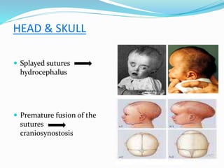
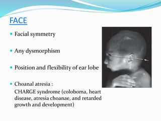
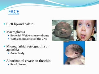
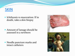
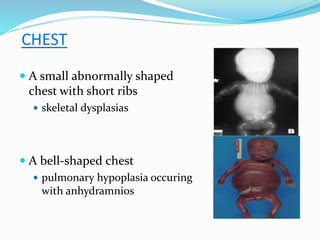
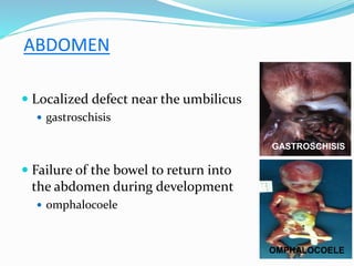
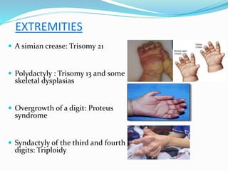
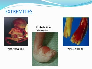
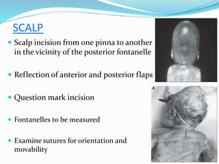
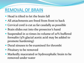
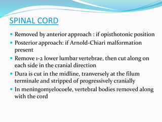
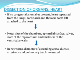
‘Oh Death Where is Thy Victory
Oh Death Where is Thy Sting.’
(I Corinthians 15:55)
PRESENTED BY : MAJ PRERNA GULERIA
GUIDE : COL V SRINIVAS
INTRODUCTION
 Pediatric age group: birth to 18 yrs of
age
 Perinatal & Neonatal
 Infant (1 yrs) to 2 yrs of age
 > 2 yrs: similar to adults
PURPOSE OF THE AUTOPSY
 Establishing the definitive cause and manner of death
 Detect deviations in growth and development
 Recognize organ and tissue pathology
 Explain clinical events and make clinicopathological
correlation
PURPOSE OF THE AUTOPSY
 The efficacy and safety of new diagnostic or
therapeutic interventions
 Identification of genetic conditions or obstetric
factors that may be of relevance to the
management of future pregnancies
 Monitor health care, provide education, direct
research and determine health care policy
PREPARATION FOR AUTOPSY
 Check the type of consent given
 Confirmation of the identity of the child
 Medical case sheet
 Consultation with clinicians
 Desirable studies
 Diagnosis considered in life
 Potential iatrogenic hazards
 Any atypical features
 Measures to prevent infection
IMAGING
Radiograph of the whole body help to
determine
 Ossification centers
 In skeletal dysplasias
 Metaphysitis of the long bones
 congenital infections such as syphilis
 Documentation of pneumothorax
 Location of lines and tubes
MRI
 Used for detecting malformations and
diseases of the central nervous system
 When consent for an autopsy is
refused, it provides a good alternative
 Potential tool in neonatal autopsy
 May replace conventional post mortem
in future
PHOTOGRAPHY
 Full body, face and facial profile photographs help
in diagnosis and can be used for consultation
 Black and white prints can be kept with the
paperwork providing for easy comparison with
reference books
 Photographs of malformations are essential
EQUIPMENT NEEDED
 Set of scales – giving
accurate weight to 1gm
 Measuring board –fixed
head and movable foot
 Autopsy instruments
 Appropriate size
 Scissors with tapered tines
 Forceps without teeth
 Small probes
EXTERNAL EXAMINATION
 Facial measurements
 Weight of the neonate
 Comparison with standard
charts available
MORPHOMETRY
MORPHOMETRY
MORPHOMETRY
 Crown rump length = 2/3 of crown heel length
 Crown rump length and head circumference do not differ by
more than 1 cm
 Foot length useful in
 Fetus of early gestational age
 Severely macerated fetus
 Anencephaly
 D&E specimens
 Facial, ear, hand and other organ measurement in cases of
suspected dysmorphology
 The infant can be classified as small, appropriate and large for
gestational age according to its weight
HEAD & SKULL
 Examination of hair and any abnormal hair patterns or
whorls
 Shape and size of skull
 Examine for abrasions, cephalhaematoma or
subaponeurotic hemorrhage
 Bulging fontanelles indicate intracranial disorder
 The presence of additional fontanelles or defects of the
skull: possibility of a chromosomal defect or Meckel–
Gruber syndrome
HEAD & SKULL
 Splayed sutures
hydrocephalus
 Premature fusion of the
sutures
craniosynostosis
FACE
 Facial symmetry
 Any dysmorphism
 Position and flexibility of ear lobe
 Choanal atresia :
CHARGE syndrome (coloboma, heart
disease, atresia choanae, and retarded
growth and development)
FACE
 Cleft lip and palate
 Macroglossia
 Beckwith Weidemann syndrome
 With abnormalities of the CNS
 Micrognathia, retrognathia or
agnathia
 Aneuploidy
 A horizontal crease on the chin
 Renal disease
SKIN
 Appearance
 Length of toe nails and finger nails
 Findings of meconium staining,
abnormal nutrition, deviations of
hydration and edema
 Skin lesions
 Multiple haemangiomas suggest Osler-
Rendu-Weber syndrome
 Leaf-shaped cafe´au lait spots suggests
tuberous sclerosis
SKIN
 Bullae, pustules or scaling
lesions
 Congenital varicella zoster
 Congenital syphillis
 Hemorrhages or blueberry
muffin
 Haematological condition
 Congenital infection
 hypoxia
SKIN
 Ichthyosis vs maceration: If in
doubt, take a skin biopsy
 Amount of lanugo should be
assessed in a newborn
 Needle puncture marks and
intact catheters
NECK
 Lateral skin webbing
 monosomy X (XO)
 multiple pterygium syndrome
 Postnuchal cystic hygroma
 XO
 Trisomy 21and 18
 A groove around the neck with
congestion of the face
 strangulation by the umbilical cord
CHEST
 A small abnormally shaped
chest with short ribs
 skeletal dysplasias
 A bell-shaped chest
 pulmonary hypoplasia occuring
with anhydramnios
CHEST
 Asymmetrical bulge
 diaphragmatic hernia
 Pneumothorax
 Size of the breast bud
 Palpable crepitus
 following difficult ventilation
ABDOMEN
 Abdominal distention due to
 ascites
 organomegaly
 gaseous distension of the bowel
 intestinal obstruction
 rarely, a tumor
 Defects of the body wall may be
related to a short umbilical cord
ABDOMEN
 Localized defect near the umbilicus
 gastroschisis
 Failure of the bowel to return into
the abdomen during development
 omphalocoele
OMPHALOCOELE
GASTROSCHISIS
GENITALIA
The external genitalia
 Descent of testes in males
 Extent of scrotal rugation
 Maturity , malformations and
ambiguity
 Vagina in females is probed
 Associated renal and anal anomalies
Male
Female
BACK
 Scoliosis
 Defects of the neural tube
 Pigmented lesions
 Abnormal tufts of hair
 Midline masses
MENINGOMYELOCOELE
EXTREMITIES
 A simian crease: Trisomy 21
 Polydactyly : Trisomy 13 and some
skeletal dysplasias
 Overgrowth of a digit: Proteus
syndrome
 Syndactyly of the third and fourth
digits: Triploidy
EXTREMITIES
Arthrogryposis
Rockerbottom
Trisomy 18
Amnion bands
INTERNAL EXAMINATION
SKIN INCISION
 Y or T- shaped incision used
 Arms of the Y to the top of the
shoulders
 Vertical incision in the midline from
xiphoid process to pubic symphysis
 Deviation around the umbilicus: to
remove it in continuity with umbilical
arteries and veins
 Girls > 5 years: incision extended only to axillary line
 With well developed breasts : initial incision to lie above
the breast tissue
 If genitourinary or anal abnormality suspected, incision
extended around the perineum to include external
genitals and anus
SKIN INCISION
BEFORE DISSECTION
 Suspected cases of pneumothorax, haemothorax, or
pleural effusion
 Look for air bubbles
 Attempt to aspirate fluid
 If present, amount aspirated measured accurately
THE ABDOMEN
 In situ examination : colour, size and relationship of organs
 Inflammation, infarction, ascites, pneumoperitoneum and
surgical wounds
 Anomalies to look for
 Malrotation
 Strictures
 Atresia
 Hernia
 Ectopic tissue
 Levels of hemidiaphragm
THE ABDOMEN
 Liver examined for symmetry
 Spleen for multiplicity
 Bladder for distention
 Stomach and gall bladder for location
 Greater omentum for transparency
 State of lymph nodes
THE ABDOMEN
 Examine kidneys, ureters, adrenals and
internal genitalia
 Look for :
 Bladder hypertrophy
 Hydronephrosis
 Renal dysplasia
 Note testicular descent
 In females, uterus, fallopian tubes and
ovaries to be identified
Lower urinary tract obstruction
THORACIC CAVITY
 Removing the chest wall
 Assessing the hemidiaphragm
 Observe position of chest tubes
 Mediastinal emphysema
 Cultures of lung tissue, pleural
fluid and heart blood
THORACIC CAVITY
 Thymus
 Atrophy : prolonged stress
 Petechial haemorrhages
 Phrenic nerve on either side
 Absence : DiGeorge syndrome
 Extent of lung distention and
pleural content
 Measure cardio-thoracic ratio
THORACIC CAVITY
 Expose great vessels by removing
pericardium and thymus
 Taussig maneuver to assess
pulmonary venous connection in
situ:
 If the heart can be lifted from the
chest without moving the lungs,
there is an anomalous
pulmonary venous connection
THORACIC CAVITY
 Evaluation of congenital anomalies : better if heart
opened in situ
 Dissection of heart and blood vessels follows the
course of blood
 Conduction system dissection : in case of arrythmias
 Ligation of arteries
 Done in older children, not required in newborns
 Right and left carotids
 Subclavian
 Common iliacs
SCALP
 Scalp incision from one pinna to another
in the vicinity of the posterior fontanelle
 Reflection of anterior and posterior flaps
 Question mark incision
 Fontanelles to be measured
 Examine sutures for orientation and
movability
OPENING OF SKULL
 Incision given along the suture
lines
 Incision kept about 1/4th inch
away from mid line to avoid
damage to falx and sinuses
 Bone flaps are reflected
downwards to expose cerebral
hemisphere (flower petal
incision)
 Inspect brain, falx and tentorium
 Falx removed along with sagittal
sinus
OPENING OF SKULL
REMOVAL OF BRAIN
 Head is tilted to let the brain fall
 All attachments are freed from front to back
 Cervical cord is cut as far caudally as possible
 Brain slides out into the prosector’s hand
 Suspended in 10 times its volume of 10% buffered
formalin (5% glacial acetic acid may be added to
promote hardening)
 Dural sinuses to be examined for thrombi
 Pituitary to be removed
 Markedly macerated or hydrocephalic brain to be
removed under water
EARS AND EYES
 Middle ears to be inspected and cultured
 Petrous temporal to be incised and ossicles and drum
inspected in situ
 Remove and process if anomalies of the ear suspected
 Remove eyes: h/o prolonged exposure to oxygen
 By removing base of anterior fossa
 Exteriorly with an ophthalmology retractor
SPINAL CORD
 Removed by anterior approach : if opisthotonic position
 Posterior approach: if Arnold-Chiari malformation
present
 Remove 1-2 lower lumbar vertebrae, then cut along on
each side in the cranial direction
 Dura is cut in the midline, tranversely at the filum
terminale and stripped of progressively cranially
 In meningomyelocoele, vertebral bodies removed along
with the cord
LIMBS
 Indicated only in few instances
 Hip dissection undertaken in severe talipes to confirm
dislocation
 Suspected congenital neuromuscular disorders
peripheral muscles should be sampled and examined
EXAMINATION OF PLACENTA
 Initially examined in unfixed state with membranes
and blood clot
 Site of rupture of sac
 Membranes are clear or cloudy
 Offensive odor &staining with meconium
 Length, site of attachment, appearance of umbilical cord
 Weight of placenta
SECTIONS FROM PLACENTA
RECOMMENDED SECTIONS
•Two sections of cord
•Membrane roll (Swiss Roll)
•Two full-thickness non-marginal normal
disc
•Areas of Abnormalities
PLACENTA
 Hypercoiling of the umbilical cord :
hypoxia
 Chorioamnionitis : most common
placental lesion associated with cerebral
palsy in term and preterm infants
 Extensive placental infarction
 ischaemic cerebral injury
 periventricular white matter necrosis in
stillbirths.
DISSECTION OF ORGANS
 Organs preferrably removed ‘en mass’
 Block of organs stretched longitudinally over a suitable
surface
 Examination of tongue
 Remnants of thyroglossal duct
 Pharynx opened first
 Inspection of tonsillar pillars, soft palate, uvula, hyoid
bone
DISSECTION OF ORGANS
 Incision proceeds caudally through the midline of
oesophagus to the level of the diaphragm close to the
oesophago-gastric junction
 Look for ectopic gastric mucosa
 Tracheo-esophageal fistula (TOF): trachea to be opened
anteriorly
 Separate oesophagus and pharynx to expose the
posterior wall of pericardium
 Interior of larynx inspected:
 Inflammation
 Ulceration
 Foreign bodies
 Level of endotracheal tube, if present
 Neck organs cut away en bloc from the main unit
 Tonsils removed with a cuff of surrounding mucosa
 Tongue with hyoid bone fixed as a whole
 Larynx, trachea and thyroid : fixed as a single unit
DISSECTION OF ORGANS
 Abdominal aorta opened: longitudinally in the midline
upto the bifurcation of the common iliac arteries
 Renal arteries, coeliac and superior mesenteric arteries
inspected
 Thoracic organs separated by cutting across IVC close
to the diaphragm
DISSECTION OF ORGANS
 If no congenital anomalies present, heart separated
from the lungs; aortic arch and thoracic aorta left
attached to the heart
 Note: sizes of the chambers, epicardial surface, valves,
state of the myocardium and thickness of the
ventricular walls
 In newborns, diameter of ascending aorta, ductus
arteriosus and pulmonary trunk measured
DISSECTION OF ORGANS: HEART
 Assess pattern of lobulation, distribution of major
bronchial branches
 Inflate the lungs with 10% buffered formalin through
the main bronchus
 Lung dissected by slicing on cutting board or opening
along pulmonary arteries
DISSECTION OF ORGANS : LUNGS
DISSECTION OF ORGANS
 IVC and renal veins opened
 Diaphragm removed with the crura in one piece
 Each adrenal removed and periadrrenal renal tissue
inspected
 Each kidney freed, hemisected in the coronal plane
and capsule removed
 Observe number and colour of pyramids, size and
shape of the pelvis
 Ureter opened from pelvis to bladder
 Bladder opened from outlet to the dome
 Inspect trigone
 Orifices
 Bladder wall thickness
DISSECTION OF ORGANS : GUT
 Remove entire intestinal tract: begin with the ligament
of Tietz and proceed distally by cutting the mesenteric
attachment
 Measure length of small and large bowel
 Bowel contents observed
 Examine serosal, muscular and mucosal surfaces,
lymphoid follicles and Peyer’s patches
DISSECTION OF ORGANS : GIT
 In suspected cases of Hirschprung’s disease, biopsy is
taken 2cm from anal verge in neonates and 3cm in
older children
 Explore biliary tree by
 Removing gall bladder from its bed
 Opening the first part of duodenum to display ampulla
of Vater
 Expressing bile by compressing the gall bladder
 Normal saline injected into the gall bladder to
demonstrate patency
DISSECTION OF ORGANS : GIT
 Duodenohepatic ligament severed to separate liver
 Examine portal and hepatic veins
 Liver sliced at 0.5 cm intervals to examine parenchyma
 Pancreas to be separated from the duodenum and
spleen, serial sectioning to be done and fixed
DISSECTION OF ORGANS : GIT
 If multiple spleens present, determine whether they lie
on each side or one side of the dorsal mesogastrium
 Linear incision of the oesophagus to be extended
along the greater curvature of the stomach
 Finally, separation of the female internal genitalia from
the urinary bladder.
DISSECTION OF ORGANS : GIT
SPECIMENS
SPECIMENS
 Specimens for Microbiology
 Heart blood, lung, liver and spleen swabs
 Aspirates from gastric, middle ear and CSF
 Viral cultures, if relevant
 Specimen for Hematology
 Hydrops foetalis: Coomb’s test
 Foetomaternal haemorrhage: Kleihauer-Betke test
 Biochemistry
 Inborn errors of metabolism
 Cytogenetic
 Chromosome analsysis can be performed on white blood cells, if
autopsy is performed within 12 hrs
 Culture of fibroblast from skin – Upto 3 days
SPECIMENS FOR HISTOLOGY
 Lungs - At least 1 block needed from each of the major
lobes
 Heart - One section is taken to include left atrium mitral
valve, left ventricle and papillary muscle
 Liver - Blocks of left and right lobes are taken
 Spleen - One block is taken
 Thymus - One block is needed
 Pancreas and umbilical cord - One block is needed
STILL BIRTHS
CLINICAL HISTORY
 Maternal age.
 Relevant medical and family history.
 Obstetric history
 History of current pregnancy:
 estimated delivery date
 antenatal infection screen, including HIV
 abnormal findings from ultrasound or other antenatal
investigations
 hypertension/bleeding/pyrexia/membrane rupture
 events leading up to intrauterine death and/or delivery
 delivery: mode, complications and use of instrumentation.
PATHOLOGY ENCOUNTERED
 Hypoxia:
 visceral petechial haemorrhages
 inhaled amniotic material
 hypoxic-ischaemic injury of brain and other internal
organs
 Growth restriction: symmetric, asymmetric
(nutritional)
 Infection:
 amniotic fluid infection (chorioamnionitis, funisitis,
pneumonia)
 haematogenous infection (including villitis)
 Malformation
 Trauma: cranial, extracranial
 Blood loss, e.g. cord, feto-maternal, internal
 Hydrops
 Maternal disease, e.g. diabetes, hypertension and pre-
eclampsia
PATHOLOGY ENCOUNTERED
 Placental and cord disease, including:
 pathology of fetoplacental and uteroplacental
circulations (e.g. fetal vessel thrombosis, placental
haemorrhage/thrombosis, placental infarction)
 features of amniotic fluid infection
(chorioamnionitis/funisitis)
 Villitis
 abnormal cord insertion, cord knots.
 Changes in the baby and placenta secondary to intrauterine
death (e.g. maceration, placental vascular involution)
PATHOLOGY ENCOUNTERED
AUTOPSY
 Measurements valuable in determining gestational age
 Foot length valuable if gestational age < 23 wks
 Macerated stillbirths : degree of maceration suggests
time since death
 <=8 hrs - Skin reddened but intact
 8 to 48-72 hrs - Skin slippage with detachment of epidermis
 >72 hrs – severe skin slippage, discoloration of internal
organs, serosanguineous effusion in the body spaces
 Spontaneous second trimester abortions :
 Infections, anomalies or abnormal karyotype
 Microscopy and culture of fetal lungs to rule out
infections
 Therapeutic abortions :
 evaluate clinical basis of termination of pregnancy
 Detect other abnormalities that may have been missed
 Assess whether death due to intrinsic abnormality or
external factors
AUTOPSY
 Lung hypoplasia :
 Compare observed with expected lung weight
 Expected ratio of lung weight with body weight
 IUGR – primary or acquired :
 Most common secondary to maternal diseases –
hypertension and diabetes mellitus
 Brain growth and development normal if cause is
maternal
 Brain growth adversely affected in primary or
constitutional IUGR
AUTOPSY
 Normally, liver weight : brain weight is < 1:2.8, in
secondary growth retardation ratio is > 1:3
 Macrosomia with visceromegaly in infants of mothers
with gestational diabetes mellitus
 Significant external dysmorphism: fibroblast culture
 Fetal skin in nonmacerated cases
 Achilles tendon or placental amnion or chorion in
macerated cases
 Minor malformations may have predictive value for
major anomaly complexes
AUTOPSY
PERINATAL DEATHS
CAUSES
 Prematurity
 Sepsis
 Complex malformations
 Acute and chronic pulmonary disease
 Infection
 Necrotising enterocolitis
 Intracranial haemorrhage
 Ischaemic encephalopathy
AUTOPSY
 Systematic sampling of baby and placenta
 Tissue sampling helpful in detection of toxoplasmosis,
CMV, HSV, syphillis, enterovirus and parvovirus
 Hydrops : severe circulatory failure
Rh factor incompatibility
arrhythmias
 Dysmorphic neonate : photographs, whole body
radiographs, fibroblast culture
 Careful examination and sampling of brain required
for delineation of acquired perinatal brain diseases
 All lines and tubes of neonatal ICU to remain in place
until their location and related complications
delineated
 Appropriate samples for neonatal sepsis :
 E. coli
 Pseudomonas aeruginosa
 Grp B streptococcus
 Staph aureus and epidermidis
 Listeria, candida, viruses
AUTOPSY
OLDER INFANTS
CAUSES
 Sequelae of perinatal diseases
 Acute infections
 Isolated malformations
 Neoplasms
 Metabolic/genetic diseases
 Accidental trauma
 Child abuse
 Sudden infant death syndrome
 History from birth
 Cultures for bacteria and viruses
 Examination for drugs or toxins
 If h/o previous unexplained crises – metabolic studies
 Retain materials for follow up studies – urine, serum,
muscle and liver
 Familial sudden deaths require biochemical evaluation
 Defects in lipid metabolism
AUTOPSY
AUTOPSY
 In suspected infections :
 Appropriate cultures to be obtained
 Responsible agent to be characterized
 In neoplasias :
 Evaluation of the extent of tumor
 Effects and complications of therapy
 Autopsy –derived tissue culture, DNA extracts or fresh
frozen tissue provide valuable resource
 In metabolic/genetic diseases :
 Determination of the extent of prior investigations and
questions to be addressed
 Expeditious collection of appropriate tissue samples
 Tissue for DNA extraction, tissue culture and
ultrastructural study
 Sudden infant death syndrome :
 Diagnosis of exclusion
 History from death scene – position of baby, bedding
materials and ambient temperature
AUTOPSY
CHILDREN > 2 yrs
CAUSES
 Trauma : a major cause
 Natural causes :
 Overwhelming infections in previously well children
 Pneumonia in children with genetic or acquired
disorders
 Unsuspected cardiac diseases – stenotic bicuspid aortic
valve, genetic cardiomyopathy
 Complications of complex cardiac , brain or intestinal
anomalies
 Neoplasms
 Diverse genetic or metabolic disorders
 Intracranial haemorrhage – vascular malformations
 Asthma
 Poorly controlled seizures
 Sudden death in child receiving medications – wrong
drug dosage and errors should be ruled out
CAUSES
AUTOPSY
 Similar to that of adults
 Thorough medical history including
 family history
 Recent medical history
 Circumstances of death
 Representative sectioning and proper sampling
REFERENCES
 Stoker J T, MacPherson T A. The Pediatric Autopsy. Pediatric Pathology;
2nd edition: 5-17.
 Bove K E and Autopsy Committee of the College of American
Pathologists. Practice guidelines for autopsy pathology – The perinatal
and pediatric autopsy. Arch Pathol Lab Med 2007; 121: 368-76.
 Gilbert-Barness E, Debich-Spicer DE. Handbook of pediatric autopsy
pathology. Humana press Totowa New Jersey, 2005.
 Cohen MC, Paley MN, Griffiths PD, Whitby EH. Less invasive autopsy:
benefits and limitations of the use of magnetic resonance imaging in the
perinatal postmortem. Pediatric Dev Pathol 2008; 11(1): 1-9.
 The Working Party on Autopsy of the Specialist Advisory Committee on
Histopathology. Guidelines on Autopsy Practice Scenario 9: Stillborn
infant (singleton). The Royal College of Pathologists 2006; 1: 1-5.
 Behrman RE, Kliegman RM, Jenson HB. Nelson Textbook of Pediatrics. W
B Saunders Company USA, 2000.
THANK YOU

Pediatric autopsy.pptx

  • 1.
    ‘Oh Death Whereis Thy Victory Oh Death Where is Thy Sting.’ (I Corinthians 15:55) PRESENTED BY : MAJ PRERNA GULERIA GUIDE : COL V SRINIVAS
  • 2.
    INTRODUCTION  Pediatric agegroup: birth to 18 yrs of age  Perinatal & Neonatal  Infant (1 yrs) to 2 yrs of age  > 2 yrs: similar to adults
  • 3.
    PURPOSE OF THEAUTOPSY  Establishing the definitive cause and manner of death  Detect deviations in growth and development  Recognize organ and tissue pathology  Explain clinical events and make clinicopathological correlation
  • 4.
    PURPOSE OF THEAUTOPSY  The efficacy and safety of new diagnostic or therapeutic interventions  Identification of genetic conditions or obstetric factors that may be of relevance to the management of future pregnancies  Monitor health care, provide education, direct research and determine health care policy
  • 5.
    PREPARATION FOR AUTOPSY Check the type of consent given  Confirmation of the identity of the child  Medical case sheet  Consultation with clinicians  Desirable studies  Diagnosis considered in life  Potential iatrogenic hazards  Any atypical features  Measures to prevent infection
  • 6.
    IMAGING Radiograph of thewhole body help to determine  Ossification centers  In skeletal dysplasias  Metaphysitis of the long bones  congenital infections such as syphilis  Documentation of pneumothorax  Location of lines and tubes
  • 7.
    MRI  Used fordetecting malformations and diseases of the central nervous system  When consent for an autopsy is refused, it provides a good alternative  Potential tool in neonatal autopsy  May replace conventional post mortem in future
  • 8.
    PHOTOGRAPHY  Full body,face and facial profile photographs help in diagnosis and can be used for consultation  Black and white prints can be kept with the paperwork providing for easy comparison with reference books  Photographs of malformations are essential
  • 9.
    EQUIPMENT NEEDED  Setof scales – giving accurate weight to 1gm  Measuring board –fixed head and movable foot  Autopsy instruments  Appropriate size  Scissors with tapered tines  Forceps without teeth  Small probes
  • 10.
  • 11.
     Facial measurements Weight of the neonate  Comparison with standard charts available MORPHOMETRY
  • 12.
  • 13.
    MORPHOMETRY  Crown rumplength = 2/3 of crown heel length  Crown rump length and head circumference do not differ by more than 1 cm  Foot length useful in  Fetus of early gestational age  Severely macerated fetus  Anencephaly  D&E specimens  Facial, ear, hand and other organ measurement in cases of suspected dysmorphology  The infant can be classified as small, appropriate and large for gestational age according to its weight
  • 14.
    HEAD & SKULL Examination of hair and any abnormal hair patterns or whorls  Shape and size of skull  Examine for abrasions, cephalhaematoma or subaponeurotic hemorrhage  Bulging fontanelles indicate intracranial disorder  The presence of additional fontanelles or defects of the skull: possibility of a chromosomal defect or Meckel– Gruber syndrome
  • 15.
    HEAD & SKULL Splayed sutures hydrocephalus  Premature fusion of the sutures craniosynostosis
  • 16.
    FACE  Facial symmetry Any dysmorphism  Position and flexibility of ear lobe  Choanal atresia : CHARGE syndrome (coloboma, heart disease, atresia choanae, and retarded growth and development)
  • 17.
    FACE  Cleft lipand palate  Macroglossia  Beckwith Weidemann syndrome  With abnormalities of the CNS  Micrognathia, retrognathia or agnathia  Aneuploidy  A horizontal crease on the chin  Renal disease
  • 18.
    SKIN  Appearance  Lengthof toe nails and finger nails  Findings of meconium staining, abnormal nutrition, deviations of hydration and edema  Skin lesions  Multiple haemangiomas suggest Osler- Rendu-Weber syndrome  Leaf-shaped cafe´au lait spots suggests tuberous sclerosis
  • 19.
    SKIN  Bullae, pustulesor scaling lesions  Congenital varicella zoster  Congenital syphillis  Hemorrhages or blueberry muffin  Haematological condition  Congenital infection  hypoxia
  • 20.
    SKIN  Ichthyosis vsmaceration: If in doubt, take a skin biopsy  Amount of lanugo should be assessed in a newborn  Needle puncture marks and intact catheters
  • 21.
    NECK  Lateral skinwebbing  monosomy X (XO)  multiple pterygium syndrome  Postnuchal cystic hygroma  XO  Trisomy 21and 18  A groove around the neck with congestion of the face  strangulation by the umbilical cord
  • 22.
    CHEST  A smallabnormally shaped chest with short ribs  skeletal dysplasias  A bell-shaped chest  pulmonary hypoplasia occuring with anhydramnios
  • 23.
    CHEST  Asymmetrical bulge diaphragmatic hernia  Pneumothorax  Size of the breast bud  Palpable crepitus  following difficult ventilation
  • 24.
    ABDOMEN  Abdominal distentiondue to  ascites  organomegaly  gaseous distension of the bowel  intestinal obstruction  rarely, a tumor  Defects of the body wall may be related to a short umbilical cord
  • 25.
    ABDOMEN  Localized defectnear the umbilicus  gastroschisis  Failure of the bowel to return into the abdomen during development  omphalocoele OMPHALOCOELE GASTROSCHISIS
  • 26.
    GENITALIA The external genitalia Descent of testes in males  Extent of scrotal rugation  Maturity , malformations and ambiguity  Vagina in females is probed  Associated renal and anal anomalies Male Female
  • 27.
    BACK  Scoliosis  Defectsof the neural tube  Pigmented lesions  Abnormal tufts of hair  Midline masses MENINGOMYELOCOELE
  • 28.
    EXTREMITIES  A simiancrease: Trisomy 21  Polydactyly : Trisomy 13 and some skeletal dysplasias  Overgrowth of a digit: Proteus syndrome  Syndactyly of the third and fourth digits: Triploidy
  • 29.
  • 30.
  • 31.
    SKIN INCISION  Yor T- shaped incision used  Arms of the Y to the top of the shoulders  Vertical incision in the midline from xiphoid process to pubic symphysis  Deviation around the umbilicus: to remove it in continuity with umbilical arteries and veins
  • 32.
     Girls >5 years: incision extended only to axillary line  With well developed breasts : initial incision to lie above the breast tissue  If genitourinary or anal abnormality suspected, incision extended around the perineum to include external genitals and anus SKIN INCISION
  • 34.
    BEFORE DISSECTION  Suspectedcases of pneumothorax, haemothorax, or pleural effusion  Look for air bubbles  Attempt to aspirate fluid  If present, amount aspirated measured accurately
  • 35.
    THE ABDOMEN  Insitu examination : colour, size and relationship of organs  Inflammation, infarction, ascites, pneumoperitoneum and surgical wounds  Anomalies to look for  Malrotation  Strictures  Atresia  Hernia  Ectopic tissue  Levels of hemidiaphragm
  • 36.
    THE ABDOMEN  Liverexamined for symmetry  Spleen for multiplicity  Bladder for distention  Stomach and gall bladder for location  Greater omentum for transparency  State of lymph nodes
  • 37.
    THE ABDOMEN  Examinekidneys, ureters, adrenals and internal genitalia  Look for :  Bladder hypertrophy  Hydronephrosis  Renal dysplasia  Note testicular descent  In females, uterus, fallopian tubes and ovaries to be identified Lower urinary tract obstruction
  • 38.
    THORACIC CAVITY  Removingthe chest wall  Assessing the hemidiaphragm  Observe position of chest tubes  Mediastinal emphysema  Cultures of lung tissue, pleural fluid and heart blood
  • 39.
    THORACIC CAVITY  Thymus Atrophy : prolonged stress  Petechial haemorrhages  Phrenic nerve on either side  Absence : DiGeorge syndrome  Extent of lung distention and pleural content  Measure cardio-thoracic ratio
  • 40.
    THORACIC CAVITY  Exposegreat vessels by removing pericardium and thymus  Taussig maneuver to assess pulmonary venous connection in situ:  If the heart can be lifted from the chest without moving the lungs, there is an anomalous pulmonary venous connection
  • 41.
    THORACIC CAVITY  Evaluationof congenital anomalies : better if heart opened in situ  Dissection of heart and blood vessels follows the course of blood  Conduction system dissection : in case of arrythmias  Ligation of arteries  Done in older children, not required in newborns  Right and left carotids  Subclavian  Common iliacs
  • 42.
    SCALP  Scalp incisionfrom one pinna to another in the vicinity of the posterior fontanelle  Reflection of anterior and posterior flaps  Question mark incision  Fontanelles to be measured  Examine sutures for orientation and movability
  • 43.
    OPENING OF SKULL Incision given along the suture lines  Incision kept about 1/4th inch away from mid line to avoid damage to falx and sinuses  Bone flaps are reflected downwards to expose cerebral hemisphere (flower petal incision)  Inspect brain, falx and tentorium  Falx removed along with sagittal sinus
  • 44.
  • 45.
    REMOVAL OF BRAIN Head is tilted to let the brain fall  All attachments are freed from front to back  Cervical cord is cut as far caudally as possible  Brain slides out into the prosector’s hand  Suspended in 10 times its volume of 10% buffered formalin (5% glacial acetic acid may be added to promote hardening)  Dural sinuses to be examined for thrombi  Pituitary to be removed  Markedly macerated or hydrocephalic brain to be removed under water
  • 46.
    EARS AND EYES Middle ears to be inspected and cultured  Petrous temporal to be incised and ossicles and drum inspected in situ  Remove and process if anomalies of the ear suspected  Remove eyes: h/o prolonged exposure to oxygen  By removing base of anterior fossa  Exteriorly with an ophthalmology retractor
  • 47.
    SPINAL CORD  Removedby anterior approach : if opisthotonic position  Posterior approach: if Arnold-Chiari malformation present  Remove 1-2 lower lumbar vertebrae, then cut along on each side in the cranial direction  Dura is cut in the midline, tranversely at the filum terminale and stripped of progressively cranially  In meningomyelocoele, vertebral bodies removed along with the cord
  • 48.
    LIMBS  Indicated onlyin few instances  Hip dissection undertaken in severe talipes to confirm dislocation  Suspected congenital neuromuscular disorders peripheral muscles should be sampled and examined
  • 49.
    EXAMINATION OF PLACENTA Initially examined in unfixed state with membranes and blood clot  Site of rupture of sac  Membranes are clear or cloudy  Offensive odor &staining with meconium  Length, site of attachment, appearance of umbilical cord  Weight of placenta
  • 50.
    SECTIONS FROM PLACENTA RECOMMENDEDSECTIONS •Two sections of cord •Membrane roll (Swiss Roll) •Two full-thickness non-marginal normal disc •Areas of Abnormalities
  • 51.
    PLACENTA  Hypercoiling ofthe umbilical cord : hypoxia  Chorioamnionitis : most common placental lesion associated with cerebral palsy in term and preterm infants  Extensive placental infarction  ischaemic cerebral injury  periventricular white matter necrosis in stillbirths.
  • 53.
  • 54.
    DISSECTION OF ORGANS Organs preferrably removed ‘en mass’  Block of organs stretched longitudinally over a suitable surface  Examination of tongue  Remnants of thyroglossal duct  Pharynx opened first  Inspection of tonsillar pillars, soft palate, uvula, hyoid bone
  • 55.
    DISSECTION OF ORGANS Incision proceeds caudally through the midline of oesophagus to the level of the diaphragm close to the oesophago-gastric junction  Look for ectopic gastric mucosa  Tracheo-esophageal fistula (TOF): trachea to be opened anteriorly  Separate oesophagus and pharynx to expose the posterior wall of pericardium
  • 56.
     Interior oflarynx inspected:  Inflammation  Ulceration  Foreign bodies  Level of endotracheal tube, if present  Neck organs cut away en bloc from the main unit  Tonsils removed with a cuff of surrounding mucosa  Tongue with hyoid bone fixed as a whole  Larynx, trachea and thyroid : fixed as a single unit DISSECTION OF ORGANS
  • 57.
     Abdominal aortaopened: longitudinally in the midline upto the bifurcation of the common iliac arteries  Renal arteries, coeliac and superior mesenteric arteries inspected  Thoracic organs separated by cutting across IVC close to the diaphragm DISSECTION OF ORGANS
  • 58.
     If nocongenital anomalies present, heart separated from the lungs; aortic arch and thoracic aorta left attached to the heart  Note: sizes of the chambers, epicardial surface, valves, state of the myocardium and thickness of the ventricular walls  In newborns, diameter of ascending aorta, ductus arteriosus and pulmonary trunk measured DISSECTION OF ORGANS: HEART
  • 59.
     Assess patternof lobulation, distribution of major bronchial branches  Inflate the lungs with 10% buffered formalin through the main bronchus  Lung dissected by slicing on cutting board or opening along pulmonary arteries DISSECTION OF ORGANS : LUNGS
  • 60.
    DISSECTION OF ORGANS IVC and renal veins opened  Diaphragm removed with the crura in one piece  Each adrenal removed and periadrrenal renal tissue inspected
  • 61.
     Each kidneyfreed, hemisected in the coronal plane and capsule removed  Observe number and colour of pyramids, size and shape of the pelvis  Ureter opened from pelvis to bladder  Bladder opened from outlet to the dome  Inspect trigone  Orifices  Bladder wall thickness DISSECTION OF ORGANS : GUT
  • 62.
     Remove entireintestinal tract: begin with the ligament of Tietz and proceed distally by cutting the mesenteric attachment  Measure length of small and large bowel  Bowel contents observed  Examine serosal, muscular and mucosal surfaces, lymphoid follicles and Peyer’s patches DISSECTION OF ORGANS : GIT
  • 63.
     In suspectedcases of Hirschprung’s disease, biopsy is taken 2cm from anal verge in neonates and 3cm in older children  Explore biliary tree by  Removing gall bladder from its bed  Opening the first part of duodenum to display ampulla of Vater  Expressing bile by compressing the gall bladder  Normal saline injected into the gall bladder to demonstrate patency DISSECTION OF ORGANS : GIT
  • 64.
     Duodenohepatic ligamentsevered to separate liver  Examine portal and hepatic veins  Liver sliced at 0.5 cm intervals to examine parenchyma  Pancreas to be separated from the duodenum and spleen, serial sectioning to be done and fixed DISSECTION OF ORGANS : GIT
  • 65.
     If multiplespleens present, determine whether they lie on each side or one side of the dorsal mesogastrium  Linear incision of the oesophagus to be extended along the greater curvature of the stomach  Finally, separation of the female internal genitalia from the urinary bladder. DISSECTION OF ORGANS : GIT
  • 66.
  • 67.
    SPECIMENS  Specimens forMicrobiology  Heart blood, lung, liver and spleen swabs  Aspirates from gastric, middle ear and CSF  Viral cultures, if relevant  Specimen for Hematology  Hydrops foetalis: Coomb’s test  Foetomaternal haemorrhage: Kleihauer-Betke test  Biochemistry  Inborn errors of metabolism  Cytogenetic  Chromosome analsysis can be performed on white blood cells, if autopsy is performed within 12 hrs  Culture of fibroblast from skin – Upto 3 days
  • 68.
    SPECIMENS FOR HISTOLOGY Lungs - At least 1 block needed from each of the major lobes  Heart - One section is taken to include left atrium mitral valve, left ventricle and papillary muscle  Liver - Blocks of left and right lobes are taken  Spleen - One block is taken  Thymus - One block is needed  Pancreas and umbilical cord - One block is needed
  • 69.
  • 70.
    CLINICAL HISTORY  Maternalage.  Relevant medical and family history.  Obstetric history  History of current pregnancy:  estimated delivery date  antenatal infection screen, including HIV  abnormal findings from ultrasound or other antenatal investigations  hypertension/bleeding/pyrexia/membrane rupture  events leading up to intrauterine death and/or delivery  delivery: mode, complications and use of instrumentation.
  • 71.
    PATHOLOGY ENCOUNTERED  Hypoxia: visceral petechial haemorrhages  inhaled amniotic material  hypoxic-ischaemic injury of brain and other internal organs  Growth restriction: symmetric, asymmetric (nutritional)  Infection:  amniotic fluid infection (chorioamnionitis, funisitis, pneumonia)  haematogenous infection (including villitis)
  • 72.
     Malformation  Trauma:cranial, extracranial  Blood loss, e.g. cord, feto-maternal, internal  Hydrops  Maternal disease, e.g. diabetes, hypertension and pre- eclampsia PATHOLOGY ENCOUNTERED
  • 73.
     Placental andcord disease, including:  pathology of fetoplacental and uteroplacental circulations (e.g. fetal vessel thrombosis, placental haemorrhage/thrombosis, placental infarction)  features of amniotic fluid infection (chorioamnionitis/funisitis)  Villitis  abnormal cord insertion, cord knots.  Changes in the baby and placenta secondary to intrauterine death (e.g. maceration, placental vascular involution) PATHOLOGY ENCOUNTERED
  • 74.
    AUTOPSY  Measurements valuablein determining gestational age  Foot length valuable if gestational age < 23 wks  Macerated stillbirths : degree of maceration suggests time since death  <=8 hrs - Skin reddened but intact  8 to 48-72 hrs - Skin slippage with detachment of epidermis  >72 hrs – severe skin slippage, discoloration of internal organs, serosanguineous effusion in the body spaces
  • 75.
     Spontaneous secondtrimester abortions :  Infections, anomalies or abnormal karyotype  Microscopy and culture of fetal lungs to rule out infections  Therapeutic abortions :  evaluate clinical basis of termination of pregnancy  Detect other abnormalities that may have been missed  Assess whether death due to intrinsic abnormality or external factors AUTOPSY
  • 76.
     Lung hypoplasia:  Compare observed with expected lung weight  Expected ratio of lung weight with body weight  IUGR – primary or acquired :  Most common secondary to maternal diseases – hypertension and diabetes mellitus  Brain growth and development normal if cause is maternal  Brain growth adversely affected in primary or constitutional IUGR AUTOPSY
  • 77.
     Normally, liverweight : brain weight is < 1:2.8, in secondary growth retardation ratio is > 1:3  Macrosomia with visceromegaly in infants of mothers with gestational diabetes mellitus  Significant external dysmorphism: fibroblast culture  Fetal skin in nonmacerated cases  Achilles tendon or placental amnion or chorion in macerated cases  Minor malformations may have predictive value for major anomaly complexes AUTOPSY
  • 78.
  • 79.
    CAUSES  Prematurity  Sepsis Complex malformations  Acute and chronic pulmonary disease  Infection  Necrotising enterocolitis  Intracranial haemorrhage  Ischaemic encephalopathy
  • 80.
    AUTOPSY  Systematic samplingof baby and placenta  Tissue sampling helpful in detection of toxoplasmosis, CMV, HSV, syphillis, enterovirus and parvovirus  Hydrops : severe circulatory failure Rh factor incompatibility arrhythmias  Dysmorphic neonate : photographs, whole body radiographs, fibroblast culture
  • 81.
     Careful examinationand sampling of brain required for delineation of acquired perinatal brain diseases  All lines and tubes of neonatal ICU to remain in place until their location and related complications delineated  Appropriate samples for neonatal sepsis :  E. coli  Pseudomonas aeruginosa  Grp B streptococcus  Staph aureus and epidermidis  Listeria, candida, viruses AUTOPSY
  • 82.
  • 83.
    CAUSES  Sequelae ofperinatal diseases  Acute infections  Isolated malformations  Neoplasms  Metabolic/genetic diseases  Accidental trauma  Child abuse  Sudden infant death syndrome
  • 84.
     History frombirth  Cultures for bacteria and viruses  Examination for drugs or toxins  If h/o previous unexplained crises – metabolic studies  Retain materials for follow up studies – urine, serum, muscle and liver  Familial sudden deaths require biochemical evaluation  Defects in lipid metabolism AUTOPSY
  • 85.
    AUTOPSY  In suspectedinfections :  Appropriate cultures to be obtained  Responsible agent to be characterized  In neoplasias :  Evaluation of the extent of tumor  Effects and complications of therapy  Autopsy –derived tissue culture, DNA extracts or fresh frozen tissue provide valuable resource
  • 86.
     In metabolic/geneticdiseases :  Determination of the extent of prior investigations and questions to be addressed  Expeditious collection of appropriate tissue samples  Tissue for DNA extraction, tissue culture and ultrastructural study  Sudden infant death syndrome :  Diagnosis of exclusion  History from death scene – position of baby, bedding materials and ambient temperature AUTOPSY
  • 87.
  • 88.
    CAUSES  Trauma :a major cause  Natural causes :  Overwhelming infections in previously well children  Pneumonia in children with genetic or acquired disorders  Unsuspected cardiac diseases – stenotic bicuspid aortic valve, genetic cardiomyopathy  Complications of complex cardiac , brain or intestinal anomalies  Neoplasms  Diverse genetic or metabolic disorders
  • 89.
     Intracranial haemorrhage– vascular malformations  Asthma  Poorly controlled seizures  Sudden death in child receiving medications – wrong drug dosage and errors should be ruled out CAUSES
  • 90.
    AUTOPSY  Similar tothat of adults  Thorough medical history including  family history  Recent medical history  Circumstances of death  Representative sectioning and proper sampling
  • 91.
    REFERENCES  Stoker JT, MacPherson T A. The Pediatric Autopsy. Pediatric Pathology; 2nd edition: 5-17.  Bove K E and Autopsy Committee of the College of American Pathologists. Practice guidelines for autopsy pathology – The perinatal and pediatric autopsy. Arch Pathol Lab Med 2007; 121: 368-76.  Gilbert-Barness E, Debich-Spicer DE. Handbook of pediatric autopsy pathology. Humana press Totowa New Jersey, 2005.  Cohen MC, Paley MN, Griffiths PD, Whitby EH. Less invasive autopsy: benefits and limitations of the use of magnetic resonance imaging in the perinatal postmortem. Pediatric Dev Pathol 2008; 11(1): 1-9.  The Working Party on Autopsy of the Specialist Advisory Committee on Histopathology. Guidelines on Autopsy Practice Scenario 9: Stillborn infant (singleton). The Royal College of Pathologists 2006; 1: 1-5.  Behrman RE, Kliegman RM, Jenson HB. Nelson Textbook of Pediatrics. W B Saunders Company USA, 2000.
  • 92.

Editor's Notes

  • #8 Vascular angiography can detect vascular patterns in abnormalities such as Sirenomelia.
  • #13 Foot length is used to determine gestational age which can then be compared with chronological age
  • #20 Bullae, pustules or scaling lesions may indicate a congenital infection such as varicella zoster or congenital syphilis, which includes the palms and soles. Hemorrhages or blueberry muffin lesions may indicate a hematological condition, congenital infection or hypoxia
  • #36 Barson states that “ the precise order and manner of dissection of the organs is of secondary importance to its thoroughness and the clarity with which it demonstrates “ pathology of a case. A fixed routine reduces the likely hood of missing important findings. Gut rotation and mesenteric attachments should always be checked by locating the caecum and appendix and following around the colon to the rectum. Liver to be examinedfor symmetry, spleen for multiplicity, bladder for distention, stomach and gall bladder for location and greater omentum for transparency and presence of lymph nodes. Umbilical vein, liver, stomach and pancreas and spleen removed in one block
  • #38 The adrenals at birth are normally 1/3rd of weight of kidney, any deviation from this should looked for. The bladder with the prostrate (or uterus & adnexa) can be removed together with rectum in most instances
  • #39 Hemidiaphragms assessed by inserting a finger upward beneath the lower margins of the ribs to measure their levels on the right and left sides and determine whether they are intact.
  • #41 Abnormalities Haemorrhages : pleural, pulmonary Pneumonia Trauma : chest tubes Aspiration : milk, meconium Congenital anomalies : hypoplasia, diaphragmatic hernia Effusions Venous anomalies : before evisceration
  • #57 Transection point : just below the isthmus of the thyroid and the bifurcation of the trachea.
  • #62 Kidneys, ureters and urinary bladder removed together with the internal genitalia attached after separation from the rectum and anus.
  • #75 Major objective is to detect abnormalities that could have implications for future pregnancies and for genetic counselling.