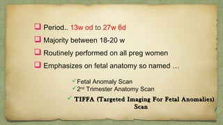
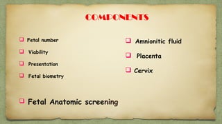
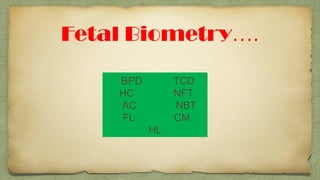
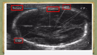
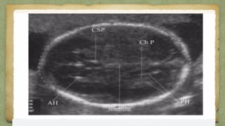
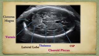
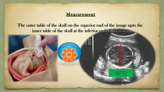
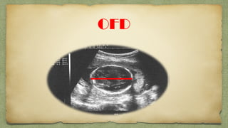
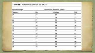
DR SOUMITRA HALDER
JUNIOR RESIDENT
DEPT. OF RADIO-DIAGNOSIS
MEDICAL COLLEGE, KOLKATA
๏ฑ Period.. 13w od to 27w 6d
๏ฑ Majority between 18-20 w
๏ฑ Routinely performed on all preg women
๏ฑ Emphasizes on fetal anatomy so named โ€ฆ
๏ƒผFetal Anomaly Scan
๏ƒผ2nd
Trimester Anatomy Scan
๏ƒผ TIFFA (Targeted Imaging For Fetal Anomalies)TIFFA (Targeted Imaging For Fetal Anomalies)
ScanScan
๏ฑ Fetal number
๏ฑ Viability
๏ฑ Presentation
๏ฑ Fetal biometry
๏ฑ Fetal Anatomic screening
๏ฑ Amnionitic fluid
๏ฑ Placenta
๏ฑ Cervix
COMPONENTS
Fetal Biometryโ€ฆ.
BPD TCD
HC NFT
AC NBT
FL CM
HL
BPD
๏ฑ Correct levels
๏ฑ Measured at a focus which shows, both the THALAMITHALAMI &
the CAVUM SEPTUM PELLUCIDUMCAVUM SEPTUM PELLUCIDUM..
SutureSuture
CSPCSPThalamusThalamus
Lateral LobeLateral Lobe
VermisVermis
CisternaCisterna
MagnaMagna
Choroid PlexusChoroid Plexus
MeasurementMeasurement
The outer table of the skull on the superior end of the image upto theThe outer table of the skull on the superior end of the image upto the
inner table of the skull at the inferior end of the imageinner table of the skull at the inferior end of the image
BPD
Not useful when the head shape is abnormal i.e, elongated or excessively roundNot useful when the head shape is abnormal i.e, elongated or excessively round
..
BPD is commonly affected by fetal position. Eg. Breech presentationBPD is commonly affected by fetal position. Eg. Breech presentation
Better to use the parameter ofBetter to use the parameter of CEPHALIC INDEX (CI)CEPHALIC INDEX (CI), instead of BPD alone., instead of BPD alone.
Also, the CI remains constant during the 3Also, the CI remains constant during the 3rdrd
trimester.trimester.
Cephalic Index (CI)ย =ย BPDย /ย  OFDย Xย 100Cephalic Index (CI)ย =ย BPDย /ย  OFDย Xย 100
OFD
H C
๏ฑ Same view as BPD
๏ฑ Outer to outer
A C
Landmark features:
๏ฑ Circular section with ribs
๏ฑ One vertebra -triangle of 3 white
spots
๏ฑ Umbilical vein - hockey stick
๏ฑ Stomach
A C
F L
TCD
๏ฑ Measured from the outerMeasured from the outer
to outerto outer
Placenta
PLACENTAL LOCALISATIONPLACENTAL LOCALISATION
MARGINALMARGINAL
PLACENTA PRAEVIAPLACENTA PRAEVIA
COMPLETE PLACENTACOMPLETE PLACENTA
PRAEVIAPRAEVIA
PSEUDO- PLACENTA PREVIAPSEUDO- PLACENTA PREVIA
Measured by adding the values of individual amniotic pocket depths (in cm's) forMeasured by adding the values of individual amniotic pocket depths (in cm's) for
each of the four quadrantseach of the four quadrants
Pocket chosen is free from the cord or any fetal part or placental tissue.Pocket chosen is free from the cord or any fetal part or placental tissue.
The normal range for amniotic fluid volumes varies with gestational age.The normal range for amniotic fluid volumes varies with gestational age.
AFI of < 5 ย OligohydramniosAFI of < 5 ย Oligohydramnios
5 - 25cm NORMAL5 - 25cm NORMAL
AFI of > 25 ย PolyhydramniosAFI of > 25 ย Polyhydramnios
MAXIMAL VERTICAL POCKETMAXIMAL VERTICAL POCKET
<<ย 2ย cm : Oligohydramniosย 2ย cm : Oligohydramnios
>ย 8ย cm : Polyhydramnios>ย 8ย cm : Polyhydramniosย 
2-8 Cm Normal
fetal anatomy assessment
Its imp to go through all-complete coverage--> then
no major abnormalities will be missed
Head & Brain
Cranial structures to Note at routine anat. Scan
1.Head shape
2.Ventricle
3.CSP
4.Thalamus
5.Cist Magna
6.Nuchal fold
7.cerebellum
DOLICOCEPHALYDOLICOCEPHALY BRACHYCEPHALYBRACHYCEPHALY
CLOVER LEAF SKULLCLOVER LEAF SKULL
ANENCEPHALYANENCEPHALY
๏ฑ No skull vault / cranium is seen.No skull vault / cranium is seen.
๏ฑ Brain tissue absentBrain tissue absent
ACRANIAACRANIA
๏ฑ Cranium is absentCranium is absent
๏ฑ Major part of the brain tissue mayMajor part of the brain tissue may
present .present .
Replaced by flattend amorphous massReplaced by flattend amorphous mass
(EXENCEPHALY)(EXENCEPHALY)
AnencephalyAnencephaly
(Frog Eye(Frog Eye
SignSign))
AcraniaAcrania
(absent Cranium(absent Cranium
But Brain tissue present)But Brain tissue present)
MENINgoCELEMENINgoCELE
ย ย When it contains only meningesWhen it contains only meninges
MENINgoENCEPHALoCELEMENINgoENCEPHALoCELE
Brain tissue is included in theBrain tissue is included in the
herniated tissue.herniated tissue.
CEPHALoCELE
Intracranial tissue herniates
Through a defect in cranium
๏ฑ May be seen as a purely cysticMay be seen as a purely cystic
mass or may contain echoes frommass or may contain echoes from
herniated brain tissue.herniated brain tissue.ย 
๏ฑ If the mass appears cystic, theIf the mass appears cystic, the
Meningoecele componentMeningoecele component
predominatespredominates, while a solid masssolid mass
indicates predominantly anindicates predominantly an
EncephaloceleEncephalocele.ย 
๏ฑ Larger encephalocoeles may showLarger encephalocoeles may show
accompanying ย microcephaly.accompanying ย microcephaly.
VENTRICULOMEGALY
Ventricular size measurementsโ€ฆ
1.Atrial width
2.V/H ratio
3.Choroid separation
4.Comb ant horn width
Atrial Width
๏ฑ Most useful and accepted
๏ฑ Must be taken on true axial view
๏ฑ Measured at widest part of ventricles
๏ฑ Inner to inner vent walls
10-12 borderline
12-15 moderate
>15 severe
Dangling Choroid
๏ฑ May be Unilateral orMay be Unilateral or
Bilateral.Bilateral.
๏ฑ Usually transient & benign.Usually transient & benign.
๏ฑ Seen in fetus normallySeen in fetus normally
between 16 โ€“ 21 weeks, afterbetween 16 โ€“ 21 weeks, after
which they start regressingwhich they start regressing..
HOLOPROCENCEPHALY
๏ฑ Resulting from incomplete separation of theResulting from incomplete separation of the
two hemisherestwo hemisheres
๏ฑ 3 typesโ€ฆ ALOBAR3 typesโ€ฆ ALOBAR
SEMILOBARSEMILOBAR
LOBARLOBAR
๏ฑ No inerhemisheric division..they
are fused
๏ฑ Absent Corpus Callosum
๏ฑ Single ventricleโ€ฆ.Horse-shoe
๏ฑ Thalami fused
๏ฑ Absent olfac tract
ALOBA
R
SEMILOBAR
๏ฑ Incomp interhemsph divisionโ€ฆ
Fused ant.
๏ฑ Rudimentary cerbral lobes
๏ฑ Partial/complele Fusion of Thallamus
๏ฑ Interhemis division fullyInterhemis division fully
formedformed
๏ฑ Fusion of Frontal horns ofFusion of Frontal horns of
lateral ventricles.lateral ventricles.
๏ฑ Midline cont. fronal neocortexMidline cont. fronal neocortex
๏ฑ Septum pellucidum is absentSeptum pellucidum is absent
๏ฑ Thalami are separateThalami are separate
๏ฑ Fully devolop cerebral lobesFully devolop cerebral lobes
LOBAR
Syntelencephaly
๏ฑ Middle interhemisheric form of holoprocencephaly
DANDY-WALKER MALFORMATION
1. Cystic dilatation of 4th
vent.
Communicating with post fossa
2. Enlarged post fossa
3. Elevated Tentorium
4. Hypoplastic or absent vermis
Dandy-Walker Variant
๏ฑ Less severe form of classic DWM
๏ฑ Partial Hypoplasia of vermis
๏ฑ Partial 4th
vent obstruction
๏ฑ Not much enlargement of post fossa
FETALFETAL
ABDOMENABDOMEN
scanscan
๏ƒผ Physiological Emptying : TransientPhysiological Emptying : Transient
๏ƒผ Lack Of Amnionitc Fluid ToLack Of Amnionitc Fluid To
SwallowSwallow
Oligohydramniosย Oligohydramniosย 
Anhydramniosย Anhydramniosย 
๏ƒผ Impaired Fetal SwallowingImpaired Fetal Swallowing
๏ƒผ Esophageal AtresiaEsophageal Atresia
๏ƒผ Obstructing Oropharyngeal Massย Obstructing Oropharyngeal Massย 
Cervical TeratomaCervical Teratoma
๏ƒผ Facial CleftsFacial Clefts
๏ƒผ CongenitalCongenital
DiaphragmaticDiaphragmatic
HerniationHerniation ::
fetal stomach may be absentfetal stomach may be absent
in an axial ultrasound scan โ€“in an axial ultrasound scan โ€“
standard view due tostandard view due to
migration into the thoraxmigration into the thorax
Absent stomach bubble
Esophageal AtresiaEsophageal Atresia
๏ฑ 2nd
bubble equal or smaller than stomach bubble
๏ฑ Shows communication with stomach
D/Dโ€ฆDuodenal atresia
Choledochal cyct
Duplication cyst
Double bubble sign
๏ฑ Another bubble adjacent to stomach bubble
Other than GB
D.atresia
Look for ant abd wall with cord insertionโ€ฆ
๏ฑ Para-umbilical abdominalPara-umbilical abdominal
wall defectwall defect..
๏ฑHerniation fetal bowel loopsHerniation fetal bowel loops
occasionally portions oroccasionally portions or
the stomach and or liver intothe stomach and or liver into
amniotic cavityamniotic cavity
GASTROSCHISIS
Free floating
Not Covered
GASTROSCHISIS
๏ฑ Midline abdominal wall defectsMidline abdominal wall defects
๏ฑ Herniation of intra abd contentsHerniation of intra abd contents
into base of cordinto base of cord
OMPHALOCELE
Gross ascites
Skin oedema
Fetal Hydrops
Dilated Bowel
>7 mm in small bowel
>20 mm in large bowel
Obstruction
High Echogenic Bowel
Chromosomal
abn
๏ฑ Fetal kidney seen from 14 wks
๏ฑ Can be identified through renal art
๏ฑ Renal agenesis..renal artery are also absent
๏ฑ B/L Ren agenesis associated with oligo /anhydramnios
๏ฑ Measurement of Renal pelvis AP diam
๏ƒผ inner to inner aspect of pelvis
๏ƒผ upper limit 4-5 mm in 2nd
trimester
๏ฑ Look for Renal cystic ds
UROGENITAL SYSTEM
๏ฑ Fetal bladder visible after 12 wk
Over distended bladder
B/L Hydro-urtero nephrosis
Obstructive uropathy
DR SOUMITRA HALDER
JUNIOR RESIDENT
DEPT. OF RADIO-DIAGNOSIS
MEDICAL COLLEGE, KOLKATA
PART - II
Important Viwes
๏ฑSaggital profile view
๏ฑAxial view Orbit
๏ฑAxial View Maxilla/Mandible
๏ฑNose-Lip view
ABNOMALITESโ€ฆ.
Retrognathia
mm=Ges age
Frontal Bossing
Slopped Forehead
Agnothia
ORBIT
๏ฑ Should be symmetrical size
๏ฑ Inner & outer distance in normal range
๏ฑ Round shape
Hypotelorism
Proboscis
Cyclopia
Hypertelorism
Micropthalmia
Anopthalmia
Dacrocystocele
Absent Nasal bone
PT/NBL ratio
NBL= Nasal Bone Length
PT= Prenasal thickness
N=0.35-0.8
CHR ABN= > 0.8
Cleft lip
Macroglossia
Lung:
Homogenous
echogenic tissue
Separated by
hypoechoic
domeshaped
diaphragm
Hypoechogenic structure
other than heartโ€ฆ.
CCAM
Pul Hypo
CDH
Pl effusion
๏ฑ Occupy 1/3 โ€“ ยฝ of thorax
๏ฑ Cardiac position & situs is
Imp
๏ƒผ Apex points toward Lt
๏ƒผ Lt atr close to spine
๏ƒผ Rt Vent close to chest wall
HEART
๏ฑ 2 ventricles/ walls of equal size
๏ฑ 2 atria / walls of equal size
๏ฑ Foramen ovale moving in left atrium
๏ฑ Pulmonary veins entering left atrium
๏ฑ Motion of mitral valves (left side) regular
๏ฑ Motion of tricuspid valves (right side) regular
๏ฑ Continuity of interventri septum
Check at a Glance
๏ฑThe ultrasound beam is directed
perpendicular to the midchest
plane at the level of the heart.
๏ฑ These chambers consist of the
right and left atrial and both
ventricular chambers
๏ฑCorresponding valves between
them
Four-Chamber View
Four-Chamber View
v
On Slight angulation
Towards rt shlder
Short-axis
view
Grt Vs
Toward Lt shoulder
Apical 5
chamber
view
Cephalad direction
3 Vs &
Trachea
view
Aortic arch
Ductus
arch
ASD
VSD
Hypo Rt Vent
TOF
TA
Double Outlet RV
TGA
Rhabdomyoma
Echogenic
intracardiac focus
SPINE
๏ฑ Coronal or Sagital of entire spine:
๏ƒผ cervical
๏ƒผ Thoracic
๏ƒผ Lumbar
๏ƒผ Sacral
๏ฑ Transverse assessment of entire spine: Triangular 3
dots form closed triangle
Spina Bifida
Spina
bifida
w
ith
m
eningom
ylocele
Overlying skin at
Sacrum is absent
Caudal
regression
Teratoma
LIMB
S๏ฑ Confirm the 3 long bones
๏ฑ Presence of Foot & Hand
๏ฑ Complete Absent....Amelia
๏ฑ Look for Phacomelia
Phacomelia
๏ฑBone scanned throughout long section
๏ฑLook for any bowing ,angulation ,#
๏ฑClub foot
๏ฑNo of fingers
Extremety measurements
๏ฑ FL routinely
๏ฑ All bones in background of family history
& strong suspicion
๏ฑ Clavice lengh in mm = Gest age in wk
Protocol
<2 SD
F/U scan after 3-4 wks
<4 SD
Sk dys
Further <1 SD
Sk dys
Pattern of limb shorting
Rhizomelia.. โ€ฆโ€ฆ.Prox
Mesoโ€ฆ โ€ฆโ€ฆโ€ฆโ€ฆ.Middle
Acroโ€ฆ. โ€ฆโ€ฆโ€ฆโ€ฆ..Distal
Microโ€ฆโ€ฆโ€ฆโ€ฆโ€ฆ..Entire limb
Femur /Foot Length ratio
N =1.
Constitutionaly small/IUGR= > 0.9
Sk dys < 0.9
2nd trimester ultrasound..

2nd trimester ultrasound..