SlideShare a Scribd company logo
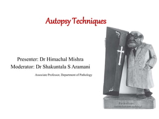
Autopsy Techniques
Presenter: Dr Himachal Mishra
Moderator: Dr Shakuntala S Aramani
Associate Professor, Department of Pathology
Overview
•Introduction
•History
•Objectives
•Various Techniques
•Examination of organs
•Removal of organs
•Summary
Introduction
• Autopsy: Greek “Autopsia” means seeing with ones eyes
• An autopsy is the examination of the dead body to figure
out the cause of death.”
• Autopsies date back to the 5th century.
Autopsy Through The Ages…
•Eristratus (310 BC) and Herophilus (335 BC)-----
searched into cause of disease and associated disease
with changes in the organs .
•Galen (129- 201AD) -Dissections on the animals /
primates .
• Frederick II- Authorised human dissection and paved way
for medical education.
• With Renaissance medicine and medical education underwent
changes
• Andrea Vesalius -given the duty of conducting public
dissections.
• Leonardo da Vinci ( 1452- 1519 ) – made drawings from about 30
human dissections .
• Giovanni Morgagni (1682-1771 )- intellectual founder of autopsy
.
• Karl Rokitansky (1804-1878 ) -performed > 30,000 autopsies.
• Rudolph Virchow ( 1821-1902 ) – founder of modern pathology.
Objectives of autopsy
•To identify the cause of death.
•To identify any hereditary disease
•To recognise any congenital anomalies
•To rule out an infectious disease.
•Education of medical students
•Information for audit.
•Material for research.
•Accurate data on death for National statistics.
Who Can Authorize Autopsy?
•Deceased person
•Surviving spouse
•Children of deceased
•Grand children of deceased
•Parents
•Brothers or sisters
•Cousins
•Friends or any person of legal age
Who can perform autopsy?
•Medical examiner
•Qualified physicians and surgeons
•Competent pathologist or toxicologist.
Biological safety
General Principles
• Recognition of risks
• Identification of hazards
• Elimination
Emerging Infections
• Tuberculosis
• Hepatitis,
• HIV
General Technical Considerations
Autopsy room:
• Well lighted
• Firm table, wooden block
• Large supply of water
• Cleanliness
• Handle with care
“Autopsy is permissible to honour the dead’’
Components of a post-mortem room
•Mortuary : For every 100 bed Hospital-4 body
storage space.
: Temp-Main cold storage40 C
-Deep freeze unit - 200 C
•Table– Stainless steel with raised edge all around
- Large deep sink at the foot with both hot
& cold water.
-Suction apparatus
Autopsy record
•Name, age, sex, colour, weight, length.
• IP No & Autopsy No.
•Date & time of death.
•Date, time & place of examination
•Name of resident & staff pathologist
•Temp & weather of room
•Name of person who identified the body
•Summary of clinical history & diagnosis
•External examination
•Internal examination
Condition and appearance of organs, tissues & cavities.
• Chemical & microscopic examination
• Diagnosis
Various Instruments
Kinds of Autopsies
There are two kinds of autopsies:
• Forensic/Medico-legal
• Clinical/Pathological Autopsy
Forensic: to find the cause of death for a police investigation.
Clinical: Performed in a hospital
Done to find the cause of death for research.
Autopsy techniques
General
1. En Masse (Le Tulle)
2. En Bloc
3. Virchow’s
4. Rokitansky
Special
1. Post operative
2. Needle
3. Endoscopic
4. Restriction of skin incisions
5. Autopsy through surgical wounds
1. En Masse (Le Tulle)
• Organs are removed as a single bulky aggregate.
•Advantages:
• Complete preservation of relationships among organs
• Organs removed and stored for later dissection
•Disadvantages:
• Difficult to handle
• Require assistant
2. En Bloc (Ghon/Zenker)
•Maintain all connections between physiologically
related organs:
• Thoracic pluck, coeliac pluck, urogenital pluck
•Advantages:
• Preserve important anatomic relations without unwieldy
mass of organs
•Disadvantages:
• Multiple organ system involvement complicates procedure
• Skill necessary to remove each block from the body intact
3. Virchow’s
• All organs examined systematically one after the other.
• Brain  spinal cord  abdominal cavity  thoracic cavity
 organs individually removed & sectioned outside body.
• Advantages:
Systematic approach
Simplicity for beginning prosecutors
• Disadvantages:
Destruction of anatomic relationships.
4. Rokitansky (in situ)
• Basic principle: Disturb the connections between organs as
little as possible.
• Dissection occurs in situ with little actual evisceration.
• If abnormality is found, regions removed intact 
combination of en bloc and in situ.
• Advantages:
Practical for single examiner
Capability of preserving abnormal anatomic relationships
• Disadvantages:
Expertise necessary to recognize abnormalities
Special autopsy techniques
1. Post operative autopsies :
• Most experienced autopsy pathologist must perform autopsies.
• Technique can be changed as required by the specific situation :
-Incision not be carried through operative wounds.
-Fistulas to be filled with a stained contrast medium.
-Don’t remove the drain before their location established.
2. Needle autopsy :
Indications
-invasive procedures are not possible
-If proper infection precautions cannot be taken
-Relatives do not give consent for a complete autopsy
3. Endoscopic autopsies :
Neoplasms & traumatic lesions can be easily identified.
4. Restriction of skin incisions :
-An autopsy permission may specify that only an abdominal
incision to be made.
• With appropriate consent, all cervical, thoracic & abdominal
organs may be removed through such an incision.
5. Autopsy through surgical wounds :
• Autopsy permission is restricted to the reopening of a surgical
wound.
External & Internal
Examinations
External Examination
•Identification
•Sex differentiation
•Age determination
•Algor Mortis
•Livor Mortis
•Rigor Mortis
•Putrefaction
Measurements
•Height
•Weight
•Head Circumference
•Chest Circumference
•Abdominal Circumference
External Examination
Sites To acess
Skull trauma / haematoma / abscess / surgical intervention / hydrocephaly &
microcephaly.
Face symmetry / cyanosis / acromegaly / cretinism & myxedema
Eyes Exophthalmos / sclera for pallor, icterus & hemorrhage
Corneas – cataract/ injury/ infections/Arcus senilitis/ KF rings
Nose deformity / gangrene/ hemorrhage / infection / malignancy.
Mouth hemorrhage / infection / malignancy/ ulcers
Tongue Geographic tongue / thick & protuberant
Ears Pus/fluid & blood
Neck Thyroid enlargement, vein prominence, lymph node enlargement
Skin Pigments, rashes, redness, nodule
Back bed sore / livor mortis / deformity
Breast Mass
External
genitalia
abnormal development, edema, local infection, testicular atrophy or enlargement
Lymph
nodes
enlarged? Matted or non matted
• 1st - External Examination
• height/weight
• Photography
• UV exam (fluids)
• 2nd - Toxicology
• Nail scrapings
• Fluid samples
• 3rd - Internal Examination
• Head (circular saw)
• Chest (incision)
• Abdomen
Steps of Autopsy
Securing CSF
Cisternal puncture
:Turn head in lateral position & flex maximally
: Sterilize the skin with iodine & acetone.
: Insert LP needle above 2nd vertebra & push it
upward in the midline.
REMOVAL OF BRAIN
• Incision of scalp :
Sawing of cranium
• Look for extradural hematoma
• Open superior sagittal sinus with scissors-
look for thrombus.
• Cut dura at the level of incision of
skull & fold dura back to the midline.
Removal of brain
Triangular incision on orbital plate
Inject full strength formalin into the eyeball
Wait for ½ hr for fixation
Expose optic N
Exert traction Removal of the eyes
cut away posterior half bulbus oculi
Replace orbit with an artificial eye.
Removal of the eyes-----A. Posterior method
Anterior removal of the eye
Different methods of Brain Fixation
1. Immersion
• Passing a thread underneath the basilar artery in front of pons.
• Pontine infarcts or other lesions- pass under ICA/ MCA.
• Thread passed through short dural flaps on either side of falx.
2. Perfusion
• Perfusion of large amount of fixative use-embalmer’s pump
• Gravity-feed method- infusion bottle raised 150–180 cm above
the specimen.
• Gross examination of brain :
• Weight in gms : Males : 1400 Females : 1275
At birth : 335
• External surface : Look for
--Symmetry
--Swelling or atrophy
--Herniation
--Softening
--Any tumors / infarcts / contusions.
--Meninges – pus / hemorrhage.
Sectioning of Brain
Approach to dissection of brain stem & cerebellum
Various sections from brain
Fourth Incision
Useful
• Death due to vehicular incidents.
• Death in suspected tortured cases
• Death due to freshly inflicted
deep-seated bruise in the
posterior aspect of the body.
Advantages
• Seepage nil
• Better acceptance
American Journal of Forensic Med Pathol. 2010; 31: 37-41.
Removal of neck organs :
•With an amputation knife cut beneath the skin & platysma
muscles of neck to mandible.
•With blunt ended scissors, define & isolate CCAs
throughout its extent. Retract them laterally.
•Cut across the top of larynx & dissect out neck organs.
•Remove tongue, tonsils & soft palate by cutting through
the floor of mouth & incising junction between the hard &
soft palate .
•Examine the teeth, mandible & buccal cavity by
palpation.
Initial Dissection & Internal Examination
A B
C D
• In situ examination :
a) Note the situs of thoracic & abdominal organs :
- Situs solitus – normal position of organs.
- Situs inversus – complete reversal of normal position.
- Situs ambiguous – situs is not clear
b) Presence & absence of spleen & their number :
-Asplenia – absence of spleen
-Polysplenia – Presence of multiple splenicules on both the sides.
c) Note direction of the apex :
-Levocardia – apex pointing to the left.
-Mesocardia - apex pointing to the midline
-Dextrocardia - apex pointing to the right.
Pericardial cavity
Pericardial Cavity
• Open the pericardial cavity with the scissors, first vertically & then
towards the apex of heart.
• Normal pericardial membrane is delicate, smooth, glistening &
transparent.
• Normally pericardial fluid: 5-50 ml, pale yellow & clear.
• Dry pericardium extreme dehydration  cholera
• Types of fluid encountered in pericardium :
-Transudate – hydropericardium
-Exudate – serous pericarditis
-Pus – purulent pericarditis
-Blood – hemopericardium
Pericarditis
Acute – fibrinous, serous, purulent, hemorhagic.
Chronic : Granulomatous, chronic adhesive, chronic
constrictive
Suspected cases of air embolism
• Ligate the root of aorta tightly.
• Make a 3 cm incision on the pericardial sac anteriorly.
• Elevate the edges of this incision & inspect the contents of the sac.
• Fill the pericardial sac with water & submerge the heart.
• Cut across the left circumflex & descending branch of LCA.
• Milk the left coronaries with the finger towards the incision.
• Bubbles will escape if air is in the coronaries.
• Repeat this maneuver with the RCA.
• Under water, incise the RA, RV & PA –Look for air bubbles.
• Examine the LA, LV, SVC, IVC & PVs in a similar manner.
Methods for demonstration of pneumothorax
Pleural cavity :
--Examine the pleural cavity for any fluid. If present collect atleast 50
ml in a clean dry vessel.
--Classification of pleural effusions :
Transudate Exudate
-Circulatory congestion - Bacterial/Mycotic/Protozoal infection
-Lymphatic obstruction - Malignancy
-Hypoproteinemia - Rheumatic fever
-Meigs syndrome - Collagen diseases
-SVC obstruction
A raised, swollen, red localised pleural area- lung infarction.
Meatstatic nodules of carcinoma & sarcoma grey white or
red subpleural nodules.
Mesothelioma of pleura – plaque on visceral/parietal pleura.
Lungs
--Fibrocaseous tuberculosis- Adhesions between the pleura & the chest
cavity in the subapical portion.
--Emphysema- Subpleural emphysematous bullae over the lung.
--Collapsed lung – small lung with wrinkling of visceral pleura.
--Bacterial pneumonia – Red to grey yellow colored foci of
consolidation, firm & rubbery in consistency.
Pulmonary metastasis – Mc site: periphery of the lung.
- Single/multiple grey white to reddish areas.
--Miliary tuberculosis –Lung studded with millet seed like foci of
lesions all over subpleural surface of lung.
Examination of abdominal cavity
Explore the cavity with the gloved hand & note the position of
omentum & the sizes of spleen, liver & kidney.
Examine omentum for fat necrosis, tumor deposits & tubercles.
Note if any fluid is present.
Search for orifices into hernial sacs.
Examine female genitalia to determine the condition of the ovaries,
adhesions about the tubes, size & position of uterus
1. Examination of Liver
• Determine the height of each dome of diaphragm with respect
to the ribs / intercostal spaces & record this finding.
• Note the downward extent of liver in the mid-clavicular line,
measure this distance in cms & record this finding.
Amyloidosis,
Fatty Liver
Storage Disorders
CVC
Malaria,
Amoebic abscess,
Tumor masses.
Small, brown to yellow
in color with wrinkling
of capsule.
Hepatomegaly-
Atrophic liver
Consistency – normally –semisolid.
It is diminished in necrosis & severe fatty change.
Increased in amyloidosis & cirrhosis.
Post mortem color of liver - deep blue - CVC
- green – if seepage of bile
Putrefaction changes - Honeycomb of gas bubbles.
Nut meg appearance  CVC
Amyloidosis  Translucent waxy appearance
Pale infarct  Obstruction of hepatic artery
Malaria  dark gray or salty appearance
Miliary TB  studded with millet seed like lesions
Amoebic abscess  large abscess
Metastasis in liver  multiple, gray white nodules
Spleen
Hemochromatosis  reddish-brown color
Chronic malaria  gray to black.
Splenic infarcts  pale
Amyloidosis  waxy translucent appearance / sago spleen.
2. G.I.T.
Examine entire GIT carefully for perforation.
Gastric ulcer perforation- a serosal fibrinous exudate, may be purulent.
Perforation of a chronic duodenal ulcer:
Anteriorly & in ‘supracolic compartment’.
Look for gangrene of small intestine & appendix.
Look for malignancies of the stomach, intestines or any other serosal
tumor deposits.
Removal of Organs
1. REMOVAL OF THORACIC ORGANS
• Remove the heart by lifting it up & cutting through the vena cavae &
pulmonary veins from beneath.
• Sever the aorta & pulmonary artery & remove the contents with a
forceps.
• Place the heart in a pan.
• Remove the lungs by cutting through the hilar structures :
2. Removal of upper abdominal organs
• Open stomach along greater curvature in continuity with esophagus.
• Incision of greater curvature is continued into duodenum & all the 4
parts are opened.
• Gall bladder is squeezed gently to look for bile coming out of the
ampulla of Vater in the 2nd part of duodenum.
Bile flows freely Bile does not flow freely
•If the bile flows freely
• Connection of the liver with the gut can be severed by cutting portal V,
CBD & hepatic artery.
Dissect hepatoduodenal ligament
• Raise left lobe & cut left triangular ligament with a scissor.
• Raise right lobe & cut right triangular ligament with a scissor.
• Cut the IVC below the diaphragm & free any remaining attachments
• Cut falciform ligament & gastro-duodenal ligament from inferior
surface of liver.
If bile does not flow freely
En block removal of upper abdominal organs must be done.
Separate diaphragm from ribs & posterior abdominal wall. Free the
lower end of oesophagus from the diaphragm.
Separate spleen & dissect pancreas out of retroperitoneum, beginning
with its tail.
Tie first part of jejunum & working towards pylorus, liberate
duodenum & head of pancreas without damaging CBD.
Lift liver & cut it free of any remaining attachments to diaphragm &
IVC.
Entire block of organs can now be shifted out.
3. Removal of abdominal & retroperitoneal organs
Removal of intestines :
Removal of intestine
Removal of genitourinary system :
Removal of male reproductive system :
Pull uterus, cervix & vagina upwards
Divide vagina as low as possible
FTs, uterus & ovaries are freed from
pelvis & are removed
Removal of female reproductive system
• Removal of bladder along with prostate & seminal vesicles &
terminal segment of rectum :
Summary
•Autopsy is examination of dead body to find the cause.
•Y is the most common primary incision given, but with
time some modification has been done.
•It includes thorough external and internal examination,
including opening of all the body cavities for proper
visualization of all the visceral organs.
•Knowledge of different diseases and their gross appearance
is must to know the leading cause of death.
References
1. Ludwig J, Handbook of autopsy practice.2002; 3 :4-302
2. Vaideeshwar P, Lanjeswar D, Autopsy practices, 2021; 2: 1-204.
3. King, Meehan. History of autopsy. American journal of
pathology,1973; 73(2):2-32.
4. Finkbeiner WE, Ursel PC. Autopsy Pathology- a manual and
atlas, 2009; 2: 2-104.
5. Dehner LP. The medical autopsy: Past, present and dubious
future. Missouri medicine. 2010; 107(2): 94-100.
6. Bauthier JP. Autopsy and identication techniques. Research
and Technology. 2011;33 :691-714.
7. Patowary A. The Fourth Incision- A Cosmetic Autopsy
Incision Technique. American Journal of Forensic Med
Pathol. 2010; 31: 37-41.
THANK YOU

More Related Content

What's hot

External examination in autopsies with pro forma
External examination in autopsies with pro formaExternal examination in autopsies with pro forma
External examination in autopsies with pro forma
Dr. Varughese George
 
Forensic Pathology
Forensic PathologyForensic Pathology
Forensic PathologyErin Mucci
 
Death Scene Investigation
Death Scene InvestigationDeath Scene Investigation
Death Scene Investigation
Francis Postrado
 
Autopsy.ppt
Autopsy.pptAutopsy.ppt
Autopsy.ppt
Dr. Armaan Singh
 
identification
identificationidentification
identification
Akash Agarwal
 
Forensic Pathology
Forensic PathologyForensic Pathology
Forensic Pathology
Christine Onwenu
 
Autopsy procedure
Autopsy procedureAutopsy procedure
Autopsy procedure
Farhan Ali
 
autopsy,Necropsy,postmortem
autopsy,Necropsy,postmortemautopsy,Necropsy,postmortem
autopsy,Necropsy,postmortem
Valliappan Medic
 
Autopsy by Dr Yaseen Mohammad for 3rd year students MBBS
Autopsy by Dr Yaseen Mohammad for 3rd year students MBBSAutopsy by Dr Yaseen Mohammad for 3rd year students MBBS
Autopsy by Dr Yaseen Mohammad for 3rd year students MBBS
Yaseen Mohammad
 
Forensic osteology
Forensic osteologyForensic osteology
Forensic osteology
ARIF MASOOD
 
Presentation autopsy incisions-.pptx. Dr. Haricharan MD RIMS Imphal . ...
  Presentation autopsy incisions-.pptx.    Dr. Haricharan MD  RIMS Imphal  . ...  Presentation autopsy incisions-.pptx.    Dr. Haricharan MD  RIMS Imphal  . ...
Presentation autopsy incisions-.pptx. Dr. Haricharan MD RIMS Imphal . ...
Dr. A. Haricharan Comrade
 
Autopsy dissection of heart and spinal cord
Autopsy dissection of heart and spinal cordAutopsy dissection of heart and spinal cord
Autopsy dissection of heart and spinal cord
Rijen Shrestha
 
External examination at autopsy
External examination at autopsyExternal examination at autopsy
External examination at autopsy
Ashish Jawarkar
 
Preservation of viscera
Preservation of visceraPreservation of viscera
Preservation of viscera
Nikhil Oza
 
Forensic Autopsy
Forensic Autopsy Forensic Autopsy
Forensic Autopsy
AbdulelahMurshid
 
Forensic Biological Sample
Forensic Biological SampleForensic Biological Sample
Forensic Biological SampleShailendra Patel
 
Postmortem artefcts
Postmortem artefctsPostmortem artefcts
Postmortem artefcts
Sujeet Samadder
 
Wounds in forensic medicine
Wounds in forensic medicineWounds in forensic medicine
Wounds in forensic medicineAhmed Almumtin
 
THE MEDICO-LEGAL AUTOPSY.pptx
THE MEDICO-LEGAL AUTOPSY.pptxTHE MEDICO-LEGAL AUTOPSY.pptx
THE MEDICO-LEGAL AUTOPSY.pptx
Balinainejoseph
 
Antemortem and Postmortem Wounds.pptx
Antemortem and Postmortem Wounds.pptxAntemortem and Postmortem Wounds.pptx
Antemortem and Postmortem Wounds.pptx
Applied Forensic Research Sciences
 

What's hot (20)

External examination in autopsies with pro forma
External examination in autopsies with pro formaExternal examination in autopsies with pro forma
External examination in autopsies with pro forma
 
Forensic Pathology
Forensic PathologyForensic Pathology
Forensic Pathology
 
Death Scene Investigation
Death Scene InvestigationDeath Scene Investigation
Death Scene Investigation
 
Autopsy.ppt
Autopsy.pptAutopsy.ppt
Autopsy.ppt
 
identification
identificationidentification
identification
 
Forensic Pathology
Forensic PathologyForensic Pathology
Forensic Pathology
 
Autopsy procedure
Autopsy procedureAutopsy procedure
Autopsy procedure
 
autopsy,Necropsy,postmortem
autopsy,Necropsy,postmortemautopsy,Necropsy,postmortem
autopsy,Necropsy,postmortem
 
Autopsy by Dr Yaseen Mohammad for 3rd year students MBBS
Autopsy by Dr Yaseen Mohammad for 3rd year students MBBSAutopsy by Dr Yaseen Mohammad for 3rd year students MBBS
Autopsy by Dr Yaseen Mohammad for 3rd year students MBBS
 
Forensic osteology
Forensic osteologyForensic osteology
Forensic osteology
 
Presentation autopsy incisions-.pptx. Dr. Haricharan MD RIMS Imphal . ...
  Presentation autopsy incisions-.pptx.    Dr. Haricharan MD  RIMS Imphal  . ...  Presentation autopsy incisions-.pptx.    Dr. Haricharan MD  RIMS Imphal  . ...
Presentation autopsy incisions-.pptx. Dr. Haricharan MD RIMS Imphal . ...
 
Autopsy dissection of heart and spinal cord
Autopsy dissection of heart and spinal cordAutopsy dissection of heart and spinal cord
Autopsy dissection of heart and spinal cord
 
External examination at autopsy
External examination at autopsyExternal examination at autopsy
External examination at autopsy
 
Preservation of viscera
Preservation of visceraPreservation of viscera
Preservation of viscera
 
Forensic Autopsy
Forensic Autopsy Forensic Autopsy
Forensic Autopsy
 
Forensic Biological Sample
Forensic Biological SampleForensic Biological Sample
Forensic Biological Sample
 
Postmortem artefcts
Postmortem artefctsPostmortem artefcts
Postmortem artefcts
 
Wounds in forensic medicine
Wounds in forensic medicineWounds in forensic medicine
Wounds in forensic medicine
 
THE MEDICO-LEGAL AUTOPSY.pptx
THE MEDICO-LEGAL AUTOPSY.pptxTHE MEDICO-LEGAL AUTOPSY.pptx
THE MEDICO-LEGAL AUTOPSY.pptx
 
Antemortem and Postmortem Wounds.pptx
Antemortem and Postmortem Wounds.pptxAntemortem and Postmortem Wounds.pptx
Antemortem and Postmortem Wounds.pptx
 

Similar to Autopsy techniques.ppt

All about Autopsy in forensic medicine .pptx
All about Autopsy in forensic medicine .pptxAll about Autopsy in forensic medicine .pptx
All about Autopsy in forensic medicine .pptx
Paul523674
 
Post Mortem Examination
Post Mortem ExaminationPost Mortem Examination
Post Mortem Examination
Applied Forensic Research Sciences
 
Tejasvi
TejasviTejasvi
Autopsy
AutopsyAutopsy
Autopsy
SMVDCoN ,J&K
 
postmortem examination.pptx
postmortem examination.pptxpostmortem examination.pptx
postmortem examination.pptx
AlinisweNgambi
 
External examination in autopsies with pro forma
External examination in autopsies with pro formaExternal examination in autopsies with pro forma
External examination in autopsies with pro forma
Dr. Varughese George
 
Autopsy.pptx
Autopsy.pptxAutopsy.pptx
Autopsy.pptx
Dr. Gurjeet Singh
 
Necropsy Of Poultry (procedure).pptx
Necropsy Of Poultry (procedure).pptxNecropsy Of Poultry (procedure).pptx
Necropsy Of Poultry (procedure).pptx
SiratAlMim
 
General physical Examination
General physical Examination General physical Examination
General physical Examination
Virendra Hindustani
 
Autopsy Lectures.pptx
Autopsy Lectures.pptxAutopsy Lectures.pptx
Autopsy Lectures.pptx
AbbaZarami Bukar
 
FORENSIC
FORENSIC FORENSIC
FORENSIC
Prashant Mehta
 
methods of clinical examination
methods of clinical examinationmethods of clinical examination
methods of clinical examination
Enas Elgendy
 
Case presentation for Reading
Case presentation for ReadingCase presentation for Reading
Case presentation for Reading
Mr.Harshad Khade
 
diagnostic Cytology introduction , Body fluids cytology
diagnostic Cytology introduction , Body fluids cytologydiagnostic Cytology introduction , Body fluids cytology
diagnostic Cytology introduction , Body fluids cytology
Aayra
 
HA unit 4.pdf
HA unit 4.pdfHA unit 4.pdf
HA unit 4.pdf
MuhammadAbbasWali
 
Histological Techniques: Section 1:Tissue collection and Grossing
Histological Techniques: Section 1:Tissue collection and GrossingHistological Techniques: Section 1:Tissue collection and Grossing
Histological Techniques: Section 1:Tissue collection and Grossing
Mathew Joseph
 
Mass disaster forensic medicine mbbs.pptx
Mass disaster forensic medicine mbbs.pptxMass disaster forensic medicine mbbs.pptx
Mass disaster forensic medicine mbbs.pptx
Drsarath2
 
Biopsy final.ppt
Biopsy final.pptBiopsy final.ppt
Biopsy final.ppt
KushPathak9
 
exfoliative cytology and fnac.pptx
exfoliative cytology and fnac.pptxexfoliative cytology and fnac.pptx
exfoliative cytology and fnac.pptx
Gourab Kuñdu
 
Autopsy Presentation.pptx
Autopsy Presentation.pptxAutopsy Presentation.pptx
Autopsy Presentation.pptx
hasnainshah59
 

Similar to Autopsy techniques.ppt (20)

All about Autopsy in forensic medicine .pptx
All about Autopsy in forensic medicine .pptxAll about Autopsy in forensic medicine .pptx
All about Autopsy in forensic medicine .pptx
 
Post Mortem Examination
Post Mortem ExaminationPost Mortem Examination
Post Mortem Examination
 
Tejasvi
TejasviTejasvi
Tejasvi
 
Autopsy
AutopsyAutopsy
Autopsy
 
postmortem examination.pptx
postmortem examination.pptxpostmortem examination.pptx
postmortem examination.pptx
 
External examination in autopsies with pro forma
External examination in autopsies with pro formaExternal examination in autopsies with pro forma
External examination in autopsies with pro forma
 
Autopsy.pptx
Autopsy.pptxAutopsy.pptx
Autopsy.pptx
 
Necropsy Of Poultry (procedure).pptx
Necropsy Of Poultry (procedure).pptxNecropsy Of Poultry (procedure).pptx
Necropsy Of Poultry (procedure).pptx
 
General physical Examination
General physical Examination General physical Examination
General physical Examination
 
Autopsy Lectures.pptx
Autopsy Lectures.pptxAutopsy Lectures.pptx
Autopsy Lectures.pptx
 
FORENSIC
FORENSIC FORENSIC
FORENSIC
 
methods of clinical examination
methods of clinical examinationmethods of clinical examination
methods of clinical examination
 
Case presentation for Reading
Case presentation for ReadingCase presentation for Reading
Case presentation for Reading
 
diagnostic Cytology introduction , Body fluids cytology
diagnostic Cytology introduction , Body fluids cytologydiagnostic Cytology introduction , Body fluids cytology
diagnostic Cytology introduction , Body fluids cytology
 
HA unit 4.pdf
HA unit 4.pdfHA unit 4.pdf
HA unit 4.pdf
 
Histological Techniques: Section 1:Tissue collection and Grossing
Histological Techniques: Section 1:Tissue collection and GrossingHistological Techniques: Section 1:Tissue collection and Grossing
Histological Techniques: Section 1:Tissue collection and Grossing
 
Mass disaster forensic medicine mbbs.pptx
Mass disaster forensic medicine mbbs.pptxMass disaster forensic medicine mbbs.pptx
Mass disaster forensic medicine mbbs.pptx
 
Biopsy final.ppt
Biopsy final.pptBiopsy final.ppt
Biopsy final.ppt
 
exfoliative cytology and fnac.pptx
exfoliative cytology and fnac.pptxexfoliative cytology and fnac.pptx
exfoliative cytology and fnac.pptx
 
Autopsy Presentation.pptx
Autopsy Presentation.pptxAutopsy Presentation.pptx
Autopsy Presentation.pptx
 

More from gimspathcme2022

Massive-transfusion.pptx
Massive-transfusion.pptxMassive-transfusion.pptx
Massive-transfusion.pptx
gimspathcme2022
 
nucleicacidamplificationtechnologynattest-190404045749.pptx
nucleicacidamplificationtechnologynattest-190404045749.pptxnucleicacidamplificationtechnologynattest-190404045749.pptx
nucleicacidamplificationtechnologynattest-190404045749.pptx
gimspathcme2022
 
Angiogenesis in health and disease.pptx
Angiogenesis in health and disease.pptxAngiogenesis in health and disease.pptx
Angiogenesis in health and disease.pptx
gimspathcme2022
 
vivek autopsy 2.pptx
vivek autopsy 2.pptxvivek autopsy 2.pptx
vivek autopsy 2.pptx
gimspathcme2022
 
liver pathgology.pptx
liver pathgology.pptxliver pathgology.pptx
liver pathgology.pptx
gimspathcme2022
 
fetalautopsy-1.pptx
fetalautopsy-1.pptxfetalautopsy-1.pptx
fetalautopsy-1.pptx
gimspathcme2022
 
urine basics.pptx
urine basics.pptxurine basics.pptx
urine basics.pptx
gimspathcme2022
 
heartfailure.pptx
heartfailure.pptxheartfailure.pptx
heartfailure.pptx
gimspathcme2022
 
Actinomycetes
ActinomycetesActinomycetes
Actinomycetes
gimspathcme2022
 
Neoplasia
NeoplasiaNeoplasia
Neoplasia
gimspathcme2022
 
Hypersensitivity
HypersensitivityHypersensitivity
Hypersensitivity
gimspathcme2022
 
Immune.pptx
Immune.pptxImmune.pptx
Immune.pptx
gimspathcme2022
 
EDEMA,TYPES.pptx
EDEMA,TYPES.pptxEDEMA,TYPES.pptx
EDEMA,TYPES.pptx
gimspathcme2022
 
csf.pptx
csf.pptxcsf.pptx
csf.pptx
gimspathcme2022
 
12- Blood Groups and Blood Transfusion 2018-converted.pptx
12- Blood Groups and Blood Transfusion 2018-converted.pptx12- Blood Groups and Blood Transfusion 2018-converted.pptx
12- Blood Groups and Blood Transfusion 2018-converted.pptx
gimspathcme2022
 
Pediatric autopsy.pptx
Pediatric autopsy.pptxPediatric autopsy.pptx
Pediatric autopsy.pptx
gimspathcme2022
 
Kidney failure.pptx
Kidney failure.pptxKidney failure.pptx
Kidney failure.pptx
gimspathcme2022
 
special stain
special stainspecial stain
special stain
gimspathcme2022
 
NEOPLASIA.pptx
NEOPLASIA.pptxNEOPLASIA.pptx
NEOPLASIA.pptx
gimspathcme2022
 
Bilirubin &jaundice.pptx
Bilirubin &jaundice.pptxBilirubin &jaundice.pptx
Bilirubin &jaundice.pptx
gimspathcme2022
 

More from gimspathcme2022 (20)

Massive-transfusion.pptx
Massive-transfusion.pptxMassive-transfusion.pptx
Massive-transfusion.pptx
 
nucleicacidamplificationtechnologynattest-190404045749.pptx
nucleicacidamplificationtechnologynattest-190404045749.pptxnucleicacidamplificationtechnologynattest-190404045749.pptx
nucleicacidamplificationtechnologynattest-190404045749.pptx
 
Angiogenesis in health and disease.pptx
Angiogenesis in health and disease.pptxAngiogenesis in health and disease.pptx
Angiogenesis in health and disease.pptx
 
vivek autopsy 2.pptx
vivek autopsy 2.pptxvivek autopsy 2.pptx
vivek autopsy 2.pptx
 
liver pathgology.pptx
liver pathgology.pptxliver pathgology.pptx
liver pathgology.pptx
 
fetalautopsy-1.pptx
fetalautopsy-1.pptxfetalautopsy-1.pptx
fetalautopsy-1.pptx
 
urine basics.pptx
urine basics.pptxurine basics.pptx
urine basics.pptx
 
heartfailure.pptx
heartfailure.pptxheartfailure.pptx
heartfailure.pptx
 
Actinomycetes
ActinomycetesActinomycetes
Actinomycetes
 
Neoplasia
NeoplasiaNeoplasia
Neoplasia
 
Hypersensitivity
HypersensitivityHypersensitivity
Hypersensitivity
 
Immune.pptx
Immune.pptxImmune.pptx
Immune.pptx
 
EDEMA,TYPES.pptx
EDEMA,TYPES.pptxEDEMA,TYPES.pptx
EDEMA,TYPES.pptx
 
csf.pptx
csf.pptxcsf.pptx
csf.pptx
 
12- Blood Groups and Blood Transfusion 2018-converted.pptx
12- Blood Groups and Blood Transfusion 2018-converted.pptx12- Blood Groups and Blood Transfusion 2018-converted.pptx
12- Blood Groups and Blood Transfusion 2018-converted.pptx
 
Pediatric autopsy.pptx
Pediatric autopsy.pptxPediatric autopsy.pptx
Pediatric autopsy.pptx
 
Kidney failure.pptx
Kidney failure.pptxKidney failure.pptx
Kidney failure.pptx
 
special stain
special stainspecial stain
special stain
 
NEOPLASIA.pptx
NEOPLASIA.pptxNEOPLASIA.pptx
NEOPLASIA.pptx
 
Bilirubin &jaundice.pptx
Bilirubin &jaundice.pptxBilirubin &jaundice.pptx
Bilirubin &jaundice.pptx
 

Recently uploaded

Cardiac Arrhythmias (2).pdf for nursing student
Cardiac Arrhythmias (2).pdf for nursing studentCardiac Arrhythmias (2).pdf for nursing student
Cardiac Arrhythmias (2).pdf for nursing student
fahmyahmed789
 
Dimensions of Healthcare Quality
Dimensions of Healthcare QualityDimensions of Healthcare Quality
Dimensions of Healthcare Quality
Naeemshahzad51
 
Cold Sores: Causes, Treatments, and Prevention Strategies | The Lifesciences ...
Cold Sores: Causes, Treatments, and Prevention Strategies | The Lifesciences ...Cold Sores: Causes, Treatments, and Prevention Strategies | The Lifesciences ...
Cold Sores: Causes, Treatments, and Prevention Strategies | The Lifesciences ...
The Lifesciences Magazine
 
Empowering ACOs: Leveraging Quality Management Tools for MIPS and Beyond
Empowering ACOs: Leveraging Quality Management Tools for MIPS and BeyondEmpowering ACOs: Leveraging Quality Management Tools for MIPS and Beyond
Empowering ACOs: Leveraging Quality Management Tools for MIPS and Beyond
Health Catalyst
 
HUMAN BRAIN.pptx.PRIYA BHOJWANI@GAMIL.COM
HUMAN BRAIN.pptx.PRIYA BHOJWANI@GAMIL.COMHUMAN BRAIN.pptx.PRIYA BHOJWANI@GAMIL.COM
HUMAN BRAIN.pptx.PRIYA BHOJWANI@GAMIL.COM
priyabhojwani1200
 
Letter to MREC - application to conduct study
Letter to MREC - application to conduct studyLetter to MREC - application to conduct study
Letter to MREC - application to conduct study
Azreen Aj
 
CHAPTER 1 SEMESTER V PREVENTIVE-PEDIATRICS.pdf
CHAPTER 1 SEMESTER V PREVENTIVE-PEDIATRICS.pdfCHAPTER 1 SEMESTER V PREVENTIVE-PEDIATRICS.pdf
CHAPTER 1 SEMESTER V PREVENTIVE-PEDIATRICS.pdf
Sachin Sharma
 
The positive impact of SGRT – The Berkshire Cancer Centre experience
The positive impact of SGRT – The Berkshire Cancer Centre experienceThe positive impact of SGRT – The Berkshire Cancer Centre experience
The positive impact of SGRT – The Berkshire Cancer Centre experience
SGRT Community
 
Trauma Outpatient Center .
Trauma Outpatient Center                       .Trauma Outpatient Center                       .
Trauma Outpatient Center .
TraumaOutpatientCent
 
Stem Cell Solutions: Dr. David Greene's Path to Non-Surgical Cardiac Care
Stem Cell Solutions: Dr. David Greene's Path to Non-Surgical Cardiac CareStem Cell Solutions: Dr. David Greene's Path to Non-Surgical Cardiac Care
Stem Cell Solutions: Dr. David Greene's Path to Non-Surgical Cardiac Care
Dr. David Greene Arizona
 
ABDOMINAL COMPARTMENT SYSNDROME
ABDOMINAL COMPARTMENT SYSNDROMEABDOMINAL COMPARTMENT SYSNDROME
ABDOMINAL COMPARTMENT SYSNDROME
Rommel Luis III Israel
 
TEST BANK For Accounting Information Systems, 3rd Edition by Vernon Richardso...
TEST BANK For Accounting Information Systems, 3rd Edition by Vernon Richardso...TEST BANK For Accounting Information Systems, 3rd Edition by Vernon Richardso...
TEST BANK For Accounting Information Systems, 3rd Edition by Vernon Richardso...
rightmanforbloodline
 
DRAFT Ventilator Rapid Reference version 2.4.pdf
DRAFT Ventilator Rapid Reference  version  2.4.pdfDRAFT Ventilator Rapid Reference  version  2.4.pdf
DRAFT Ventilator Rapid Reference version 2.4.pdf
Robert Cole
 
Deep Leg Vein Thrombosis (DVT): Meaning, Causes, Symptoms, Treatment, and Mor...
Deep Leg Vein Thrombosis (DVT): Meaning, Causes, Symptoms, Treatment, and Mor...Deep Leg Vein Thrombosis (DVT): Meaning, Causes, Symptoms, Treatment, and Mor...
Deep Leg Vein Thrombosis (DVT): Meaning, Causes, Symptoms, Treatment, and Mor...
The Lifesciences Magazine
 
Bringing AI into a Mid-Sized Company: A structured Approach
Bringing AI into a Mid-Sized Company: A structured ApproachBringing AI into a Mid-Sized Company: A structured Approach
Bringing AI into a Mid-Sized Company: A structured Approach
Brian Frerichs
 
LGBTQ+ Adults: Unique Opportunities and Inclusive Approaches to Care
LGBTQ+ Adults: Unique Opportunities and Inclusive Approaches to CareLGBTQ+ Adults: Unique Opportunities and Inclusive Approaches to Care
LGBTQ+ Adults: Unique Opportunities and Inclusive Approaches to Care
VITASAuthor
 
PET CT beginners Guide covers some of the underrepresented topics in PET CT
PET CT  beginners Guide  covers some of the underrepresented topics  in PET CTPET CT  beginners Guide  covers some of the underrepresented topics  in PET CT
PET CT beginners Guide covers some of the underrepresented topics in PET CT
MiadAlsulami
 
Under Pressure : Kenneth Kruk's Strategy
Under Pressure : Kenneth Kruk's StrategyUnder Pressure : Kenneth Kruk's Strategy
Under Pressure : Kenneth Kruk's Strategy
Kenneth Kruk
 
CANSA support - Caring for Cancer Patients' Caregivers
CANSA support - Caring for Cancer Patients' CaregiversCANSA support - Caring for Cancer Patients' Caregivers
CANSA support - Caring for Cancer Patients' Caregivers
CANSA The Cancer Association of South Africa
 
Tips for Pet Care in winters How to take care of pets.
Tips for Pet Care in winters How to take care of pets.Tips for Pet Care in winters How to take care of pets.
Tips for Pet Care in winters How to take care of pets.
Dinesh Chauhan
 

Recently uploaded (20)

Cardiac Arrhythmias (2).pdf for nursing student
Cardiac Arrhythmias (2).pdf for nursing studentCardiac Arrhythmias (2).pdf for nursing student
Cardiac Arrhythmias (2).pdf for nursing student
 
Dimensions of Healthcare Quality
Dimensions of Healthcare QualityDimensions of Healthcare Quality
Dimensions of Healthcare Quality
 
Cold Sores: Causes, Treatments, and Prevention Strategies | The Lifesciences ...
Cold Sores: Causes, Treatments, and Prevention Strategies | The Lifesciences ...Cold Sores: Causes, Treatments, and Prevention Strategies | The Lifesciences ...
Cold Sores: Causes, Treatments, and Prevention Strategies | The Lifesciences ...
 
Empowering ACOs: Leveraging Quality Management Tools for MIPS and Beyond
Empowering ACOs: Leveraging Quality Management Tools for MIPS and BeyondEmpowering ACOs: Leveraging Quality Management Tools for MIPS and Beyond
Empowering ACOs: Leveraging Quality Management Tools for MIPS and Beyond
 
HUMAN BRAIN.pptx.PRIYA BHOJWANI@GAMIL.COM
HUMAN BRAIN.pptx.PRIYA BHOJWANI@GAMIL.COMHUMAN BRAIN.pptx.PRIYA BHOJWANI@GAMIL.COM
HUMAN BRAIN.pptx.PRIYA BHOJWANI@GAMIL.COM
 
Letter to MREC - application to conduct study
Letter to MREC - application to conduct studyLetter to MREC - application to conduct study
Letter to MREC - application to conduct study
 
CHAPTER 1 SEMESTER V PREVENTIVE-PEDIATRICS.pdf
CHAPTER 1 SEMESTER V PREVENTIVE-PEDIATRICS.pdfCHAPTER 1 SEMESTER V PREVENTIVE-PEDIATRICS.pdf
CHAPTER 1 SEMESTER V PREVENTIVE-PEDIATRICS.pdf
 
The positive impact of SGRT – The Berkshire Cancer Centre experience
The positive impact of SGRT – The Berkshire Cancer Centre experienceThe positive impact of SGRT – The Berkshire Cancer Centre experience
The positive impact of SGRT – The Berkshire Cancer Centre experience
 
Trauma Outpatient Center .
Trauma Outpatient Center                       .Trauma Outpatient Center                       .
Trauma Outpatient Center .
 
Stem Cell Solutions: Dr. David Greene's Path to Non-Surgical Cardiac Care
Stem Cell Solutions: Dr. David Greene's Path to Non-Surgical Cardiac CareStem Cell Solutions: Dr. David Greene's Path to Non-Surgical Cardiac Care
Stem Cell Solutions: Dr. David Greene's Path to Non-Surgical Cardiac Care
 
ABDOMINAL COMPARTMENT SYSNDROME
ABDOMINAL COMPARTMENT SYSNDROMEABDOMINAL COMPARTMENT SYSNDROME
ABDOMINAL COMPARTMENT SYSNDROME
 
TEST BANK For Accounting Information Systems, 3rd Edition by Vernon Richardso...
TEST BANK For Accounting Information Systems, 3rd Edition by Vernon Richardso...TEST BANK For Accounting Information Systems, 3rd Edition by Vernon Richardso...
TEST BANK For Accounting Information Systems, 3rd Edition by Vernon Richardso...
 
DRAFT Ventilator Rapid Reference version 2.4.pdf
DRAFT Ventilator Rapid Reference  version  2.4.pdfDRAFT Ventilator Rapid Reference  version  2.4.pdf
DRAFT Ventilator Rapid Reference version 2.4.pdf
 
Deep Leg Vein Thrombosis (DVT): Meaning, Causes, Symptoms, Treatment, and Mor...
Deep Leg Vein Thrombosis (DVT): Meaning, Causes, Symptoms, Treatment, and Mor...Deep Leg Vein Thrombosis (DVT): Meaning, Causes, Symptoms, Treatment, and Mor...
Deep Leg Vein Thrombosis (DVT): Meaning, Causes, Symptoms, Treatment, and Mor...
 
Bringing AI into a Mid-Sized Company: A structured Approach
Bringing AI into a Mid-Sized Company: A structured ApproachBringing AI into a Mid-Sized Company: A structured Approach
Bringing AI into a Mid-Sized Company: A structured Approach
 
LGBTQ+ Adults: Unique Opportunities and Inclusive Approaches to Care
LGBTQ+ Adults: Unique Opportunities and Inclusive Approaches to CareLGBTQ+ Adults: Unique Opportunities and Inclusive Approaches to Care
LGBTQ+ Adults: Unique Opportunities and Inclusive Approaches to Care
 
PET CT beginners Guide covers some of the underrepresented topics in PET CT
PET CT  beginners Guide  covers some of the underrepresented topics  in PET CTPET CT  beginners Guide  covers some of the underrepresented topics  in PET CT
PET CT beginners Guide covers some of the underrepresented topics in PET CT
 
Under Pressure : Kenneth Kruk's Strategy
Under Pressure : Kenneth Kruk's StrategyUnder Pressure : Kenneth Kruk's Strategy
Under Pressure : Kenneth Kruk's Strategy
 
CANSA support - Caring for Cancer Patients' Caregivers
CANSA support - Caring for Cancer Patients' CaregiversCANSA support - Caring for Cancer Patients' Caregivers
CANSA support - Caring for Cancer Patients' Caregivers
 
Tips for Pet Care in winters How to take care of pets.
Tips for Pet Care in winters How to take care of pets.Tips for Pet Care in winters How to take care of pets.
Tips for Pet Care in winters How to take care of pets.
 

Autopsy techniques.ppt

  • 1. Autopsy Techniques Presenter: Dr Himachal Mishra Moderator: Dr Shakuntala S Aramani Associate Professor, Department of Pathology
  • 3. Introduction • Autopsy: Greek “Autopsia” means seeing with ones eyes • An autopsy is the examination of the dead body to figure out the cause of death.” • Autopsies date back to the 5th century.
  • 4. Autopsy Through The Ages… •Eristratus (310 BC) and Herophilus (335 BC)----- searched into cause of disease and associated disease with changes in the organs . •Galen (129- 201AD) -Dissections on the animals / primates .
  • 5. • Frederick II- Authorised human dissection and paved way for medical education. • With Renaissance medicine and medical education underwent changes • Andrea Vesalius -given the duty of conducting public dissections.
  • 6. • Leonardo da Vinci ( 1452- 1519 ) – made drawings from about 30 human dissections . • Giovanni Morgagni (1682-1771 )- intellectual founder of autopsy . • Karl Rokitansky (1804-1878 ) -performed > 30,000 autopsies. • Rudolph Virchow ( 1821-1902 ) – founder of modern pathology.
  • 7. Objectives of autopsy •To identify the cause of death. •To identify any hereditary disease •To recognise any congenital anomalies •To rule out an infectious disease.
  • 8. •Education of medical students •Information for audit. •Material for research. •Accurate data on death for National statistics.
  • 9. Who Can Authorize Autopsy? •Deceased person •Surviving spouse •Children of deceased •Grand children of deceased •Parents •Brothers or sisters •Cousins •Friends or any person of legal age
  • 10. Who can perform autopsy? •Medical examiner •Qualified physicians and surgeons •Competent pathologist or toxicologist.
  • 11. Biological safety General Principles • Recognition of risks • Identification of hazards • Elimination Emerging Infections • Tuberculosis • Hepatitis, • HIV
  • 12. General Technical Considerations Autopsy room: • Well lighted • Firm table, wooden block • Large supply of water • Cleanliness • Handle with care “Autopsy is permissible to honour the dead’’
  • 13. Components of a post-mortem room •Mortuary : For every 100 bed Hospital-4 body storage space. : Temp-Main cold storage40 C -Deep freeze unit - 200 C •Table– Stainless steel with raised edge all around - Large deep sink at the foot with both hot & cold water. -Suction apparatus
  • 14. Autopsy record •Name, age, sex, colour, weight, length. • IP No & Autopsy No. •Date & time of death. •Date, time & place of examination •Name of resident & staff pathologist •Temp & weather of room •Name of person who identified the body
  • 15. •Summary of clinical history & diagnosis •External examination •Internal examination Condition and appearance of organs, tissues & cavities. • Chemical & microscopic examination • Diagnosis
  • 17. Kinds of Autopsies There are two kinds of autopsies: • Forensic/Medico-legal • Clinical/Pathological Autopsy Forensic: to find the cause of death for a police investigation. Clinical: Performed in a hospital Done to find the cause of death for research.
  • 18. Autopsy techniques General 1. En Masse (Le Tulle) 2. En Bloc 3. Virchow’s 4. Rokitansky Special 1. Post operative 2. Needle 3. Endoscopic 4. Restriction of skin incisions 5. Autopsy through surgical wounds
  • 19. 1. En Masse (Le Tulle) • Organs are removed as a single bulky aggregate. •Advantages: • Complete preservation of relationships among organs • Organs removed and stored for later dissection •Disadvantages: • Difficult to handle • Require assistant
  • 20. 2. En Bloc (Ghon/Zenker) •Maintain all connections between physiologically related organs: • Thoracic pluck, coeliac pluck, urogenital pluck •Advantages: • Preserve important anatomic relations without unwieldy mass of organs •Disadvantages: • Multiple organ system involvement complicates procedure • Skill necessary to remove each block from the body intact
  • 21. 3. Virchow’s • All organs examined systematically one after the other. • Brain  spinal cord  abdominal cavity  thoracic cavity  organs individually removed & sectioned outside body. • Advantages: Systematic approach Simplicity for beginning prosecutors • Disadvantages: Destruction of anatomic relationships.
  • 22. 4. Rokitansky (in situ) • Basic principle: Disturb the connections between organs as little as possible. • Dissection occurs in situ with little actual evisceration. • If abnormality is found, regions removed intact  combination of en bloc and in situ.
  • 23. • Advantages: Practical for single examiner Capability of preserving abnormal anatomic relationships • Disadvantages: Expertise necessary to recognize abnormalities
  • 24. Special autopsy techniques 1. Post operative autopsies : • Most experienced autopsy pathologist must perform autopsies. • Technique can be changed as required by the specific situation : -Incision not be carried through operative wounds. -Fistulas to be filled with a stained contrast medium. -Don’t remove the drain before their location established.
  • 25. 2. Needle autopsy : Indications -invasive procedures are not possible -If proper infection precautions cannot be taken -Relatives do not give consent for a complete autopsy 3. Endoscopic autopsies : Neoplasms & traumatic lesions can be easily identified.
  • 26. 4. Restriction of skin incisions : -An autopsy permission may specify that only an abdominal incision to be made. • With appropriate consent, all cervical, thoracic & abdominal organs may be removed through such an incision. 5. Autopsy through surgical wounds : • Autopsy permission is restricted to the reopening of a surgical wound.
  • 28. External Examination •Identification •Sex differentiation •Age determination •Algor Mortis •Livor Mortis •Rigor Mortis •Putrefaction
  • 30. External Examination Sites To acess Skull trauma / haematoma / abscess / surgical intervention / hydrocephaly & microcephaly. Face symmetry / cyanosis / acromegaly / cretinism & myxedema Eyes Exophthalmos / sclera for pallor, icterus & hemorrhage Corneas – cataract/ injury/ infections/Arcus senilitis/ KF rings Nose deformity / gangrene/ hemorrhage / infection / malignancy. Mouth hemorrhage / infection / malignancy/ ulcers Tongue Geographic tongue / thick & protuberant Ears Pus/fluid & blood Neck Thyroid enlargement, vein prominence, lymph node enlargement Skin Pigments, rashes, redness, nodule Back bed sore / livor mortis / deformity Breast Mass External genitalia abnormal development, edema, local infection, testicular atrophy or enlargement Lymph nodes enlarged? Matted or non matted
  • 31. • 1st - External Examination • height/weight • Photography • UV exam (fluids) • 2nd - Toxicology • Nail scrapings • Fluid samples • 3rd - Internal Examination • Head (circular saw) • Chest (incision) • Abdomen Steps of Autopsy
  • 32. Securing CSF Cisternal puncture :Turn head in lateral position & flex maximally : Sterilize the skin with iodine & acetone. : Insert LP needle above 2nd vertebra & push it upward in the midline.
  • 33. REMOVAL OF BRAIN • Incision of scalp :
  • 34. Sawing of cranium • Look for extradural hematoma • Open superior sagittal sinus with scissors- look for thrombus. • Cut dura at the level of incision of skull & fold dura back to the midline.
  • 36. Triangular incision on orbital plate Inject full strength formalin into the eyeball Wait for ½ hr for fixation Expose optic N Exert traction Removal of the eyes cut away posterior half bulbus oculi Replace orbit with an artificial eye. Removal of the eyes-----A. Posterior method
  • 38. Different methods of Brain Fixation 1. Immersion • Passing a thread underneath the basilar artery in front of pons. • Pontine infarcts or other lesions- pass under ICA/ MCA. • Thread passed through short dural flaps on either side of falx. 2. Perfusion • Perfusion of large amount of fixative use-embalmer’s pump • Gravity-feed method- infusion bottle raised 150–180 cm above the specimen.
  • 39. • Gross examination of brain : • Weight in gms : Males : 1400 Females : 1275 At birth : 335 • External surface : Look for --Symmetry --Swelling or atrophy --Herniation --Softening --Any tumors / infarcts / contusions. --Meninges – pus / hemorrhage.
  • 41. Approach to dissection of brain stem & cerebellum
  • 43.
  • 44. Fourth Incision Useful • Death due to vehicular incidents. • Death in suspected tortured cases • Death due to freshly inflicted deep-seated bruise in the posterior aspect of the body. Advantages • Seepage nil • Better acceptance American Journal of Forensic Med Pathol. 2010; 31: 37-41.
  • 45. Removal of neck organs : •With an amputation knife cut beneath the skin & platysma muscles of neck to mandible. •With blunt ended scissors, define & isolate CCAs throughout its extent. Retract them laterally. •Cut across the top of larynx & dissect out neck organs. •Remove tongue, tonsils & soft palate by cutting through the floor of mouth & incising junction between the hard & soft palate . •Examine the teeth, mandible & buccal cavity by palpation.
  • 46.
  • 47. Initial Dissection & Internal Examination A B C D
  • 48. • In situ examination : a) Note the situs of thoracic & abdominal organs : - Situs solitus – normal position of organs. - Situs inversus – complete reversal of normal position. - Situs ambiguous – situs is not clear b) Presence & absence of spleen & their number : -Asplenia – absence of spleen -Polysplenia – Presence of multiple splenicules on both the sides.
  • 49. c) Note direction of the apex : -Levocardia – apex pointing to the left. -Mesocardia - apex pointing to the midline -Dextrocardia - apex pointing to the right.
  • 51. Pericardial Cavity • Open the pericardial cavity with the scissors, first vertically & then towards the apex of heart. • Normal pericardial membrane is delicate, smooth, glistening & transparent. • Normally pericardial fluid: 5-50 ml, pale yellow & clear. • Dry pericardium extreme dehydration  cholera
  • 52. • Types of fluid encountered in pericardium : -Transudate – hydropericardium -Exudate – serous pericarditis -Pus – purulent pericarditis -Blood – hemopericardium Pericarditis Acute – fibrinous, serous, purulent, hemorhagic. Chronic : Granulomatous, chronic adhesive, chronic constrictive
  • 53. Suspected cases of air embolism • Ligate the root of aorta tightly. • Make a 3 cm incision on the pericardial sac anteriorly. • Elevate the edges of this incision & inspect the contents of the sac. • Fill the pericardial sac with water & submerge the heart. • Cut across the left circumflex & descending branch of LCA.
  • 54. • Milk the left coronaries with the finger towards the incision. • Bubbles will escape if air is in the coronaries. • Repeat this maneuver with the RCA. • Under water, incise the RA, RV & PA –Look for air bubbles. • Examine the LA, LV, SVC, IVC & PVs in a similar manner.
  • 55. Methods for demonstration of pneumothorax
  • 56. Pleural cavity : --Examine the pleural cavity for any fluid. If present collect atleast 50 ml in a clean dry vessel. --Classification of pleural effusions : Transudate Exudate -Circulatory congestion - Bacterial/Mycotic/Protozoal infection -Lymphatic obstruction - Malignancy -Hypoproteinemia - Rheumatic fever -Meigs syndrome - Collagen diseases -SVC obstruction
  • 57. A raised, swollen, red localised pleural area- lung infarction. Meatstatic nodules of carcinoma & sarcoma grey white or red subpleural nodules. Mesothelioma of pleura – plaque on visceral/parietal pleura.
  • 58. Lungs --Fibrocaseous tuberculosis- Adhesions between the pleura & the chest cavity in the subapical portion. --Emphysema- Subpleural emphysematous bullae over the lung. --Collapsed lung – small lung with wrinkling of visceral pleura. --Bacterial pneumonia – Red to grey yellow colored foci of consolidation, firm & rubbery in consistency.
  • 59. Pulmonary metastasis – Mc site: periphery of the lung. - Single/multiple grey white to reddish areas. --Miliary tuberculosis –Lung studded with millet seed like foci of lesions all over subpleural surface of lung.
  • 60. Examination of abdominal cavity Explore the cavity with the gloved hand & note the position of omentum & the sizes of spleen, liver & kidney. Examine omentum for fat necrosis, tumor deposits & tubercles. Note if any fluid is present. Search for orifices into hernial sacs. Examine female genitalia to determine the condition of the ovaries, adhesions about the tubes, size & position of uterus
  • 61. 1. Examination of Liver • Determine the height of each dome of diaphragm with respect to the ribs / intercostal spaces & record this finding. • Note the downward extent of liver in the mid-clavicular line, measure this distance in cms & record this finding. Amyloidosis, Fatty Liver Storage Disorders CVC Malaria, Amoebic abscess, Tumor masses. Small, brown to yellow in color with wrinkling of capsule. Hepatomegaly- Atrophic liver
  • 62. Consistency – normally –semisolid. It is diminished in necrosis & severe fatty change. Increased in amyloidosis & cirrhosis. Post mortem color of liver - deep blue - CVC - green – if seepage of bile Putrefaction changes - Honeycomb of gas bubbles. Nut meg appearance  CVC
  • 63. Amyloidosis  Translucent waxy appearance Pale infarct  Obstruction of hepatic artery Malaria  dark gray or salty appearance Miliary TB  studded with millet seed like lesions Amoebic abscess  large abscess Metastasis in liver  multiple, gray white nodules
  • 64. Spleen Hemochromatosis  reddish-brown color Chronic malaria  gray to black. Splenic infarcts  pale Amyloidosis  waxy translucent appearance / sago spleen.
  • 65. 2. G.I.T. Examine entire GIT carefully for perforation. Gastric ulcer perforation- a serosal fibrinous exudate, may be purulent. Perforation of a chronic duodenal ulcer: Anteriorly & in ‘supracolic compartment’. Look for gangrene of small intestine & appendix. Look for malignancies of the stomach, intestines or any other serosal tumor deposits.
  • 67. 1. REMOVAL OF THORACIC ORGANS • Remove the heart by lifting it up & cutting through the vena cavae & pulmonary veins from beneath. • Sever the aorta & pulmonary artery & remove the contents with a forceps. • Place the heart in a pan.
  • 68. • Remove the lungs by cutting through the hilar structures :
  • 69. 2. Removal of upper abdominal organs • Open stomach along greater curvature in continuity with esophagus. • Incision of greater curvature is continued into duodenum & all the 4 parts are opened. • Gall bladder is squeezed gently to look for bile coming out of the ampulla of Vater in the 2nd part of duodenum. Bile flows freely Bile does not flow freely
  • 70. •If the bile flows freely • Connection of the liver with the gut can be severed by cutting portal V, CBD & hepatic artery. Dissect hepatoduodenal ligament • Raise left lobe & cut left triangular ligament with a scissor. • Raise right lobe & cut right triangular ligament with a scissor. • Cut the IVC below the diaphragm & free any remaining attachments • Cut falciform ligament & gastro-duodenal ligament from inferior surface of liver.
  • 71. If bile does not flow freely En block removal of upper abdominal organs must be done. Separate diaphragm from ribs & posterior abdominal wall. Free the lower end of oesophagus from the diaphragm. Separate spleen & dissect pancreas out of retroperitoneum, beginning with its tail. Tie first part of jejunum & working towards pylorus, liberate duodenum & head of pancreas without damaging CBD. Lift liver & cut it free of any remaining attachments to diaphragm & IVC. Entire block of organs can now be shifted out.
  • 72. 3. Removal of abdominal & retroperitoneal organs Removal of intestines :
  • 75. Removal of male reproductive system :
  • 76. Pull uterus, cervix & vagina upwards Divide vagina as low as possible FTs, uterus & ovaries are freed from pelvis & are removed Removal of female reproductive system
  • 77. • Removal of bladder along with prostate & seminal vesicles & terminal segment of rectum :
  • 78. Summary •Autopsy is examination of dead body to find the cause. •Y is the most common primary incision given, but with time some modification has been done. •It includes thorough external and internal examination, including opening of all the body cavities for proper visualization of all the visceral organs. •Knowledge of different diseases and their gross appearance is must to know the leading cause of death.
  • 79. References 1. Ludwig J, Handbook of autopsy practice.2002; 3 :4-302 2. Vaideeshwar P, Lanjeswar D, Autopsy practices, 2021; 2: 1-204. 3. King, Meehan. History of autopsy. American journal of pathology,1973; 73(2):2-32. 4. Finkbeiner WE, Ursel PC. Autopsy Pathology- a manual and atlas, 2009; 2: 2-104.
  • 80. 5. Dehner LP. The medical autopsy: Past, present and dubious future. Missouri medicine. 2010; 107(2): 94-100. 6. Bauthier JP. Autopsy and identication techniques. Research and Technology. 2011;33 :691-714. 7. Patowary A. The Fourth Incision- A Cosmetic Autopsy Incision Technique. American Journal of Forensic Med Pathol. 2010; 31: 37-41.