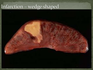
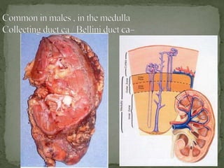
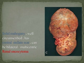
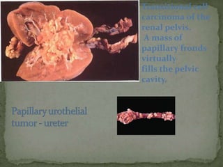
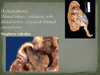
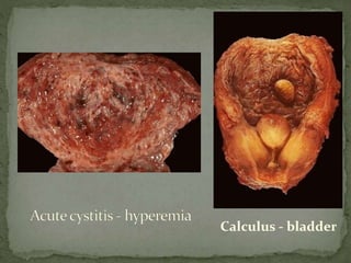
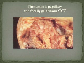
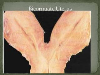
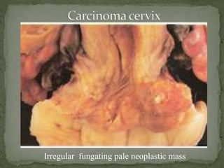
MODERATOR: DR RAJESH
PRESENTER: DR VIVEK TG
 Penis and male urethra are not dissected routinely
unless indicated in condition like
◦ Cong Urethral Valve
◦ Stricture
◦ Tumors
 The penis with or without surrounding skin should be
left attached to urinary bladder. Which is either done by
sawing out a portion of pubic bone or pulling out
through the pubic arch
• In the region of inguinal canal the inner surface of
abdominal wall is incised & spermatic cord located.
• This incision should permit the introduction of 2 fingers
of the left hand into the scrotal sac.
• Right hand is used to locate the testis & epidiymis into
the view.
• When these structure appear in the inguinal region they
are cut off from the scrotum with a knife(damage to the
scrotal skin should be looked for)
 Removal of male reproductive system :
• Removal of female reproductive system :
Pull the uterus, cervix & vagina upwards
Divide the vagina as low as possible
The FTs, uterus & ovaries are freed from the pelvis &
are removed
 Removal of bladder along with prostate & seminal vesicles &
terminal segment of rectum :
 Kidneys Should be dissected from its fatty capsule. Weight
should be taken with ureters attached
 Weight- Adults- 160 gms
At birth- 26gms
 Size 11x6x3cm
 Ureters : length 25cm
KIDNEY
 Kidneys should be held in the left hand, Should be cut
from the convex surface to hilum with a single stroke of
long knife.
 Fibrous capsule has to be stripped with a toothed forceps.
 Any amount of pus noted – between capsule & kidney /
capsule of kidney & surrounding tissue
 The ureter from the pelvis to the bladder should be opened
with a scissors
 Thickness of the renal cortex & circumference of the
renal arteries should be measured.
 Papillae should be examined for the areas of necrosis,
crystals or minute stones.
 The block should be taken from renal cortex, medulla with
a papilla & portion of the renal pelvis
• Inferior venacava opened (rt renal artery shouldn’t be
cut) :- tumor embolus / thrombus
• Renal vein – checked for thrombus
• Lower abdominal aorta – opened dorsally
• Renal arteries – opened & inspected for patency
/plaques / thrombi
• Atypical vessels – noted crossing pelvis / ureters
(responsible for hydronephrosis)
 Urinary bladder
 By placing a scissors blade into the urethra the bladder is
opened from the anterior wall & block is taken for fixation
 Capacity 120-320ml
Urethra:
 Male 18-20cm
Female 4cm
Prostate :
 It is cone shaped with
 Base measuring 4cm
 Length 3cm
 Thickness 2cm
 Weight 8gm
 Prostate is incised transversely at several levels
sectioned to include the posterior portion
 Seminal Vesicles
 Size 5cm
 Cut each vesicle longitudinally or transversely. Fluid
content to include of the wall is to be noted. Block is
to be taken
 Testis & Epididymis
 Size 4-5x3.5-2x2.7cm
 Weight 10-12 gms
 Vas deference 18 Inches
 With a sliding motion of the long knife the testis should be
cut through,
 The surface should be observed to see the tubules “string”
out normally
 One block each has to be taken from testis & epididymis
• Uterus , cervix & Vagina
Uterus
– Size 7.5x5x2.5cm
– Weight 30.40 gms
• Vagina : Anterior wall- 8cm
Posterior wall 10 cm
• With a scissor blade placed in the cervix the uterus should be opened
anteriorly
• Tubes & Ovaries
Tube measure 10cm
• The pointed end of the blade of the scissor should be put in the
fimbriated end and opened longtudianally.
• A block should be taken
• Ovaries : Should be sectioned length wise and a block should be taken
 Size- Hypoplasia / hypertrophy / atrophy /enlargement
 Shape
 External surface – scars , abscesses , tubercles ,
petechiae ,cysts , granularity
 Cut surface - corticomedullar differentiation (1:3
ratio) / cysts / abscess/ mass
 Pelvis - calculi , tumor , dilatation ,hydronephrosis
 Papillae - necrosis
 Consistency - soft – ATN / Rubbery - amyloidosis
 Capsule – thickening/ scarring
 Vessels
 Scars
 V- shaped scars- infarcts and hypertensive renal
disease
 U- shaped scars- Reflux nephropathy
• Capsule – stripped off with difficulty in
- Chronic pyelonephritis
- Benign nephrosclerosis
- Chronic glomerulonephritis
- Surface of the infarcts
Capsule should not be stripped – if
paediatric lesions suspected
eg : nephrogenic rests – located sub capsularly
• Ureters – length (25 cms) , palpated for stones , strictures
,inflammation , dilatation & tumors
• Urinary bladder :
Observe – absent anterior wall / residual urine / thickness
of muscle / polyps or diverticuli / trabeculations /
growth / stones / inflammation/ discoloration of mucosa /
rupture (trauma)
• Urethra : partial obliteration /inflammation / strictures /
caruncle / tumour growths
Prostate : inflammation / abscesses / TB foci / infarction /
median lobe enlargement / firm gland with yellowish cut
surface (carcinoma)
CONGENITAL ANOMALIES :
1 .Agenesis : absence of one kidney
2. Hypoplasia : 5 or less calyces
3. Distopia : congenital misplaced kidney (pelvis)
4. Congenital HORSE SHOE KIDNEY – both the kidneys united at the
lower pole
5 . Supernumerary kidneys
6 . Persistent fetal lobulation
7 . Atresia / stenosis of ureter / double ureters
8. Congenital megalo ureter – enters perpendicularly in to
bladder
9 .NEPHROPTOSIS –kidney easily movable due to absence of
fat tissue & may be displaced to iliac fossa
10.Congenital cysts of the kidney – uni /bilateral , in cortex /
medulla / both , communication with pelvis , contents of cysts
, external surface
1 . Bilateral large cysts & bosselated surface, not communicating
with pelvis:
ADULT POLYCYSTIC KIDNEY DISEASE , each weighing
2-4 kg, not communicating with pelvis (Potter’s type III)
2 . Bilateral large kidneys with smooth external surface :
C/S : cylindrical cysts, radiating appearance
AUTOSOMAL RECESSIVE CHILDHOOD POLYCYSTIC
KIDNEY (Potter’s type I)
3 . Cysts in the medulla : MEDULLARY SPONGE
KIDNEY
4 . Occassional cysts in cortex : Simple renal cysts
5. Normal sized / small contracted kidneys with cysts m/s
1-15mm in the corticomedullary junction :
nephronophthisis –medullary cystic disease complex
6. Reniform mass of cysts of varying sizes obscures
renal parenchyma , uni / bilateral –
normal /enlarged / small : renal cystic dysplasia
a ) Multicystic renal dysplasia : unilateral , enlarged kidneys
with cysts of varying sizes , not associated with congenital
anomalies
b) Diffuse type : bilateral , maintains shape , associated with
other abnormalities
7) Cyst with laminated membrane : HYDATID CYST
ARPKD
Due to : petechial hemorrhages over the external surface
conditions of flea – bitten kidney :
Acute post streptococcal glomerulonephritis
Malignant nephrosclerosis
Subacute bacterial endocarditis
Hemolytic uremic syndrome
Thrombotic thrombocytopenic purpura
Henoch –schonlein purpura
• Anaemia: clay coloured and firm.
• c/s pyramid light than cortex.
• Old case fatty degeneration and soft in consistency.
• Mercury poisoning- cloudy swelling and necrosis of
renal epithelium.
• Carbontetrachloride poisoning- necrotizing
nephrosis, Kidney enlarged and yellowish.
• Diaxane poisoning- hydropic degeneration with
necrosis, Kidney enlarged, pale and yellowish gray.
• 1.Chronic glomerulonephritis : b/l symmetrical small ,
contracted kidneys , with a diffusely coarsely
granular surface
• 2.Chronic pyelonephritis : Smallest of the contracted
kidneys , unilateral ,asymmetrical ,
weighing < 100 gms, surface irregularly scarred ,
capsule stripped off with difficulty due to adherence
to U -shaped scars
( Scars seen at both the poles in reflux nephropathy)
Granularity- Fine- Benign nephrosclerosis
-Coarse- Chronic glomerulonephritis
• 3. Benign nephrosclerosis :
b/l symmetrical small contracted kidneys ,
weighing < 100 gms
Capsule – adherent to surface
Surface – finely granular , with V-SHAPED scars
Other conditions of small , contracted kidney :
diabetes , amyloidosis , myeloma kidney
• Tuberculous pyelonephritis :
Often bilateral , involves medulla , papillae
replaced by caseous tissue
With obstruction  calyces filled with caseous
material , pelvis dilated  thin rim of
parenchyma surrounds pelvis ( Putty kidney /
caseous kidney)
• Tuberculosis bladder : reduced capacity due
to extensive sub mucous fibrosis (Thimble
bladder)
• Tuberculosis ureter : dilated , wide mouthed
• Renal infarction : it is a pale / anemic infarct ,
wedge shaped– base resting on cortex, apex pointing
towards medulla (occluded artery)
A rim of renal tissue preserved under the capsule
(becoz of capsular vessels)
C/S : red yellow  pale in a week & becomes
depressed below the surface
• Centered in the cortex ,well delineated , sometimes as extra
renal mass, satellite nodules may be present
• C/S : solid, at either pole (usually upper) sharply seperated by a
pseudocapsule, hemorrhages, necrosis, cystic change &
calcification-VARIEGATED APPEARNCE
Sometimes a mural nodule formation – extensive necrosis
Papillary type
Collecting duct
carcinoma
Medullary renal ca
Chromophobe renal
ca
Sarcomatoid
Other types
Solitary
angiomyolipoma -
sporadic
Transitional cell
carcinoma of the
renal pelvis.
A mass of
papillary fronds
virtually
fills the pelvic
cavity.
Staghorn calculus
1. Red hyperemic mucosa : acute cystitis
2. Solitary polypoidal lesion lobulated / smooth :
inverted papilloma / transitional ca / van brunn’s
nests
3. Confluent papillary tissue with cauliflower
appearance , multiple lesions in surrounding
mucosa : papillary transitional ca
4. Bulky polypoidal tumors with full thickness
involvement & nodular appearance :
sarcomatoid ca/ adeno ca / squamous cell ca/
poorly diff transitional ca
5 . A cluster of edematous smooth surfaced polypoid fronds
protrude in to bladder lumen : Sarcoma botroides /
hamartoma ( particularly in children)
6 . Black nodular mass : Malignant melanoma
7 . Cystic neoplasm dark blue : Hemangioma
8. Black & hemorrhagic mucosa : hemorrhagic cystitis due
to adminstration of chemotherapeutic drugs (
cyclophosphamide)
Calculus - bladder
Cystitis with malakoplakia Hypertrophy & trabeculation of bladder
The tumor is papillary
and focally gelatinous:TCC
Melanoma - urethra
The large polypoid
tumor is focally black, suggesting the
diagnosis.
Prostate:- median lobe of prostate should be examined
mild enlargement cause urinary obstruction.
Sections are made through lateral and median lobe with
knife.
Acute prostatitis- enlarged, soft in consistency small abcess
may be present.
Chronic:- large, soft, few firm areas.
TB:- enlarged, soft, military nodule, caseous foci, fistulous
tract to rectum.
Carcinoma: 20% of men after 50 shows ca
prostate.Posterior lobe is involved, firm.
 Section of prostate showing a irregular shaped
yellowish mass with a small foci of necrosis
 Malformations- Cryptorchidism
 Acute orchitis & epididymitis: swollen and firm C/S –
Abscess. Gonorrhea
 Chronic orchitis epididymitis: Smaller & firmer than
normal
 Tuberculosis: - affects epididymis- larger & soft , contains
caseous material, miliary tubercles may be found.
 Syphilis- affects testis – small, white streaks or larger with
elevated well demarcated nodules of rubbery consistency
 Tumours
 Spermatic cord: Varicocele, Tumours
 Vas deferens & seminal vesicle: Acute inflammation,
Tuberculosis
 Penis: Cong. Anomalies- Hypo/ Epispadias,
Inflammation- Posthitis, balanitis, Chancroid- round
ulcer with red soft margin, Syphilis-Chancre,
Condylomas, gummas- solitary , rubbery , elastic
 Carcinoma- Cauliflower like mass
Large tumor, bulges on c/s
Solid nodular areas of necrosis and Hemorrhage
Granular part corresponds to EC, Pearly nodules correspond to with
cartilage
Replaced testis – granular yellowish area
Well circumscribed mass multicystic yellowish keratinous
debris
Ovaries, Fallopian tubes, uterus,
Cervix and , Vagina
 Abnormalities of ovary: Senile changes, cysts, tumours
Multiloculated smooth cyst , cyst lining multiple smoth
locules
Multiple papillae locules containg viscid
Large uniloculated tumour
Well circumscribed solid
Well Circumscribed Yellow within which are cystic
foci
Fibrous , yellowish
Sebaceous material , matted hair
Fibrous and whorled – Meig syndrome
Peritonitis or gangrene rapidly develop
Infiltrating pattern of growth, that invades the myometrium
Grape like hydropic villi
Diffuse replacement of myometrium extensive hemorrhage
Irregular fungating pale neoplastic mass
Arising in post vaginal wall
• Autopsy diagnosis and technique-Otto Saphir ,4th edi
1961.
• Handbook of autopsy practise, Jurgen Ludwig, 3rd edn
• The essentials of forensic medicine and toxicology – K S
Narayana Reddy.5th ed
• National autopsy workshop GMC Mumbai, 2005.
• Internet sources
vivek autopsy 2.pptx

vivek autopsy 2.pptx

  • 1.
  • 4.
     Penis andmale urethra are not dissected routinely unless indicated in condition like ◦ Cong Urethral Valve ◦ Stricture ◦ Tumors  The penis with or without surrounding skin should be left attached to urinary bladder. Which is either done by sawing out a portion of pubic bone or pulling out through the pubic arch
  • 5.
    • In theregion of inguinal canal the inner surface of abdominal wall is incised & spermatic cord located. • This incision should permit the introduction of 2 fingers of the left hand into the scrotal sac. • Right hand is used to locate the testis & epidiymis into the view. • When these structure appear in the inguinal region they are cut off from the scrotum with a knife(damage to the scrotal skin should be looked for)
  • 6.
     Removal ofmale reproductive system :
  • 7.
    • Removal offemale reproductive system : Pull the uterus, cervix & vagina upwards Divide the vagina as low as possible The FTs, uterus & ovaries are freed from the pelvis & are removed
  • 8.
     Removal ofbladder along with prostate & seminal vesicles & terminal segment of rectum :
  • 10.
     Kidneys Shouldbe dissected from its fatty capsule. Weight should be taken with ureters attached  Weight- Adults- 160 gms At birth- 26gms  Size 11x6x3cm  Ureters : length 25cm KIDNEY
  • 11.
     Kidneys shouldbe held in the left hand, Should be cut from the convex surface to hilum with a single stroke of long knife.  Fibrous capsule has to be stripped with a toothed forceps.  Any amount of pus noted – between capsule & kidney / capsule of kidney & surrounding tissue  The ureter from the pelvis to the bladder should be opened with a scissors
  • 12.
     Thickness ofthe renal cortex & circumference of the renal arteries should be measured.  Papillae should be examined for the areas of necrosis, crystals or minute stones.  The block should be taken from renal cortex, medulla with a papilla & portion of the renal pelvis
  • 13.
    • Inferior venacavaopened (rt renal artery shouldn’t be cut) :- tumor embolus / thrombus • Renal vein – checked for thrombus • Lower abdominal aorta – opened dorsally • Renal arteries – opened & inspected for patency /plaques / thrombi • Atypical vessels – noted crossing pelvis / ureters (responsible for hydronephrosis)
  • 14.
     Urinary bladder By placing a scissors blade into the urethra the bladder is opened from the anterior wall & block is taken for fixation  Capacity 120-320ml Urethra:  Male 18-20cm Female 4cm
  • 15.
    Prostate :  Itis cone shaped with  Base measuring 4cm  Length 3cm  Thickness 2cm  Weight 8gm  Prostate is incised transversely at several levels sectioned to include the posterior portion  Seminal Vesicles  Size 5cm  Cut each vesicle longitudinally or transversely. Fluid content to include of the wall is to be noted. Block is to be taken
  • 16.
     Testis &Epididymis  Size 4-5x3.5-2x2.7cm  Weight 10-12 gms  Vas deference 18 Inches  With a sliding motion of the long knife the testis should be cut through,  The surface should be observed to see the tubules “string” out normally  One block each has to be taken from testis & epididymis
  • 17.
    • Uterus ,cervix & Vagina Uterus – Size 7.5x5x2.5cm – Weight 30.40 gms • Vagina : Anterior wall- 8cm Posterior wall 10 cm • With a scissor blade placed in the cervix the uterus should be opened anteriorly • Tubes & Ovaries Tube measure 10cm • The pointed end of the blade of the scissor should be put in the fimbriated end and opened longtudianally. • A block should be taken • Ovaries : Should be sectioned length wise and a block should be taken
  • 20.
     Size- Hypoplasia/ hypertrophy / atrophy /enlargement  Shape  External surface – scars , abscesses , tubercles , petechiae ,cysts , granularity  Cut surface - corticomedullar differentiation (1:3 ratio) / cysts / abscess/ mass  Pelvis - calculi , tumor , dilatation ,hydronephrosis  Papillae - necrosis  Consistency - soft – ATN / Rubbery - amyloidosis  Capsule – thickening/ scarring  Vessels
  • 21.
     Scars  V-shaped scars- infarcts and hypertensive renal disease  U- shaped scars- Reflux nephropathy
  • 22.
    • Capsule –stripped off with difficulty in - Chronic pyelonephritis - Benign nephrosclerosis - Chronic glomerulonephritis - Surface of the infarcts Capsule should not be stripped – if paediatric lesions suspected eg : nephrogenic rests – located sub capsularly
  • 23.
    • Ureters –length (25 cms) , palpated for stones , strictures ,inflammation , dilatation & tumors • Urinary bladder : Observe – absent anterior wall / residual urine / thickness of muscle / polyps or diverticuli / trabeculations / growth / stones / inflammation/ discoloration of mucosa / rupture (trauma)
  • 24.
    • Urethra :partial obliteration /inflammation / strictures / caruncle / tumour growths Prostate : inflammation / abscesses / TB foci / infarction / median lobe enlargement / firm gland with yellowish cut surface (carcinoma)
  • 26.
    CONGENITAL ANOMALIES : 1.Agenesis : absence of one kidney 2. Hypoplasia : 5 or less calyces 3. Distopia : congenital misplaced kidney (pelvis) 4. Congenital HORSE SHOE KIDNEY – both the kidneys united at the lower pole 5 . Supernumerary kidneys 6 . Persistent fetal lobulation
  • 29.
    7 . Atresia/ stenosis of ureter / double ureters 8. Congenital megalo ureter – enters perpendicularly in to bladder 9 .NEPHROPTOSIS –kidney easily movable due to absence of fat tissue & may be displaced to iliac fossa 10.Congenital cysts of the kidney – uni /bilateral , in cortex / medulla / both , communication with pelvis , contents of cysts , external surface
  • 30.
    1 . Bilaterallarge cysts & bosselated surface, not communicating with pelvis: ADULT POLYCYSTIC KIDNEY DISEASE , each weighing 2-4 kg, not communicating with pelvis (Potter’s type III) 2 . Bilateral large kidneys with smooth external surface : C/S : cylindrical cysts, radiating appearance AUTOSOMAL RECESSIVE CHILDHOOD POLYCYSTIC KIDNEY (Potter’s type I)
  • 31.
    3 . Cystsin the medulla : MEDULLARY SPONGE KIDNEY 4 . Occassional cysts in cortex : Simple renal cysts 5. Normal sized / small contracted kidneys with cysts m/s 1-15mm in the corticomedullary junction : nephronophthisis –medullary cystic disease complex
  • 32.
    6. Reniform massof cysts of varying sizes obscures renal parenchyma , uni / bilateral – normal /enlarged / small : renal cystic dysplasia a ) Multicystic renal dysplasia : unilateral , enlarged kidneys with cysts of varying sizes , not associated with congenital anomalies b) Diffuse type : bilateral , maintains shape , associated with other abnormalities 7) Cyst with laminated membrane : HYDATID CYST
  • 36.
  • 38.
    Due to :petechial hemorrhages over the external surface conditions of flea – bitten kidney : Acute post streptococcal glomerulonephritis Malignant nephrosclerosis Subacute bacterial endocarditis Hemolytic uremic syndrome Thrombotic thrombocytopenic purpura Henoch –schonlein purpura
  • 40.
    • Anaemia: claycoloured and firm. • c/s pyramid light than cortex. • Old case fatty degeneration and soft in consistency. • Mercury poisoning- cloudy swelling and necrosis of renal epithelium. • Carbontetrachloride poisoning- necrotizing nephrosis, Kidney enlarged and yellowish. • Diaxane poisoning- hydropic degeneration with necrosis, Kidney enlarged, pale and yellowish gray.
  • 41.
    • 1.Chronic glomerulonephritis: b/l symmetrical small , contracted kidneys , with a diffusely coarsely granular surface • 2.Chronic pyelonephritis : Smallest of the contracted kidneys , unilateral ,asymmetrical , weighing < 100 gms, surface irregularly scarred , capsule stripped off with difficulty due to adherence to U -shaped scars ( Scars seen at both the poles in reflux nephropathy)
  • 42.
    Granularity- Fine- Benignnephrosclerosis -Coarse- Chronic glomerulonephritis
  • 44.
    • 3. Benignnephrosclerosis : b/l symmetrical small contracted kidneys , weighing < 100 gms Capsule – adherent to surface Surface – finely granular , with V-SHAPED scars Other conditions of small , contracted kidney : diabetes , amyloidosis , myeloma kidney
  • 46.
    • Tuberculous pyelonephritis: Often bilateral , involves medulla , papillae replaced by caseous tissue With obstruction  calyces filled with caseous material , pelvis dilated  thin rim of parenchyma surrounds pelvis ( Putty kidney / caseous kidney) • Tuberculosis bladder : reduced capacity due to extensive sub mucous fibrosis (Thimble bladder) • Tuberculosis ureter : dilated , wide mouthed
  • 48.
    • Renal infarction: it is a pale / anemic infarct , wedge shaped– base resting on cortex, apex pointing towards medulla (occluded artery) A rim of renal tissue preserved under the capsule (becoz of capsular vessels) C/S : red yellow  pale in a week & becomes depressed below the surface
  • 52.
    • Centered inthe cortex ,well delineated , sometimes as extra renal mass, satellite nodules may be present • C/S : solid, at either pole (usually upper) sharply seperated by a pseudocapsule, hemorrhages, necrosis, cystic change & calcification-VARIEGATED APPEARNCE Sometimes a mural nodule formation – extensive necrosis
  • 54.
    Papillary type Collecting duct carcinoma Medullaryrenal ca Chromophobe renal ca Sarcomatoid Other types
  • 58.
  • 61.
    Transitional cell carcinoma ofthe renal pelvis. A mass of papillary fronds virtually fills the pelvic cavity.
  • 62.
  • 64.
    1. Red hyperemicmucosa : acute cystitis 2. Solitary polypoidal lesion lobulated / smooth : inverted papilloma / transitional ca / van brunn’s nests 3. Confluent papillary tissue with cauliflower appearance , multiple lesions in surrounding mucosa : papillary transitional ca 4. Bulky polypoidal tumors with full thickness involvement & nodular appearance : sarcomatoid ca/ adeno ca / squamous cell ca/ poorly diff transitional ca
  • 65.
    5 . Acluster of edematous smooth surfaced polypoid fronds protrude in to bladder lumen : Sarcoma botroides / hamartoma ( particularly in children) 6 . Black nodular mass : Malignant melanoma 7 . Cystic neoplasm dark blue : Hemangioma 8. Black & hemorrhagic mucosa : hemorrhagic cystitis due to adminstration of chemotherapeutic drugs ( cyclophosphamide)
  • 66.
  • 67.
    Cystitis with malakoplakiaHypertrophy & trabeculation of bladder
  • 68.
    The tumor ispapillary and focally gelatinous:TCC
  • 69.
    Melanoma - urethra Thelarge polypoid tumor is focally black, suggesting the diagnosis.
  • 71.
    Prostate:- median lobeof prostate should be examined mild enlargement cause urinary obstruction. Sections are made through lateral and median lobe with knife. Acute prostatitis- enlarged, soft in consistency small abcess may be present. Chronic:- large, soft, few firm areas. TB:- enlarged, soft, military nodule, caseous foci, fistulous tract to rectum. Carcinoma: 20% of men after 50 shows ca prostate.Posterior lobe is involved, firm.
  • 72.
     Section ofprostate showing a irregular shaped yellowish mass with a small foci of necrosis
  • 75.
     Malformations- Cryptorchidism Acute orchitis & epididymitis: swollen and firm C/S – Abscess. Gonorrhea  Chronic orchitis epididymitis: Smaller & firmer than normal  Tuberculosis: - affects epididymis- larger & soft , contains caseous material, miliary tubercles may be found.  Syphilis- affects testis – small, white streaks or larger with elevated well demarcated nodules of rubbery consistency  Tumours
  • 76.
     Spermatic cord:Varicocele, Tumours  Vas deferens & seminal vesicle: Acute inflammation, Tuberculosis  Penis: Cong. Anomalies- Hypo/ Epispadias, Inflammation- Posthitis, balanitis, Chancroid- round ulcer with red soft margin, Syphilis-Chancre, Condylomas, gummas- solitary , rubbery , elastic  Carcinoma- Cauliflower like mass
  • 78.
  • 79.
    Solid nodular areasof necrosis and Hemorrhage
  • 80.
    Granular part correspondsto EC, Pearly nodules correspond to with cartilage
  • 81.
    Replaced testis –granular yellowish area
  • 82.
    Well circumscribed massmulticystic yellowish keratinous debris
  • 84.
    Ovaries, Fallopian tubes,uterus, Cervix and , Vagina
  • 85.
     Abnormalities ofovary: Senile changes, cysts, tumours
  • 86.
    Multiloculated smooth cyst, cyst lining multiple smoth locules
  • 87.
  • 88.
  • 89.
  • 90.
    Well Circumscribed Yellowwithin which are cystic foci
  • 91.
  • 93.
  • 94.
    Fibrous and whorled– Meig syndrome
  • 97.
    Peritonitis or gangrenerapidly develop
  • 100.
    Infiltrating pattern ofgrowth, that invades the myometrium
  • 101.
  • 102.
    Diffuse replacement ofmyometrium extensive hemorrhage
  • 103.
    Irregular fungating paleneoplastic mass
  • 104.
    Arising in postvaginal wall
  • 105.
    • Autopsy diagnosisand technique-Otto Saphir ,4th edi 1961. • Handbook of autopsy practise, Jurgen Ludwig, 3rd edn • The essentials of forensic medicine and toxicology – K S Narayana Reddy.5th ed • National autopsy workshop GMC Mumbai, 2005. • Internet sources

Editor's Notes

  • #75 Irregular focally necrotic
  • #76 C/o of multiple yellowish white nodules
  • #80 WC lobulated pin- wh tu
  • #81 Large t, bulges on c/s
  • #82 Solid nodular areas of nec n hmmrr
  • #83 Granular part corresp to EC, Pearly nodules correspond to wd cartilage
  • #84 Replaced testis – granular yellowish area
  • #85 WC mass multicystic yellowish keratinous debris
  • #89 Multiloculated smooth cyst , cyst lining multiple sm locules
  • #90 Mult papillae locules containg viscid
  • #91 Large uniloclated tumour
  • #92 Wc solid
  • #93 WC Yellow t within which are cystic foci
  • #94 Fibrous , yellowish
  • #96 Sebaceous material , matted hair
  • #97 Fibrous and whorled – Meig syndrome
  • #98 Endomet, git breast
  • #99 9 wks wall is thickened and hmmr
  • #100 Peritonitis or gangrene rapidly develop
  • #103 Polypoidal focally necrotic
  • #104 Grape like hydropic villi
  • #105 Diffuse replacement of myometrium extensiv hmmrr
  • #106 Irre fungating pale neopl
  • #107 Arising in post vg wall