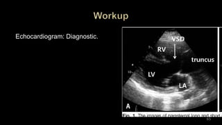
SlideShare a Scribd company logo
Dr. Walinjom Joshua
supervisor
Pr CHELO
1
Persistant truncus arteriosus
Truncus Arteriosus communis
Common aortico pulmonary trunk
2
Plan
• Definition
• Epidemiology
• History
• Embryology
• Anatomy and classification
• Pathophysiology
• Presentation
• Workup
• Treatment
• Conclusion 3
• Congenital cyanotic cardiac defect with a
single common arterial trunk giving rise to
systemic, pulmonary and coronary circulations
proximal to brachiocephalic branches
• Associated with a large perimembranous VSD
below the truncus
4
Epidemiology
TA is responsible for 0.21%-0.34 % of congenital heart
defects
Incidence has been found to be 0.03-0.05/1,000 live
births.
5
Epidemiology
risk factors:
Maternal cigarette smoking during gestation
Advanced maternal age
2q11.2 deletions (DiGeorge syndrome)
6
7
8
1798 – Wilson documents 1st case
1942 –Basic morphologic criteria - Lev
and Safir 1949 – Collet & Edwards
Classification
1962 – Ist Intracardiac repair conduit University of
Michigan
1967 – Ascending aortic allograft and valved conduit -
McGoon et al.
1971 – first conduit repair in infancy by Barratt-Boyes 9
10
• incomplete or failed septation of the embryonic truncus
arteriosus
• Or abnormality of conotruncal septation
11
Heart & great
vessels
Primitive mesoderm & neural crest cells
Give
rise
to
12
Blood islands of cardiogenic
plate
Left and right endocardial tubes
Intra embryonic coelom (early
pericardial cavity)
Coalesc
e at
20
days
within
13
Left & right endocardial
tubes
Bulbous cordis
Fus
e at
23
days
14
15
At this period, bulbo ventricular structures rotate anteriorly and
to the right
to form the heart loop
16
17
Trunco-conal swellings
Trunco-conal ridges
Truncal septum
Fus
e
18
Truncal septum divides
aorta from
Pulmonary artery and Conal
septum
Supraventricular crest and
subpulmonic infundibulum
19
Day
37Fusion of conal septum
with
endocardial cushions
establishes
ventricular separation
20
Anatomic Defects
• Single aortopulmonary trunk from base of heart
• Large perimembranous VSD (obligatory) below truncus
• Truncal valve – bi, tri or quadricuspid and often incompetent.
• Pulmonary artery arise in several patterns
• Truncal overriding equally in 60 – 80%,
to right in 10-30%,
left in 4 – 6%
21
• Anatomic Defects
• There is a single truncal valve with two, three,
or four leaflets. It is often incompetent,
resulting in regurgitation (backflow of blood).
• A large perimembranous ventricular septal
defect (VSD) is present directly below the
truncus in all cases. This allows for mixing of the
pulmonary and systemic venous blood and
equal pressures in both ventricles.
22
• The coronary arteries are frequently abnormal.
• 30% have a right aortic arch
• 33% of individuals with truncus have DiGeorge
syndrome.
23
• Anatomic Defects
• Truncal valve – bi, tri or quadricuspid and often incompetent.
24
25
Coronary anomalies
• Stenotic ostia,
• Single ostium
• high & low take off,
• abnormal branching & course
anterior descending from RCA & cross RV
circumflex from RCA
RCA from LAD
intramuscular course
26
• Right aortic arch – 30%
• Interrupted aortic arch – 10% (distal to left common carotid)
• Di George syndrome with hypocalcemia - 33%
• PFO
• OS-ASD
• Tricuspid valve lesions
• 22q11 chromosome deletion
27
Classification
Type 1
single pulmonary trunk from the left lateral aspect of the
common trunk,
with branching of the left and right pulmonary arteries from the
pulmonary trunk
28
29
Type 2
separate but proximale origins of the left and right pulmonary
arterial branches from the posterolateral aspect of the
common trunk
30
31
Type 3
branch pulmonary arteries originate independently from the
common trunk
32
33
Type 4
Pseudo-truncus;
TOF with pulm. Atresia with MAPCA
major aorto pulmonary collateral
arteries
34
35
36
Type A1
Identical to the type I of Collett and Edwards
37
38
Type A2
Collett and Edwards type II and most cases of type III
39
40
Type A3 (hemitruncus)
• one branch pulmonary artery (usually the right) from the
common trunk
• The other branch pulmonary artery from the aortic arch (a
subtype of Collett and Edwards type III) or by systemic to
pulmonary arterial collaterals
41
42
Type A4
coexistence of an interrupted aortic arch
not by the pattern of origin of branch pulmonary arteries
43
44
Cyanotic al heart disease with increased pulmonary blood
flow
Fetal pulmonary blood flow less than 10%
PVR falls in early infancy improving PBF hence good
oxygen saturation
Hypoxia in this period implies pulmonary arterial narrowing
Equilibration of RV LV pressures
45
The physiology of TA is largely related to the
volume of blood flowing to the pulmonary circuit.
This is affected by:
1.The degree of pulmonary vascular resistance
2.The degree of truncal valvular insufficiency
3.The severity of any aortic arch abnormalities
46
1. Pulmonary vascular resistance
•At birth the pulmonary vascular resistance is high
enough to prevent left-to-right shunting, which
restricts the amount of blood that can flow to the
pulmonary system.
•As this resistance drops more blood flows to the
pulmonary system, the amount of fluid overloads the
system, and heart failure begins. 47
Heart failure can begin within several
weeks of birth and present by
6 months of age in patients with TA.
48
2 . Truncal valvular insufficiency
•The degree of regurgitation or stenosis of
the truncal valve causes additional ventricular
volume.
•The myocardium then requires greater amounts
of oxygen, which leads to coronary artery
ischemia and ventricular dysfunction.
•Together, this also contributes to the onset and
severity of heart failure. 49
3. Aortic arch abnormalities
•Patients with interrupted aortic
• arches or coarctation require
•the ductus arteriosus for
• alleviating pressure overload
•in the ventricles and allowing for
• blood flow distally
50
• The magnitude of pulmonary blood flow (PBF) is
determined by the size of the pulmonary artery.
• If PBF is excessive, congestive heart failure
(CHF) may occur as a result of volume overload
placed on the ventricle.
• If PBF is small, the infant may appear more
cyanotic (blue) with no CHF symptoms.
51
• Most infants present with cyanosis or symptoms
of CHF within the first two weeks of life if not
diagnosed prenatally.
• Truncus arteriosus occurs in less than 1% of
congenital heart defects.
52
Cyanotic congenital heart disease with increased pulmonary
blood flow
Fetal pulmonary blood flow less than 10%
PVR falls in early infancy improving PBF hence good
oxygen saturation
Hypoxia in this period implies pulmonary arterial narrowing
Equilibration of RV LV pressures
53
As the PBF increases PAH
Increased pulmonary venous return
CCF
54
History
• Cyanosis at birth
• Early CCF
• Failure to thrive,
• Respiratory tract infections
55
. Cyanosis in patients with TA is due to the mixing of pulmonary
and systemic blood.
. Most patients with TA are diagnosed within one week of birth –
after pulmonary vascular resistance decreases and the ductus
arteriosus closes.
Patients present with cyanosis and respiratory distress from
pulmonary congestion and onset of heart failure.
56
57
.Some patients also present with a murmur.
.Pulmonary congestion and heart failure can manifest as:
.Poor feeding
.Lethargy
.Respiratory distress (tachypnea, subcostal retractions, nasal
flaring, grunting)
.Tachycardia
.Hyperdynamic precordium
. Hepatomegaly
58
Bounding peripheral pulses and a wide pulse pressure are
present.
An early diastolic murmur of truncal regurgitation may be heard.
Occasionally,
a harsh, regurgitant systolic VSD murmur may be heard along
the left sternal border.
59
A systolic click
may be heard at the apex and upper left sternal border and S2
is single.
If pulmonary blood flow is excessive, an apical diastolic
rumble with or without gallop rhythm may be present.
60
Physical examination
• Cyanosis
• Signs of CCF
• Bounding peripheral pulses, wide pulse pressure
• Single S2
• Harsh systolic regurgitant murmur – VSD
• EDM – truncal valve regurgitation
61
Survival
50% survival in 1 month
18% survival in 6 months
12% survival in 1 year
Modes of death
. Congestive heart failure in early life
. SBE, cerebral abscess → Eisenmenger syndrome (death in 3rd
decade)
. Adversely affected by truncal regurgitation, IAA, CoA
. Survival is favorably affected by PS
62
Prenatal and early postnatal diagnosis common
Proper evaluation of cyanotic infants clinches diagnosis
ABG
Pulse oximetry
ECG
CXR
TTE,
TOE
63
Diagnostics:
Chest X-ray: Cardiomegaly (enlarged heart) with
increased pulmonary vascular markings.
EKG: Normal QRS axis. Biventricular hypertrophy is
present in 70% of children.
Echocardiogram: Diagnostic.
64
Echocardiogram: Diagnostic.
65
ECG
• Normal QRS axis
• Bi-ventricular hypertrophy – 70%
66
CXR
• Cardiomegaly
• Pulmonary plethora
• Right aortic arch –
30%
67
CARDIAC CATHETERISATION
• Assess PVR in late presentations.
68
69
Differential
•Large VSD
•Pulmonary atresia with VSD
•Univentricular heart
Medical
Surgical
• Palliative
• Defintive
70
71
Treatment for TA is surgical intervention.
72
Patients with TA are initially medically managed to stabilize
them for surgery.
Medications include:
• Diuretics to manage volume overload
• Inotropes (e.g. dobutamine or dopamine) for cardiac
contractility,
• ACE inhibitors to reduce afterload
Ventilation
• Prostaglandin E1 is given to patients with severe aortic
coarctation to maintain patency of the ductus arteriosus.
73
Surgery is usually performed within 30 days of birth.
A typical procedure:
1.Removes the pulmonary arteries from the truncus and
reattaches them to the right ventricle
2. Opens and repairs the truncus
3. Closes the VSD..
74
Perioperative mortality is 10%
The risks of not intervening are so great that most patients
undergo primary surgical repair.
Long-term survival:
90% at 5 years after primary repair
85% at 10 years
83% at 15 years
75
After surgery, all patients with TA are followed by pediatric
cardiologists.
Prognosis for patients with unrepaired TA is poor and
patients usually do not survive without surgical
intervention.
For patients without repair, the mean age of survival is 5
weeks and 85% do not survive to 12 months of age.
Thank you
76

More Related Content

What's hot

Pediatric pulmonary hypertension
Pediatric pulmonary hypertension Pediatric pulmonary hypertension
Pediatric pulmonary hypertension
Nagendra prasad Kulari
 
ATRIOVENTRICULAR SEPTAL DEFECT
ATRIOVENTRICULAR SEPTAL DEFECTATRIOVENTRICULAR SEPTAL DEFECT
ATRIOVENTRICULAR SEPTAL DEFECT
Dr. Murtaza Kamal MD,DNB,DrNB Ped Cardiology
 
D TGA
D TGAD TGA
AV septal defects (AVCD)
AV septal defects (AVCD)AV septal defects (AVCD)
AV septal defects (AVCD)
Malleswara rao Dangeti
 
Total anomalous pulmonary venous connections seminar ppt.
Total anomalous pulmonary venous connections seminar ppt.Total anomalous pulmonary venous connections seminar ppt.
Total anomalous pulmonary venous connections seminar ppt.
Pawan Ola
 
Approach to cyanotic congenital heart disease
Approach to cyanotic congenital heart diseaseApproach to cyanotic congenital heart disease
Approach to cyanotic congenital heart disease
ikramdr01
 
Functional echocardiography ppt
Functional echocardiography   pptFunctional echocardiography   ppt
Functional echocardiography ppt
rajasthan govt
 
Truncus Arteriosus
Truncus Arteriosus Truncus Arteriosus
Truncus Arteriosus
Vishwanath Hesarur
 
Dr ranjith mp av canal defect
Dr ranjith mp av canal defectDr ranjith mp av canal defect
Dr ranjith mp av canal defectdrranjithmp
 
Truncus arteriosus
Truncus arteriosusTruncus arteriosus
Truncus arteriosusgisa_legal
 
L-TGA or CCTGA
L-TGA or CCTGA L-TGA or CCTGA
L-TGA or CCTGA
Malleswara rao Dangeti
 
EBSTEIN ANOMALY
EBSTEIN ANOMALYEBSTEIN ANOMALY
Approach to Cyanotic Congenital Heart Diseases
Approach to Cyanotic Congenital Heart DiseasesApproach to Cyanotic Congenital Heart Diseases
Approach to Cyanotic Congenital Heart DiseasesCSN Vittal
 
Truncus arteriosus
Truncus arteriosusTruncus arteriosus
Truncus arteriosus
Aliaa Shaban
 
Truncus arteriosus Dr Shiva CTVS JIPMER
Truncus arteriosus Dr Shiva CTVS JIPMERTruncus arteriosus Dr Shiva CTVS JIPMER
Truncus arteriosus Dr Shiva CTVS JIPMERShivashankar Sadasivam
 
Hypoplastic left heart syndrome
Hypoplastic left heart syndromeHypoplastic left heart syndrome
Hypoplastic left heart syndrome
Drhunny88
 
Truncus arteriosus - Hemitruncus - Pseudotruncus CT role
Truncus arteriosus - Hemitruncus - Pseudotruncus CT roleTruncus arteriosus - Hemitruncus - Pseudotruncus CT role
Truncus arteriosus - Hemitruncus - Pseudotruncus CT role
Mohamed Gibreel
 
single ventricle physiology
single ventricle physiologysingle ventricle physiology
single ventricle physiology
richamalik99
 

What's hot (20)

Pediatric pulmonary hypertension
Pediatric pulmonary hypertension Pediatric pulmonary hypertension
Pediatric pulmonary hypertension
 
ATRIOVENTRICULAR SEPTAL DEFECT
ATRIOVENTRICULAR SEPTAL DEFECTATRIOVENTRICULAR SEPTAL DEFECT
ATRIOVENTRICULAR SEPTAL DEFECT
 
D TGA
D TGAD TGA
D TGA
 
AV septal defects (AVCD)
AV septal defects (AVCD)AV septal defects (AVCD)
AV septal defects (AVCD)
 
Total anomalous pulmonary venous connections seminar ppt.
Total anomalous pulmonary venous connections seminar ppt.Total anomalous pulmonary venous connections seminar ppt.
Total anomalous pulmonary venous connections seminar ppt.
 
Approach to cyanotic congenital heart disease
Approach to cyanotic congenital heart diseaseApproach to cyanotic congenital heart disease
Approach to cyanotic congenital heart disease
 
Functional echocardiography ppt
Functional echocardiography   pptFunctional echocardiography   ppt
Functional echocardiography ppt
 
Truncus Arteriosus
Truncus Arteriosus Truncus Arteriosus
Truncus Arteriosus
 
Dr ranjith mp av canal defect
Dr ranjith mp av canal defectDr ranjith mp av canal defect
Dr ranjith mp av canal defect
 
Truncus arteriosus
Truncus arteriosusTruncus arteriosus
Truncus arteriosus
 
L-TGA or CCTGA
L-TGA or CCTGA L-TGA or CCTGA
L-TGA or CCTGA
 
EBSTEIN ANOMALY
EBSTEIN ANOMALYEBSTEIN ANOMALY
EBSTEIN ANOMALY
 
Approach to Cyanotic Congenital Heart Diseases
Approach to Cyanotic Congenital Heart DiseasesApproach to Cyanotic Congenital Heart Diseases
Approach to Cyanotic Congenital Heart Diseases
 
Truncus arteriosus
Truncus arteriosusTruncus arteriosus
Truncus arteriosus
 
Truncus arteriosus Dr Shiva CTVS JIPMER
Truncus arteriosus Dr Shiva CTVS JIPMERTruncus arteriosus Dr Shiva CTVS JIPMER
Truncus arteriosus Dr Shiva CTVS JIPMER
 
Hypoplastic left heart syndrome
Hypoplastic left heart syndromeHypoplastic left heart syndrome
Hypoplastic left heart syndrome
 
Truncus arteriosus - Hemitruncus - Pseudotruncus CT role
Truncus arteriosus - Hemitruncus - Pseudotruncus CT roleTruncus arteriosus - Hemitruncus - Pseudotruncus CT role
Truncus arteriosus - Hemitruncus - Pseudotruncus CT role
 
single ventricle physiology
single ventricle physiologysingle ventricle physiology
single ventricle physiology
 
Fontan
FontanFontan
Fontan
 
CONGENITAL CORONARY ANOMALIES AND VARIANTS, ALCAPA
CONGENITAL CORONARY ANOMALIES AND VARIANTS, ALCAPACONGENITAL CORONARY ANOMALIES AND VARIANTS, ALCAPA
CONGENITAL CORONARY ANOMALIES AND VARIANTS, ALCAPA
 

Similar to Truncus arteriosus by Dr Wali

1)Congenital HD 2009.ppt
1)Congenital HD 2009.ppt1)Congenital HD 2009.ppt
1)Congenital HD 2009.ppt
FenembarMekonnen
 
Tricuspid atresia
Tricuspid atresiaTricuspid atresia
Tricuspid atresia
Dina Mostafa
 
Cchd
CchdCchd
congenital heart diseases.pptx
congenital heart diseases.pptxcongenital heart diseases.pptx
congenital heart diseases.pptx
CHANDAN PADHAN
 
Approach to cyanotic congenital heart disease in new born
Approach to cyanotic congenital heart disease in new bornApproach to cyanotic congenital heart disease in new born
Approach to cyanotic congenital heart disease in new born
Jigar Patel
 
Pulmonary arterial hypertension (PAH) in ccongenital heart diseases
Pulmonary arterial hypertension (PAH) in ccongenital heart diseasesPulmonary arterial hypertension (PAH) in ccongenital heart diseases
Pulmonary arterial hypertension (PAH) in ccongenital heart diseases
Malleswara rao Dangeti
 
congenital heart disease_january2011_final
congenital heart disease_january2011_finalcongenital heart disease_january2011_final
congenital heart disease_january2011_final
Engidaw Ambelu
 
Persistent pulmonary hypertension of newborn PPHN
Persistent pulmonary hypertension of newborn PPHNPersistent pulmonary hypertension of newborn PPHN
Persistent pulmonary hypertension of newborn PPHN
Chandan Gowda
 
Congenital heart diseases.pptx
Congenital heart diseases.pptxCongenital heart diseases.pptx
Congenital heart diseases.pptx
maneeshsen2
 
Endocardial Cushion Defect / AVSD
Endocardial Cushion Defect / AVSDEndocardial Cushion Defect / AVSD
Endocardial Cushion Defect / AVSD
Harshitha
 
Asd and vsd
Asd and vsdAsd and vsd
Asd and vsd
Mohit Aggarwal
 
CONGENITAL HEART DISEASE LECTURE NOTES MD3.pptx
CONGENITAL HEART DISEASE LECTURE NOTES MD3.pptxCONGENITAL HEART DISEASE LECTURE NOTES MD3.pptx
CONGENITAL HEART DISEASE LECTURE NOTES MD3.pptx
ErhardRutakulemberwa
 
Cyan
CyanCyan
Cyan
sangpeds
 
Aortic regurgitation
Aortic regurgitationAortic regurgitation
Aortic regurgitation
Vitrag Shah
 
Cyanotic heart disease complete ppts
Cyanotic heart disease complete pptsCyanotic heart disease complete ppts
Cyanotic heart disease complete ppts
DrMuddasarHussain
 
HEMODYNAMICS & NATURAL HISTORY OF PS.pptx
HEMODYNAMICS & NATURAL HISTORY OF PS.pptxHEMODYNAMICS & NATURAL HISTORY OF PS.pptx
HEMODYNAMICS & NATURAL HISTORY OF PS.pptx
Aparanji Gopidi
 
Pavm
PavmPavm
CHD cvs.pdf
CHD cvs.pdfCHD cvs.pdf
CHD cvs.pdf
RyanKhan40
 
Congenital Heart Disorders (TOF, TGV, COA)
Congenital Heart Disorders (TOF, TGV, COA) Congenital Heart Disorders (TOF, TGV, COA)
Congenital Heart Disorders (TOF, TGV, COA)
Kishore Rajan
 

Similar to Truncus arteriosus by Dr Wali (20)

1)Congenital HD 2009.ppt
1)Congenital HD 2009.ppt1)Congenital HD 2009.ppt
1)Congenital HD 2009.ppt
 
Ppt ph delay
Ppt ph delayPpt ph delay
Ppt ph delay
 
Tricuspid atresia
Tricuspid atresiaTricuspid atresia
Tricuspid atresia
 
Cchd
CchdCchd
Cchd
 
congenital heart diseases.pptx
congenital heart diseases.pptxcongenital heart diseases.pptx
congenital heart diseases.pptx
 
Approach to cyanotic congenital heart disease in new born
Approach to cyanotic congenital heart disease in new bornApproach to cyanotic congenital heart disease in new born
Approach to cyanotic congenital heart disease in new born
 
Pulmonary arterial hypertension (PAH) in ccongenital heart diseases
Pulmonary arterial hypertension (PAH) in ccongenital heart diseasesPulmonary arterial hypertension (PAH) in ccongenital heart diseases
Pulmonary arterial hypertension (PAH) in ccongenital heart diseases
 
congenital heart disease_january2011_final
congenital heart disease_january2011_finalcongenital heart disease_january2011_final
congenital heart disease_january2011_final
 
Persistent pulmonary hypertension of newborn PPHN
Persistent pulmonary hypertension of newborn PPHNPersistent pulmonary hypertension of newborn PPHN
Persistent pulmonary hypertension of newborn PPHN
 
Congenital heart diseases.pptx
Congenital heart diseases.pptxCongenital heart diseases.pptx
Congenital heart diseases.pptx
 
Endocardial Cushion Defect / AVSD
Endocardial Cushion Defect / AVSDEndocardial Cushion Defect / AVSD
Endocardial Cushion Defect / AVSD
 
Asd and vsd
Asd and vsdAsd and vsd
Asd and vsd
 
CONGENITAL HEART DISEASE LECTURE NOTES MD3.pptx
CONGENITAL HEART DISEASE LECTURE NOTES MD3.pptxCONGENITAL HEART DISEASE LECTURE NOTES MD3.pptx
CONGENITAL HEART DISEASE LECTURE NOTES MD3.pptx
 
Cyan
CyanCyan
Cyan
 
Aortic regurgitation
Aortic regurgitationAortic regurgitation
Aortic regurgitation
 
Cyanotic heart disease complete ppts
Cyanotic heart disease complete pptsCyanotic heart disease complete ppts
Cyanotic heart disease complete ppts
 
HEMODYNAMICS & NATURAL HISTORY OF PS.pptx
HEMODYNAMICS & NATURAL HISTORY OF PS.pptxHEMODYNAMICS & NATURAL HISTORY OF PS.pptx
HEMODYNAMICS & NATURAL HISTORY OF PS.pptx
 
Pavm
PavmPavm
Pavm
 
CHD cvs.pdf
CHD cvs.pdfCHD cvs.pdf
CHD cvs.pdf
 
Congenital Heart Disorders (TOF, TGV, COA)
Congenital Heart Disorders (TOF, TGV, COA) Congenital Heart Disorders (TOF, TGV, COA)
Congenital Heart Disorders (TOF, TGV, COA)
 

More from Dr. Joshua WALINJOM

Atrial Fibrillation By Dr Joshua Walinjom.ppt
Atrial Fibrillation By Dr Joshua Walinjom.pptAtrial Fibrillation By Dr Joshua Walinjom.ppt
Atrial Fibrillation By Dr Joshua Walinjom.ppt
Dr. Joshua WALINJOM
 
Differential Diagnosis of chest pain. by Dr Joshua Walinjom
Differential Diagnosis of chest pain.  by Dr Joshua WalinjomDifferential Diagnosis of chest pain.  by Dr Joshua Walinjom
Differential Diagnosis of chest pain. by Dr Joshua Walinjom
Dr. Joshua WALINJOM
 
Conduction disorders with sinoatrial blocks Dr Walinjom
Conduction disorders with sinoatrial blocks Dr WalinjomConduction disorders with sinoatrial blocks Dr Walinjom
Conduction disorders with sinoatrial blocks Dr Walinjom
Dr. Joshua WALINJOM
 
Aortic stenosis
Aortic stenosis Aortic stenosis
Aortic stenosis
Dr. Joshua WALINJOM
 
Urgene hypertensive
Urgene hypertensiveUrgene hypertensive
Urgene hypertensive
Dr. Joshua WALINJOM
 
Transposition of the great arteries
Transposition of the great arteriesTransposition of the great arteries
Transposition of the great arteries
Dr. Joshua WALINJOM
 

More from Dr. Joshua WALINJOM (6)

Atrial Fibrillation By Dr Joshua Walinjom.ppt
Atrial Fibrillation By Dr Joshua Walinjom.pptAtrial Fibrillation By Dr Joshua Walinjom.ppt
Atrial Fibrillation By Dr Joshua Walinjom.ppt
 
Differential Diagnosis of chest pain. by Dr Joshua Walinjom
Differential Diagnosis of chest pain.  by Dr Joshua WalinjomDifferential Diagnosis of chest pain.  by Dr Joshua Walinjom
Differential Diagnosis of chest pain. by Dr Joshua Walinjom
 
Conduction disorders with sinoatrial blocks Dr Walinjom
Conduction disorders with sinoatrial blocks Dr WalinjomConduction disorders with sinoatrial blocks Dr Walinjom
Conduction disorders with sinoatrial blocks Dr Walinjom
 
Aortic stenosis
Aortic stenosis Aortic stenosis
Aortic stenosis
 
Urgene hypertensive
Urgene hypertensiveUrgene hypertensive
Urgene hypertensive
 
Transposition of the great arteries
Transposition of the great arteriesTransposition of the great arteries
Transposition of the great arteries
 

Recently uploaded

Pulmonary Thromboembolism - etilogy, types, medical- Surgical and nursing man...
Pulmonary Thromboembolism - etilogy, types, medical- Surgical and nursing man...Pulmonary Thromboembolism - etilogy, types, medical- Surgical and nursing man...
Pulmonary Thromboembolism - etilogy, types, medical- Surgical and nursing man...
VarunMahajani
 
ANATOMY AND PHYSIOLOGY OF URINARY SYSTEM.pptx
ANATOMY AND PHYSIOLOGY OF URINARY SYSTEM.pptxANATOMY AND PHYSIOLOGY OF URINARY SYSTEM.pptx
ANATOMY AND PHYSIOLOGY OF URINARY SYSTEM.pptx
Swetaba Besh
 
How to Give Better Lectures: Some Tips for Doctors
How to Give Better Lectures: Some Tips for DoctorsHow to Give Better Lectures: Some Tips for Doctors
How to Give Better Lectures: Some Tips for Doctors
LanceCatedral
 
ACUTE SCROTUM.....pdf. ACUTE SCROTAL CONDITIOND
ACUTE SCROTUM.....pdf. ACUTE SCROTAL CONDITIONDACUTE SCROTUM.....pdf. ACUTE SCROTAL CONDITIOND
ACUTE SCROTUM.....pdf. ACUTE SCROTAL CONDITIOND
DR SETH JOTHAM
 
24 Upakrama.pptx class ppt useful in all
24 Upakrama.pptx class ppt useful in all24 Upakrama.pptx class ppt useful in all
24 Upakrama.pptx class ppt useful in all
DrSathishMS1
 
Ocular injury ppt Upendra pal optometrist upums saifai etawah
Ocular injury  ppt  Upendra pal  optometrist upums saifai etawahOcular injury  ppt  Upendra pal  optometrist upums saifai etawah
Ocular injury ppt Upendra pal optometrist upums saifai etawah
pal078100
 
basicmodesofventilation2022-220313203758.pdf
basicmodesofventilation2022-220313203758.pdfbasicmodesofventilation2022-220313203758.pdf
basicmodesofventilation2022-220313203758.pdf
aljamhori teaching hospital
 
Surat @ℂall @Girls ꧁❤8527049040❤꧂@ℂall @Girls Service Vip Top Model Safe
Surat @ℂall @Girls ꧁❤8527049040❤꧂@ℂall @Girls Service Vip Top Model SafeSurat @ℂall @Girls ꧁❤8527049040❤꧂@ℂall @Girls Service Vip Top Model Safe
Surat @ℂall @Girls ꧁❤8527049040❤꧂@ℂall @Girls Service Vip Top Model Safe
Savita Shen $i11
 
Alcohol_Dr. Jeenal Mistry MD Pharmacology.pdf
Alcohol_Dr. Jeenal Mistry MD Pharmacology.pdfAlcohol_Dr. Jeenal Mistry MD Pharmacology.pdf
Alcohol_Dr. Jeenal Mistry MD Pharmacology.pdf
Dr Jeenal Mistry
 
Physiology of Special Chemical Sensation of Taste
Physiology of Special Chemical Sensation of TastePhysiology of Special Chemical Sensation of Taste
Physiology of Special Chemical Sensation of Taste
MedicoseAcademics
 
Non-respiratory Functions of the Lungs.pdf
Non-respiratory Functions of the Lungs.pdfNon-respiratory Functions of the Lungs.pdf
Non-respiratory Functions of the Lungs.pdf
MedicoseAcademics
 
ARTIFICIAL INTELLIGENCE IN HEALTHCARE.pdf
ARTIFICIAL INTELLIGENCE IN  HEALTHCARE.pdfARTIFICIAL INTELLIGENCE IN  HEALTHCARE.pdf
ARTIFICIAL INTELLIGENCE IN HEALTHCARE.pdf
Anujkumaranit
 
Maxilla, Mandible & Hyoid Bone & Clinical Correlations by Dr. RIG.pptx
Maxilla, Mandible & Hyoid Bone & Clinical Correlations by Dr. RIG.pptxMaxilla, Mandible & Hyoid Bone & Clinical Correlations by Dr. RIG.pptx
Maxilla, Mandible & Hyoid Bone & Clinical Correlations by Dr. RIG.pptx
Dr. Rabia Inam Gandapore
 
Ophthalmology Clinical Tests for OSCE exam
Ophthalmology Clinical Tests for OSCE examOphthalmology Clinical Tests for OSCE exam
Ophthalmology Clinical Tests for OSCE exam
KafrELShiekh University
 
Flu Vaccine Alert in Bangalore Karnataka
Flu Vaccine Alert in Bangalore KarnatakaFlu Vaccine Alert in Bangalore Karnataka
Flu Vaccine Alert in Bangalore Karnataka
addon Scans
 
How STIs Influence the Development of Pelvic Inflammatory Disease.pptx
How STIs Influence the Development of Pelvic Inflammatory Disease.pptxHow STIs Influence the Development of Pelvic Inflammatory Disease.pptx
How STIs Influence the Development of Pelvic Inflammatory Disease.pptx
FFragrant
 
Lung Cancer: Artificial Intelligence, Synergetics, Complex System Analysis, S...
Lung Cancer: Artificial Intelligence, Synergetics, Complex System Analysis, S...Lung Cancer: Artificial Intelligence, Synergetics, Complex System Analysis, S...
Lung Cancer: Artificial Intelligence, Synergetics, Complex System Analysis, S...
Oleg Kshivets
 
ARTHROLOGY PPT NCISM SYLLABUS AYURVEDA STUDENTS
ARTHROLOGY PPT NCISM SYLLABUS AYURVEDA STUDENTSARTHROLOGY PPT NCISM SYLLABUS AYURVEDA STUDENTS
ARTHROLOGY PPT NCISM SYLLABUS AYURVEDA STUDENTS
Dr. Vinay Pareek
 
Hemodialysis: Chapter 3, Dialysis Water Unit - Dr.Gawad
Hemodialysis: Chapter 3, Dialysis Water Unit - Dr.GawadHemodialysis: Chapter 3, Dialysis Water Unit - Dr.Gawad
Hemodialysis: Chapter 3, Dialysis Water Unit - Dr.Gawad
NephroTube - Dr.Gawad
 
BRACHYTHERAPY OVERVIEW AND APPLICATORS
BRACHYTHERAPY OVERVIEW  AND  APPLICATORSBRACHYTHERAPY OVERVIEW  AND  APPLICATORS
BRACHYTHERAPY OVERVIEW AND APPLICATORS
Krishan Murari
 

Recently uploaded (20)

Pulmonary Thromboembolism - etilogy, types, medical- Surgical and nursing man...
Pulmonary Thromboembolism - etilogy, types, medical- Surgical and nursing man...Pulmonary Thromboembolism - etilogy, types, medical- Surgical and nursing man...
Pulmonary Thromboembolism - etilogy, types, medical- Surgical and nursing man...
 
ANATOMY AND PHYSIOLOGY OF URINARY SYSTEM.pptx
ANATOMY AND PHYSIOLOGY OF URINARY SYSTEM.pptxANATOMY AND PHYSIOLOGY OF URINARY SYSTEM.pptx
ANATOMY AND PHYSIOLOGY OF URINARY SYSTEM.pptx
 
How to Give Better Lectures: Some Tips for Doctors
How to Give Better Lectures: Some Tips for DoctorsHow to Give Better Lectures: Some Tips for Doctors
How to Give Better Lectures: Some Tips for Doctors
 
ACUTE SCROTUM.....pdf. ACUTE SCROTAL CONDITIOND
ACUTE SCROTUM.....pdf. ACUTE SCROTAL CONDITIONDACUTE SCROTUM.....pdf. ACUTE SCROTAL CONDITIOND
ACUTE SCROTUM.....pdf. ACUTE SCROTAL CONDITIOND
 
24 Upakrama.pptx class ppt useful in all
24 Upakrama.pptx class ppt useful in all24 Upakrama.pptx class ppt useful in all
24 Upakrama.pptx class ppt useful in all
 
Ocular injury ppt Upendra pal optometrist upums saifai etawah
Ocular injury  ppt  Upendra pal  optometrist upums saifai etawahOcular injury  ppt  Upendra pal  optometrist upums saifai etawah
Ocular injury ppt Upendra pal optometrist upums saifai etawah
 
basicmodesofventilation2022-220313203758.pdf
basicmodesofventilation2022-220313203758.pdfbasicmodesofventilation2022-220313203758.pdf
basicmodesofventilation2022-220313203758.pdf
 
Surat @ℂall @Girls ꧁❤8527049040❤꧂@ℂall @Girls Service Vip Top Model Safe
Surat @ℂall @Girls ꧁❤8527049040❤꧂@ℂall @Girls Service Vip Top Model SafeSurat @ℂall @Girls ꧁❤8527049040❤꧂@ℂall @Girls Service Vip Top Model Safe
Surat @ℂall @Girls ꧁❤8527049040❤꧂@ℂall @Girls Service Vip Top Model Safe
 
Alcohol_Dr. Jeenal Mistry MD Pharmacology.pdf
Alcohol_Dr. Jeenal Mistry MD Pharmacology.pdfAlcohol_Dr. Jeenal Mistry MD Pharmacology.pdf
Alcohol_Dr. Jeenal Mistry MD Pharmacology.pdf
 
Physiology of Special Chemical Sensation of Taste
Physiology of Special Chemical Sensation of TastePhysiology of Special Chemical Sensation of Taste
Physiology of Special Chemical Sensation of Taste
 
Non-respiratory Functions of the Lungs.pdf
Non-respiratory Functions of the Lungs.pdfNon-respiratory Functions of the Lungs.pdf
Non-respiratory Functions of the Lungs.pdf
 
ARTIFICIAL INTELLIGENCE IN HEALTHCARE.pdf
ARTIFICIAL INTELLIGENCE IN  HEALTHCARE.pdfARTIFICIAL INTELLIGENCE IN  HEALTHCARE.pdf
ARTIFICIAL INTELLIGENCE IN HEALTHCARE.pdf
 
Maxilla, Mandible & Hyoid Bone & Clinical Correlations by Dr. RIG.pptx
Maxilla, Mandible & Hyoid Bone & Clinical Correlations by Dr. RIG.pptxMaxilla, Mandible & Hyoid Bone & Clinical Correlations by Dr. RIG.pptx
Maxilla, Mandible & Hyoid Bone & Clinical Correlations by Dr. RIG.pptx
 
Ophthalmology Clinical Tests for OSCE exam
Ophthalmology Clinical Tests for OSCE examOphthalmology Clinical Tests for OSCE exam
Ophthalmology Clinical Tests for OSCE exam
 
Flu Vaccine Alert in Bangalore Karnataka
Flu Vaccine Alert in Bangalore KarnatakaFlu Vaccine Alert in Bangalore Karnataka
Flu Vaccine Alert in Bangalore Karnataka
 
How STIs Influence the Development of Pelvic Inflammatory Disease.pptx
How STIs Influence the Development of Pelvic Inflammatory Disease.pptxHow STIs Influence the Development of Pelvic Inflammatory Disease.pptx
How STIs Influence the Development of Pelvic Inflammatory Disease.pptx
 
Lung Cancer: Artificial Intelligence, Synergetics, Complex System Analysis, S...
Lung Cancer: Artificial Intelligence, Synergetics, Complex System Analysis, S...Lung Cancer: Artificial Intelligence, Synergetics, Complex System Analysis, S...
Lung Cancer: Artificial Intelligence, Synergetics, Complex System Analysis, S...
 
ARTHROLOGY PPT NCISM SYLLABUS AYURVEDA STUDENTS
ARTHROLOGY PPT NCISM SYLLABUS AYURVEDA STUDENTSARTHROLOGY PPT NCISM SYLLABUS AYURVEDA STUDENTS
ARTHROLOGY PPT NCISM SYLLABUS AYURVEDA STUDENTS
 
Hemodialysis: Chapter 3, Dialysis Water Unit - Dr.Gawad
Hemodialysis: Chapter 3, Dialysis Water Unit - Dr.GawadHemodialysis: Chapter 3, Dialysis Water Unit - Dr.Gawad
Hemodialysis: Chapter 3, Dialysis Water Unit - Dr.Gawad
 
BRACHYTHERAPY OVERVIEW AND APPLICATORS
BRACHYTHERAPY OVERVIEW  AND  APPLICATORSBRACHYTHERAPY OVERVIEW  AND  APPLICATORS
BRACHYTHERAPY OVERVIEW AND APPLICATORS
 

Truncus arteriosus by Dr Wali

  • 2. Persistant truncus arteriosus Truncus Arteriosus communis Common aortico pulmonary trunk 2
  • 3. Plan • Definition • Epidemiology • History • Embryology • Anatomy and classification • Pathophysiology • Presentation • Workup • Treatment • Conclusion 3
  • 4. • Congenital cyanotic cardiac defect with a single common arterial trunk giving rise to systemic, pulmonary and coronary circulations proximal to brachiocephalic branches • Associated with a large perimembranous VSD below the truncus 4
  • 5. Epidemiology TA is responsible for 0.21%-0.34 % of congenital heart defects Incidence has been found to be 0.03-0.05/1,000 live births. 5
  • 6. Epidemiology risk factors: Maternal cigarette smoking during gestation Advanced maternal age 2q11.2 deletions (DiGeorge syndrome) 6
  • 7. 7
  • 8. 8
  • 9. 1798 – Wilson documents 1st case 1942 –Basic morphologic criteria - Lev and Safir 1949 – Collet & Edwards Classification 1962 – Ist Intracardiac repair conduit University of Michigan 1967 – Ascending aortic allograft and valved conduit - McGoon et al. 1971 – first conduit repair in infancy by Barratt-Boyes 9
  • 10. 10
  • 11. • incomplete or failed septation of the embryonic truncus arteriosus • Or abnormality of conotruncal septation 11
  • 12. Heart & great vessels Primitive mesoderm & neural crest cells Give rise to 12
  • 13. Blood islands of cardiogenic plate Left and right endocardial tubes Intra embryonic coelom (early pericardial cavity) Coalesc e at 20 days within 13
  • 14. Left & right endocardial tubes Bulbous cordis Fus e at 23 days 14
  • 15. 15
  • 16. At this period, bulbo ventricular structures rotate anteriorly and to the right to form the heart loop 16
  • 17. 17
  • 19. Truncal septum divides aorta from Pulmonary artery and Conal septum Supraventricular crest and subpulmonic infundibulum 19
  • 20. Day 37Fusion of conal septum with endocardial cushions establishes ventricular separation 20
  • 21. Anatomic Defects • Single aortopulmonary trunk from base of heart • Large perimembranous VSD (obligatory) below truncus • Truncal valve – bi, tri or quadricuspid and often incompetent. • Pulmonary artery arise in several patterns • Truncal overriding equally in 60 – 80%, to right in 10-30%, left in 4 – 6% 21
  • 22. • Anatomic Defects • There is a single truncal valve with two, three, or four leaflets. It is often incompetent, resulting in regurgitation (backflow of blood). • A large perimembranous ventricular septal defect (VSD) is present directly below the truncus in all cases. This allows for mixing of the pulmonary and systemic venous blood and equal pressures in both ventricles. 22
  • 23. • The coronary arteries are frequently abnormal. • 30% have a right aortic arch • 33% of individuals with truncus have DiGeorge syndrome. 23
  • 24. • Anatomic Defects • Truncal valve – bi, tri or quadricuspid and often incompetent. 24
  • 25. 25
  • 26. Coronary anomalies • Stenotic ostia, • Single ostium • high & low take off, • abnormal branching & course anterior descending from RCA & cross RV circumflex from RCA RCA from LAD intramuscular course 26
  • 27. • Right aortic arch – 30% • Interrupted aortic arch – 10% (distal to left common carotid) • Di George syndrome with hypocalcemia - 33% • PFO • OS-ASD • Tricuspid valve lesions • 22q11 chromosome deletion 27
  • 28. Classification Type 1 single pulmonary trunk from the left lateral aspect of the common trunk, with branching of the left and right pulmonary arteries from the pulmonary trunk 28
  • 29. 29
  • 30. Type 2 separate but proximale origins of the left and right pulmonary arterial branches from the posterolateral aspect of the common trunk 30
  • 31. 31
  • 32. Type 3 branch pulmonary arteries originate independently from the common trunk 32
  • 33. 33
  • 34. Type 4 Pseudo-truncus; TOF with pulm. Atresia with MAPCA major aorto pulmonary collateral arteries 34
  • 35. 35
  • 36. 36
  • 37. Type A1 Identical to the type I of Collett and Edwards 37
  • 38. 38
  • 39. Type A2 Collett and Edwards type II and most cases of type III 39
  • 40. 40
  • 41. Type A3 (hemitruncus) • one branch pulmonary artery (usually the right) from the common trunk • The other branch pulmonary artery from the aortic arch (a subtype of Collett and Edwards type III) or by systemic to pulmonary arterial collaterals 41
  • 42. 42
  • 43. Type A4 coexistence of an interrupted aortic arch not by the pattern of origin of branch pulmonary arteries 43
  • 44. 44
  • 45. Cyanotic al heart disease with increased pulmonary blood flow Fetal pulmonary blood flow less than 10% PVR falls in early infancy improving PBF hence good oxygen saturation Hypoxia in this period implies pulmonary arterial narrowing Equilibration of RV LV pressures 45
  • 46. The physiology of TA is largely related to the volume of blood flowing to the pulmonary circuit. This is affected by: 1.The degree of pulmonary vascular resistance 2.The degree of truncal valvular insufficiency 3.The severity of any aortic arch abnormalities 46
  • 47. 1. Pulmonary vascular resistance •At birth the pulmonary vascular resistance is high enough to prevent left-to-right shunting, which restricts the amount of blood that can flow to the pulmonary system. •As this resistance drops more blood flows to the pulmonary system, the amount of fluid overloads the system, and heart failure begins. 47
  • 48. Heart failure can begin within several weeks of birth and present by 6 months of age in patients with TA. 48
  • 49. 2 . Truncal valvular insufficiency •The degree of regurgitation or stenosis of the truncal valve causes additional ventricular volume. •The myocardium then requires greater amounts of oxygen, which leads to coronary artery ischemia and ventricular dysfunction. •Together, this also contributes to the onset and severity of heart failure. 49
  • 50. 3. Aortic arch abnormalities •Patients with interrupted aortic • arches or coarctation require •the ductus arteriosus for • alleviating pressure overload •in the ventricles and allowing for • blood flow distally 50
  • 51. • The magnitude of pulmonary blood flow (PBF) is determined by the size of the pulmonary artery. • If PBF is excessive, congestive heart failure (CHF) may occur as a result of volume overload placed on the ventricle. • If PBF is small, the infant may appear more cyanotic (blue) with no CHF symptoms. 51
  • 52. • Most infants present with cyanosis or symptoms of CHF within the first two weeks of life if not diagnosed prenatally. • Truncus arteriosus occurs in less than 1% of congenital heart defects. 52
  • 53. Cyanotic congenital heart disease with increased pulmonary blood flow Fetal pulmonary blood flow less than 10% PVR falls in early infancy improving PBF hence good oxygen saturation Hypoxia in this period implies pulmonary arterial narrowing Equilibration of RV LV pressures 53
  • 54. As the PBF increases PAH Increased pulmonary venous return CCF 54
  • 55. History • Cyanosis at birth • Early CCF • Failure to thrive, • Respiratory tract infections 55
  • 56. . Cyanosis in patients with TA is due to the mixing of pulmonary and systemic blood. . Most patients with TA are diagnosed within one week of birth – after pulmonary vascular resistance decreases and the ductus arteriosus closes. Patients present with cyanosis and respiratory distress from pulmonary congestion and onset of heart failure. 56
  • 57. 57
  • 58. .Some patients also present with a murmur. .Pulmonary congestion and heart failure can manifest as: .Poor feeding .Lethargy .Respiratory distress (tachypnea, subcostal retractions, nasal flaring, grunting) .Tachycardia .Hyperdynamic precordium . Hepatomegaly 58
  • 59. Bounding peripheral pulses and a wide pulse pressure are present. An early diastolic murmur of truncal regurgitation may be heard. Occasionally, a harsh, regurgitant systolic VSD murmur may be heard along the left sternal border. 59
  • 60. A systolic click may be heard at the apex and upper left sternal border and S2 is single. If pulmonary blood flow is excessive, an apical diastolic rumble with or without gallop rhythm may be present. 60
  • 61. Physical examination • Cyanosis • Signs of CCF • Bounding peripheral pulses, wide pulse pressure • Single S2 • Harsh systolic regurgitant murmur – VSD • EDM – truncal valve regurgitation 61
  • 62. Survival 50% survival in 1 month 18% survival in 6 months 12% survival in 1 year Modes of death . Congestive heart failure in early life . SBE, cerebral abscess → Eisenmenger syndrome (death in 3rd decade) . Adversely affected by truncal regurgitation, IAA, CoA . Survival is favorably affected by PS 62
  • 63. Prenatal and early postnatal diagnosis common Proper evaluation of cyanotic infants clinches diagnosis ABG Pulse oximetry ECG CXR TTE, TOE 63
  • 64. Diagnostics: Chest X-ray: Cardiomegaly (enlarged heart) with increased pulmonary vascular markings. EKG: Normal QRS axis. Biventricular hypertrophy is present in 70% of children. Echocardiogram: Diagnostic. 64
  • 66. ECG • Normal QRS axis • Bi-ventricular hypertrophy – 70% 66
  • 67. CXR • Cardiomegaly • Pulmonary plethora • Right aortic arch – 30% 67
  • 68. CARDIAC CATHETERISATION • Assess PVR in late presentations. 68
  • 69. 69 Differential •Large VSD •Pulmonary atresia with VSD •Univentricular heart
  • 71. 71 Treatment for TA is surgical intervention.
  • 72. 72 Patients with TA are initially medically managed to stabilize them for surgery. Medications include: • Diuretics to manage volume overload • Inotropes (e.g. dobutamine or dopamine) for cardiac contractility, • ACE inhibitors to reduce afterload Ventilation • Prostaglandin E1 is given to patients with severe aortic coarctation to maintain patency of the ductus arteriosus.
  • 73. 73 Surgery is usually performed within 30 days of birth. A typical procedure: 1.Removes the pulmonary arteries from the truncus and reattaches them to the right ventricle 2. Opens and repairs the truncus 3. Closes the VSD..
  • 74. 74 Perioperative mortality is 10% The risks of not intervening are so great that most patients undergo primary surgical repair. Long-term survival: 90% at 5 years after primary repair 85% at 10 years 83% at 15 years
  • 75. 75 After surgery, all patients with TA are followed by pediatric cardiologists. Prognosis for patients with unrepaired TA is poor and patients usually do not survive without surgical intervention. For patients without repair, the mean age of survival is 5 weeks and 85% do not survive to 12 months of age.