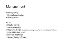
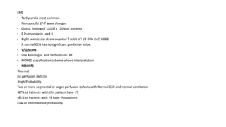
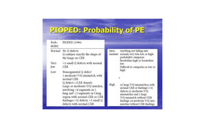
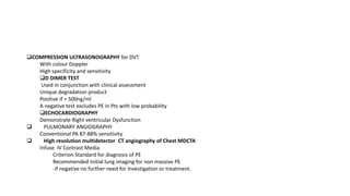
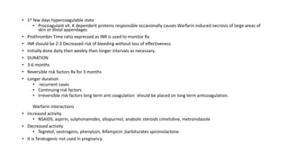
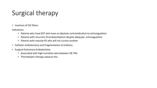
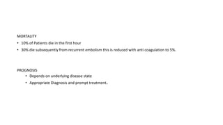
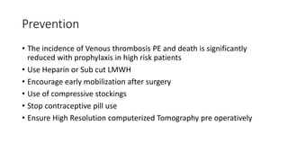
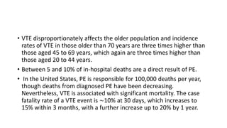
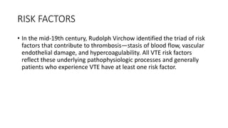
PULMONARY EMBOLISM
OUTLINE
• Introduction
• Definition
• Epidemiology
• Risk factors
• Pathophysiology
• Classification
• Clinical features
• Management
• Prevention
• Complications
• Prognosis
Introduction
• Pulmonary Embolism(PE) is the 3rd most common cause of
cardiovascular death after acute myocardial infarction and stroke.
• Pulmonary Embolism is a significant obstruction in the flow of blood
in the pulmonary arterial tree which may be partial or total
• PE is a common and potentially lethal condition affecting all age
groups.
• Prompt diagnosis and treatment dramatically reduce the morbidity
and mortality of the disease.
• PE is not a disease itself it is a complication of underlying venous
thrombosis.
Definition
• Pulmonary Embolism refers to the obstruction of the pulmonary artery or one of
its branches by a thrombus(or thrombi) that originates somewhere in the venous
system.
• PE is not a disease but a complication of underlying venous thrombosis. Most
often venous thrombosis arises from the deep vein of the leg: hence the name
deep venous thrombosis. The thrombus may remain localized to the leg veins or
may embolize through the circulation to result in a pulmonary embolism.
• If thrombus remains confined to the calf veins it is called a calf or distal DVT.
Untreated, the thrombus may extend proximally and reach the popliteal vein or
above, this is called a proximal DVT. Thrombi at this level are larger and more
likely to embolize. It may be transported through the large veins of the pelvis and
abdomen to the right atrium and ventricle. From there, they are pumped into the
pulmonary arteries, which progressively divide into smaller arteries as they
course through the lungs to supply the alveoli. The emboli stops in the pulmonary
arteries, where there are no longer able to progress and in doing so obstruct the
flow of blood distally.
EPIDEMIOLOGY
• A significant global health burden, venous thromboembolism accounts for
approximately 10 million cases annually and carries a significant morbidity
and mortality risk. Although the actual prevalence of PE is unknown, up to
600,000 cases of VTE are diagnosed annually in the US, accounting for
100,000 fatalities from the illness. Approximately one-third of hospitalized
patients are thought to be at risk of developing VTE. The estimated
incidence of diagnosed VTE in the United States is 117 per 100,000, but
since these diseases are often undetected or only discovered after death,
the true prevalence is probably higher. Reviewing national inpatient data
revealed that admissions for PE rose from almost 60,000 in 1993 (23 per
100,000) to over 60,000 in the current year.Despite the increased incidence
of PE, there was a decreased incidence of massive PE and hospital
mortality over the same time period.
• VTE disproportionately affects the older population and incidence
rates of VTE in those older than 70 years are three times higher than
those aged 45 to 69 years, which again are three times higher than
those aged 20 to 44 years.
• Between 5 and 10% of in-hospital deaths are a direct result of PE.
• In the United States, PE is responsible for 100,000 deaths per year,
though deaths from diagnosed PE have been decreasing.
Nevertheless, VTE is associated with significant mortality. The case
fatality rate of a VTE event is ∼10% at 30 days, which increases to
15% within 3 months, with a further increase up to 20% by 1 year.
RISK FACTORS
• In the mid-19th century, Rudolph Virchow identified the triad of risk
factors that contribute to thrombosis—stasis of blood flow, vascular
endothelial damage, and hypercoagulability. All VTE risk factors
reflect these underlying pathophysiologic processes and generally
patients who experience VTE have at least one risk factor.
Virchow’s Triad
Risk factors can be divided into inherited and
acquired factors:
PATHOPHYSIOLOGY
• Under normal circumstances micro thrombi are formed and lysed
continually within the venous system
• Under pathological conditions clots grow and propagate and may
break loose leading to formation of emboli
• The Pathophysiology depends on:
• Number and size of emboli: When large pulmonary vessels are involved, it could cause severe hemodynamic
instability including right ventricular (RV) pressure overload, RV failure, and eventually death.
• Leading to reduction in vascular bed
• The pre-existing state of the heart and Lungs.
• Small emboli may have no acute physiologic effects and may begin to lyse immediately
and resolve within hours or days. Larger emboli can cause a reflex increase in ventilation
(tachypnea), hypoxemia due to ventilation/perfusion (V/Q) mismatch, low mixed venous
oxygen content as a result of low cardiac output, atelectasis due to alveolar hypocapnia
and abnormalities in surfactant, and an increase in pulmonary vascular resistance caused
by mechanical obstruction and vasoconstriction resulting in tachycardia and hypotension.
Endogenous lysis reduces most emboli, even those of moderate size, and physiologic
alterations decrease over hours or days. Some emboli resist lysis and may organize and
persist and sometimes cause chronic thromboembolic pulmonary hypertension (CTEPH).
• When a large embolus acutely occludes major pulmonary arteries or when many smaller
emboli combine to occlude a significant proportion of the more distal vessels, RV
pressure increases, which may lead to acute RV failure, shock, or sudden death. The risk
of death depends on the degree and rate of rise of right-sided pressures and on the
patient’s underlying cardiopulmonary status. Patients with preexisting cardiopulmonary
disease are at higher risk of death, but young and/or otherwise healthy patients may
survive a PE that occludes > 50% of the pulmonary bed.
• Saddle pulmonary embolism describes a pulmonary embolus that
lodges in the bifurcation of the main pulmonary artery and into the
right and left pulmonary arteries; saddle emboli are usually, but not
always, intermediate risk or high risk. A saddle configuration does not
dictate a specific therapeutic approach. Although saddle emboli are
often large, causing near-complete or complete obstruction, they may
also be a relatively thin, nonobstructive embolus.
• Pulmonary infarction (interruption of pulmonary artery blood flow
leading to ischemia of lung tissue, sometimes represented by a
pleural-based [peripherally located], often wedge-shaped pattern on
chest x-ray [Hampton hump] or other imaging modalities) occurs in <
10% of patients diagnosed with PE. This low rate has been attributed
to the dual blood supply to the lung (ie, bronchial and pulmonary).
Generally, pulmonary infarction is due to smaller emboli that become
lodged in more distal pulmonary arteries and is nearly always
completely reversible; pulmonary infarction is recognized early, often
before necrosis occurs.
Respiratory effects of pulmonary embolism
• Increase in Alveolar dead space
• Bronchoconstriction
• Hyperventilation
• V/Q mismatch
• Loss of surfactant
• Alveolar instability.
• Atelectasis/collapse
• Infarction(Uncommon <10%)
Hemodynamic effects
• PE reduces the area of the vascular bed leading to increase in
pulmonary vascular resistance
• Increased Right ventricular after load
• Reduced pulmonary blood flow
• Reduced filling of the Left ventricle
• Reduced output causing hypotension
• Embolic fragments maybe trapped in Right Atrium
Clinical features
Clinical presentation of PE depends on
i. the acuity and
ii. severity of pulmonary artery occlusion,
This varies from catastrophic hemodynamic collapse to gradually progressive
dyspnea
• MASSIVE EMBOLISM
• Systolic pressure less than 90mmHg
• Circulatory collapse and shock and death.
• Chest pain central cyanosis dyspnea
• Syncope ,pallor restlessness confusion hypo tension
• Tachycardia, sweating
Medium and small sized emboli
• Acute onset of chest pain pleuritic
• Dyspnea, cough, hemoptysis
• May have non specific symptoms
Multiple pulmonary emboli
• Repeated episodes or no documented episode
• Present with exertional dyspnea chest pain
• Features of pulmonary hypertension and cor pulmonale
• Other clinical features include:
Fever
Pleural effusion
Decreased air entry, crepitations, localized wheeze
Pleural rub
Raised JVP
Engorged neck veins
Clinical scoring system
• Pt has clinical features compatible with PE
• Tachypnea, breathlessness, chest pain, haemoptysis
• Plus two other factors sought
• a/ The absence of another reasonable clinical explanation
• b/ Presence of a major risk factor
• If one and two are true high probability
• If only one is true intermediate
• If none low probability
• Other scoring systems available
• Modified Wells’ Score, Revised Geneva, PERC rule, Charlotte criteria
Well’s score
Modified well’s score
Management
• History taking
• Physical examination
• Investigations :
• CXR
o May be normal
o linear atelectasis
o Westermark Sign: Dilatation of vessel proximal to the embolus distal collapse
o Pleural Effusion- small
o Elevated diaphragm
o Wedge shaped infiltrate
ECG
• Tachycardia most common
• Non specific ST-T wave changes
• Classic finding of S1Q3T3 20% of patients
• P Pulmonale in Lead II
• Right ventricular strain inverted T in V1 V2 V3 RVH RAD RBBB
• A normal ECG has no significant predictive value.
• V/Q Scans
• Use Xenon gas and Technetium- 99
• PIOPED classification scheme allows interpretation
• RESULTS
-Normal
no perfusion deficits
-High Probability
Two or more segmental or larger perfusion defects with Normal CXR and normal ventilation
-87% of Patients with this pattern have PE
-41% of Patients with PE have this pattern
Low or intermediate probability
COMPRESSION ULTRASONOGRAPHY for DVT
With colour Doppler
High specificity and sensitivity
D DIMER TEST
Used in conjunction with clinical assessment
Unique degradation product
Positive if > 500ng/ml
A negative test excludes PE in Pts with low probability
ECHOCARDIOGRAPHY
Demonstrate Right ventricular Dysfunction
 PULMONARY ANGIOGRAPHY
Conventional PA 87-88% sensitivity
 High resolution multidetector CT angiography of Chest MDCTA
Infuse IV Contrast Media
Criterion Standard for diagnosis of PE
Recommended initial lung imaging for non massive PE
-if negative no further need for Investigation or treatment.
Other investigations
 Cardiac troponin levels
 Arterial Blood Gases
• Decreased PaO2 and PaCO2
 Clotting profiles
 MRI
 BNP
 Ischaemia-modified albumin (IMA) Level 93%sensitive 75% specific for PE
 FBC
• Wbc normal or elevated
Treatment
• Supportive therapy
• Oxygen
• Plasma expanders and inotropic support in hypotensive
• Pain relief
• Immediate full anticoagulation is mandatory for all patients suspected to have PE or DVT
• Avoid Delay
• Initial Anticoagulation with IV Heparin or LMWH
• Activates anti thrombin III
• Does not dissolve clot but slows or prevents progression and recurrence of thrombosis so reduces embolism
Unfractionated heparin preferred in
• First dose bolus
• Massive PE
• Patients who may need rapid reversal
• LMWH has equal efficacy and safety with Unfractionated Heparin and is easier to use
• Side Effects
• Bleeding
• Osteoporosis
• Alopecia
• To assess the efficacy of RX
• Use aPTT aim for 1.5x control
• Give loading Dose followed by continuous or bolus doses
• 10000 bolus followed by 20 units/kg/hr IV adjust as necessary
• If a PTT very high clip off drip
Thrombolytic therapy
• Those hemodynamically unstable-Massive PE
• Right ventricular dysfunction
• Limited cardiopulmonary reserve
• DRUGS
• Urokinase Streptokinase
• Tissue Plasminogen activators
• Alteplase (100mgs) over 2 hours, Reteplase(20u) IV bolus
• WAFARIN Oral anticoagulation
• Commenced one to three days of effective heparinization and after it has achieved a therapeutic
level for 2 days Heparin is discontinued. This is usually after 5 to 7 days
• Coumarin Derivative similar structure to Vitamin K
• Inhibits Vitamin K dependent factors II VII IX X
• 1st few days hypercoagulable state
• Procoagulant vit. K dependent proteins responsible occasionally causes Warfarin induced necrosis of large areas of
skin or distal appendages
• Prothrombin Time ratio expressed as INR is used to monitor Rx
• INR should be 2-3 Decreased risk of bleeding without loss of effectiveness
• Initially done daily then weekly then longer intervals as necessary.
• DURATION
• 3-6 months
• Reversible risk factors Rx for 3 months
• Longer duration
• recurrent cases
• Continuing risk factors
• Irreversible risk factors long term anti coagulation should be placed on long term anticoagulation.
Warfarin interactions
• Increased activity
• NSAIDS, aspirin, sulphonamides, allopurinol, anabolic steroids cimetidine, metronidazole
• Decreased activity
• Tegretol, oestrogens, phenytoin, Rifampicin ,barbiturates spironolactone
• It is Teratogenic not used in pregnancy.
Surgical therapy
• Insertion of IVC filters.
Indications
• Patients who have DVT who have an absolute contraindication to anticoagulation
• Patients with recurrent thromboembolism despite adequate anticoagulation
• Patients with massive PE who will not survive another.
• Catheter embolectomy and Fragmentation of embolus
• Surgical Pulmonary Embolectomy
• Associated with high mortality rates between 28-74%
• Thrombolytic therapy replaces this
MORTALITY
• 10% of Patients die in the first hour
• 30% die subsequently from recurrent embolism this is reduced with anti coagulation to 5%.
PROGNOSIS
• Depends on underlying disease state
• Appropriate Diagnosis and prompt treatment.
Prevention
• The incidence of Venous thrombosis PE and death is significantly
reduced with prophylaxis in high risk patients
• Use Heparin or Sub cut LMWH
• Encourage early mobilization after surgery
• Use of compressive stockings
• Stop contraceptive pill use
• Ensure High Resolution computerized Tomography pre operatively
Differential diagnosis
• Myocardial Infarction
• Cardiogenic Shock
• COPD
• Pneumonia
• Cor pulmonale
• Pleuritis
• pericarditis
Special Consideration
• Pregnancy
• Warfarin teratogenic avoid
• Use LMWH
• Near delivery use UFH
• Continue anticoagulation till 3 months after delivery
• Malignancy
• 3-6 months Rx or indefinite
• Right heart Thrombi
• Early mortality in Pts with acute PE
• Thrombolysis and embolectomy
Complications
• Sudden death
• Arrhythmias
• Pulmonary hypertension
• Lung infarction
• Pleural effusion
• Cor pulmonale
• Respiratory Failure
1. Which of the following is a common symptom of pulmonary embolism?
A) Chest pain
B) B) Nausea
C) C) Back pain
D) D) Headache
2. What is the most common cause of pulmonary embolism?
A) Atherosclerosis
B) Deep vein thrombosis (DVT)
C) Pneumonia
D) Asthma
3. Which imaging test is often used to diagnose pulmonary embolism?
A) X-ray
B) B) MRI
C) C) CT scan
D) D) Ultrasound
4. : Which of the following is a risk factor for pulmonary embolism?
A) Smoking
B) Regular exercise
C) Low cholesterol levels
D) High blood pressure
5. What is the most common site for a deep vein thrombosis (DVT) that can lead to a pulmonary embolism?
• A) Arm
• B) Leg
• C) Abdomen
• D) Neck
• 6. Which laboratory test is commonly elevated in pulmonary embolism?
• A) Troponin
• B) D-dimer
• C) Creatinine
• D) BNP
• 7. . What is the most common complication of pulmonary embolism?
• A) Pneumothorax
• B) Pulmonary hypertension
• C) Pleural effusion
• D) Atelectasis
• 8. Which of the following medications is often used as a first-line treatment for pulmonary embolism?
• A) Aspirin
• B) Warfarin
• C) Heparin
• D) Metoprolol
• 9. Which echocardiographic finding is characteristic of severe pulmonary embolism?
• A) Left ventricular hypertrophy
• B) Right atrial enlargement
• C) Aortic stenosis
• D) Mitral regurgitation
• Answers:
• 1. A) Chest pain
• 2. B) Deep vein thrombosis (DVT)
• 3. C) CT scan
• 4. A) Smoking
• 5. B) Leg
• 6. B) D-dimer
• 7. B) Pulmonary hypertension
• 8 C) Heparin
• 9. B) Right atrial enlargement
References
• Kumar and Clark’s clinical medicine.
• Medscape: pulmonary Embolism

Pulmonary embolism slide presentation. pptx

  • 1.
  • 2.
    OUTLINE • Introduction • Definition •Epidemiology • Risk factors • Pathophysiology • Classification • Clinical features • Management • Prevention • Complications • Prognosis
  • 3.
    Introduction • Pulmonary Embolism(PE)is the 3rd most common cause of cardiovascular death after acute myocardial infarction and stroke. • Pulmonary Embolism is a significant obstruction in the flow of blood in the pulmonary arterial tree which may be partial or total • PE is a common and potentially lethal condition affecting all age groups. • Prompt diagnosis and treatment dramatically reduce the morbidity and mortality of the disease. • PE is not a disease itself it is a complication of underlying venous thrombosis.
  • 4.
    Definition • Pulmonary Embolismrefers to the obstruction of the pulmonary artery or one of its branches by a thrombus(or thrombi) that originates somewhere in the venous system. • PE is not a disease but a complication of underlying venous thrombosis. Most often venous thrombosis arises from the deep vein of the leg: hence the name deep venous thrombosis. The thrombus may remain localized to the leg veins or may embolize through the circulation to result in a pulmonary embolism. • If thrombus remains confined to the calf veins it is called a calf or distal DVT. Untreated, the thrombus may extend proximally and reach the popliteal vein or above, this is called a proximal DVT. Thrombi at this level are larger and more likely to embolize. It may be transported through the large veins of the pelvis and abdomen to the right atrium and ventricle. From there, they are pumped into the pulmonary arteries, which progressively divide into smaller arteries as they course through the lungs to supply the alveoli. The emboli stops in the pulmonary arteries, where there are no longer able to progress and in doing so obstruct the flow of blood distally.
  • 5.
    EPIDEMIOLOGY • A significantglobal health burden, venous thromboembolism accounts for approximately 10 million cases annually and carries a significant morbidity and mortality risk. Although the actual prevalence of PE is unknown, up to 600,000 cases of VTE are diagnosed annually in the US, accounting for 100,000 fatalities from the illness. Approximately one-third of hospitalized patients are thought to be at risk of developing VTE. The estimated incidence of diagnosed VTE in the United States is 117 per 100,000, but since these diseases are often undetected or only discovered after death, the true prevalence is probably higher. Reviewing national inpatient data revealed that admissions for PE rose from almost 60,000 in 1993 (23 per 100,000) to over 60,000 in the current year.Despite the increased incidence of PE, there was a decreased incidence of massive PE and hospital mortality over the same time period.
  • 6.
    • VTE disproportionatelyaffects the older population and incidence rates of VTE in those older than 70 years are three times higher than those aged 45 to 69 years, which again are three times higher than those aged 20 to 44 years. • Between 5 and 10% of in-hospital deaths are a direct result of PE. • In the United States, PE is responsible for 100,000 deaths per year, though deaths from diagnosed PE have been decreasing. Nevertheless, VTE is associated with significant mortality. The case fatality rate of a VTE event is ∼10% at 30 days, which increases to 15% within 3 months, with a further increase up to 20% by 1 year.
  • 7.
    RISK FACTORS • Inthe mid-19th century, Rudolph Virchow identified the triad of risk factors that contribute to thrombosis—stasis of blood flow, vascular endothelial damage, and hypercoagulability. All VTE risk factors reflect these underlying pathophysiologic processes and generally patients who experience VTE have at least one risk factor.
  • 8.
  • 9.
    Risk factors canbe divided into inherited and acquired factors:
  • 10.
    PATHOPHYSIOLOGY • Under normalcircumstances micro thrombi are formed and lysed continually within the venous system • Under pathological conditions clots grow and propagate and may break loose leading to formation of emboli • The Pathophysiology depends on: • Number and size of emboli: When large pulmonary vessels are involved, it could cause severe hemodynamic instability including right ventricular (RV) pressure overload, RV failure, and eventually death. • Leading to reduction in vascular bed • The pre-existing state of the heart and Lungs.
  • 11.
    • Small embolimay have no acute physiologic effects and may begin to lyse immediately and resolve within hours or days. Larger emboli can cause a reflex increase in ventilation (tachypnea), hypoxemia due to ventilation/perfusion (V/Q) mismatch, low mixed venous oxygen content as a result of low cardiac output, atelectasis due to alveolar hypocapnia and abnormalities in surfactant, and an increase in pulmonary vascular resistance caused by mechanical obstruction and vasoconstriction resulting in tachycardia and hypotension. Endogenous lysis reduces most emboli, even those of moderate size, and physiologic alterations decrease over hours or days. Some emboli resist lysis and may organize and persist and sometimes cause chronic thromboembolic pulmonary hypertension (CTEPH). • When a large embolus acutely occludes major pulmonary arteries or when many smaller emboli combine to occlude a significant proportion of the more distal vessels, RV pressure increases, which may lead to acute RV failure, shock, or sudden death. The risk of death depends on the degree and rate of rise of right-sided pressures and on the patient’s underlying cardiopulmonary status. Patients with preexisting cardiopulmonary disease are at higher risk of death, but young and/or otherwise healthy patients may survive a PE that occludes > 50% of the pulmonary bed.
  • 12.
    • Saddle pulmonaryembolism describes a pulmonary embolus that lodges in the bifurcation of the main pulmonary artery and into the right and left pulmonary arteries; saddle emboli are usually, but not always, intermediate risk or high risk. A saddle configuration does not dictate a specific therapeutic approach. Although saddle emboli are often large, causing near-complete or complete obstruction, they may also be a relatively thin, nonobstructive embolus.
  • 13.
    • Pulmonary infarction(interruption of pulmonary artery blood flow leading to ischemia of lung tissue, sometimes represented by a pleural-based [peripherally located], often wedge-shaped pattern on chest x-ray [Hampton hump] or other imaging modalities) occurs in < 10% of patients diagnosed with PE. This low rate has been attributed to the dual blood supply to the lung (ie, bronchial and pulmonary). Generally, pulmonary infarction is due to smaller emboli that become lodged in more distal pulmonary arteries and is nearly always completely reversible; pulmonary infarction is recognized early, often before necrosis occurs.
  • 14.
    Respiratory effects ofpulmonary embolism • Increase in Alveolar dead space • Bronchoconstriction • Hyperventilation • V/Q mismatch • Loss of surfactant • Alveolar instability. • Atelectasis/collapse • Infarction(Uncommon <10%)
  • 15.
    Hemodynamic effects • PEreduces the area of the vascular bed leading to increase in pulmonary vascular resistance • Increased Right ventricular after load • Reduced pulmonary blood flow • Reduced filling of the Left ventricle • Reduced output causing hypotension • Embolic fragments maybe trapped in Right Atrium
  • 16.
    Clinical features Clinical presentationof PE depends on i. the acuity and ii. severity of pulmonary artery occlusion, This varies from catastrophic hemodynamic collapse to gradually progressive dyspnea • MASSIVE EMBOLISM • Systolic pressure less than 90mmHg • Circulatory collapse and shock and death. • Chest pain central cyanosis dyspnea • Syncope ,pallor restlessness confusion hypo tension • Tachycardia, sweating
  • 17.
    Medium and smallsized emboli • Acute onset of chest pain pleuritic • Dyspnea, cough, hemoptysis • May have non specific symptoms Multiple pulmonary emboli • Repeated episodes or no documented episode • Present with exertional dyspnea chest pain • Features of pulmonary hypertension and cor pulmonale • Other clinical features include: Fever Pleural effusion Decreased air entry, crepitations, localized wheeze Pleural rub Raised JVP Engorged neck veins
  • 18.
    Clinical scoring system •Pt has clinical features compatible with PE • Tachypnea, breathlessness, chest pain, haemoptysis • Plus two other factors sought • a/ The absence of another reasonable clinical explanation • b/ Presence of a major risk factor • If one and two are true high probability • If only one is true intermediate • If none low probability • Other scoring systems available • Modified Wells’ Score, Revised Geneva, PERC rule, Charlotte criteria
  • 19.
  • 20.
  • 22.
    Management • History taking •Physical examination • Investigations : • CXR o May be normal o linear atelectasis o Westermark Sign: Dilatation of vessel proximal to the embolus distal collapse o Pleural Effusion- small o Elevated diaphragm o Wedge shaped infiltrate
  • 23.
    ECG • Tachycardia mostcommon • Non specific ST-T wave changes • Classic finding of S1Q3T3 20% of patients • P Pulmonale in Lead II • Right ventricular strain inverted T in V1 V2 V3 RVH RAD RBBB • A normal ECG has no significant predictive value. • V/Q Scans • Use Xenon gas and Technetium- 99 • PIOPED classification scheme allows interpretation • RESULTS -Normal no perfusion deficits -High Probability Two or more segmental or larger perfusion defects with Normal CXR and normal ventilation -87% of Patients with this pattern have PE -41% of Patients with PE have this pattern Low or intermediate probability
  • 25.
    COMPRESSION ULTRASONOGRAPHY forDVT With colour Doppler High specificity and sensitivity D DIMER TEST Used in conjunction with clinical assessment Unique degradation product Positive if > 500ng/ml A negative test excludes PE in Pts with low probability ECHOCARDIOGRAPHY Demonstrate Right ventricular Dysfunction  PULMONARY ANGIOGRAPHY Conventional PA 87-88% sensitivity  High resolution multidetector CT angiography of Chest MDCTA Infuse IV Contrast Media Criterion Standard for diagnosis of PE Recommended initial lung imaging for non massive PE -if negative no further need for Investigation or treatment.
  • 26.
    Other investigations  Cardiactroponin levels  Arterial Blood Gases • Decreased PaO2 and PaCO2  Clotting profiles  MRI  BNP  Ischaemia-modified albumin (IMA) Level 93%sensitive 75% specific for PE  FBC • Wbc normal or elevated
  • 27.
    Treatment • Supportive therapy •Oxygen • Plasma expanders and inotropic support in hypotensive • Pain relief • Immediate full anticoagulation is mandatory for all patients suspected to have PE or DVT • Avoid Delay • Initial Anticoagulation with IV Heparin or LMWH • Activates anti thrombin III • Does not dissolve clot but slows or prevents progression and recurrence of thrombosis so reduces embolism Unfractionated heparin preferred in • First dose bolus • Massive PE • Patients who may need rapid reversal • LMWH has equal efficacy and safety with Unfractionated Heparin and is easier to use
  • 28.
    • Side Effects •Bleeding • Osteoporosis • Alopecia • To assess the efficacy of RX • Use aPTT aim for 1.5x control • Give loading Dose followed by continuous or bolus doses • 10000 bolus followed by 20 units/kg/hr IV adjust as necessary • If a PTT very high clip off drip
  • 29.
    Thrombolytic therapy • Thosehemodynamically unstable-Massive PE • Right ventricular dysfunction • Limited cardiopulmonary reserve • DRUGS • Urokinase Streptokinase • Tissue Plasminogen activators • Alteplase (100mgs) over 2 hours, Reteplase(20u) IV bolus • WAFARIN Oral anticoagulation • Commenced one to three days of effective heparinization and after it has achieved a therapeutic level for 2 days Heparin is discontinued. This is usually after 5 to 7 days • Coumarin Derivative similar structure to Vitamin K • Inhibits Vitamin K dependent factors II VII IX X
  • 30.
    • 1st fewdays hypercoagulable state • Procoagulant vit. K dependent proteins responsible occasionally causes Warfarin induced necrosis of large areas of skin or distal appendages • Prothrombin Time ratio expressed as INR is used to monitor Rx • INR should be 2-3 Decreased risk of bleeding without loss of effectiveness • Initially done daily then weekly then longer intervals as necessary. • DURATION • 3-6 months • Reversible risk factors Rx for 3 months • Longer duration • recurrent cases • Continuing risk factors • Irreversible risk factors long term anti coagulation should be placed on long term anticoagulation. Warfarin interactions • Increased activity • NSAIDS, aspirin, sulphonamides, allopurinol, anabolic steroids cimetidine, metronidazole • Decreased activity • Tegretol, oestrogens, phenytoin, Rifampicin ,barbiturates spironolactone • It is Teratogenic not used in pregnancy.
  • 31.
    Surgical therapy • Insertionof IVC filters. Indications • Patients who have DVT who have an absolute contraindication to anticoagulation • Patients with recurrent thromboembolism despite adequate anticoagulation • Patients with massive PE who will not survive another. • Catheter embolectomy and Fragmentation of embolus • Surgical Pulmonary Embolectomy • Associated with high mortality rates between 28-74% • Thrombolytic therapy replaces this
  • 32.
    MORTALITY • 10% ofPatients die in the first hour • 30% die subsequently from recurrent embolism this is reduced with anti coagulation to 5%. PROGNOSIS • Depends on underlying disease state • Appropriate Diagnosis and prompt treatment.
  • 33.
    Prevention • The incidenceof Venous thrombosis PE and death is significantly reduced with prophylaxis in high risk patients • Use Heparin or Sub cut LMWH • Encourage early mobilization after surgery • Use of compressive stockings • Stop contraceptive pill use • Ensure High Resolution computerized Tomography pre operatively
  • 34.
    Differential diagnosis • MyocardialInfarction • Cardiogenic Shock • COPD • Pneumonia • Cor pulmonale • Pleuritis • pericarditis
  • 35.
    Special Consideration • Pregnancy •Warfarin teratogenic avoid • Use LMWH • Near delivery use UFH • Continue anticoagulation till 3 months after delivery • Malignancy • 3-6 months Rx or indefinite • Right heart Thrombi • Early mortality in Pts with acute PE • Thrombolysis and embolectomy
  • 36.
    Complications • Sudden death •Arrhythmias • Pulmonary hypertension • Lung infarction • Pleural effusion • Cor pulmonale • Respiratory Failure
  • 37.
    1. Which ofthe following is a common symptom of pulmonary embolism? A) Chest pain B) B) Nausea C) C) Back pain D) D) Headache 2. What is the most common cause of pulmonary embolism? A) Atherosclerosis B) Deep vein thrombosis (DVT) C) Pneumonia D) Asthma 3. Which imaging test is often used to diagnose pulmonary embolism? A) X-ray B) B) MRI C) C) CT scan D) D) Ultrasound
  • 38.
    4. : Whichof the following is a risk factor for pulmonary embolism? A) Smoking B) Regular exercise C) Low cholesterol levels D) High blood pressure 5. What is the most common site for a deep vein thrombosis (DVT) that can lead to a pulmonary embolism? • A) Arm • B) Leg • C) Abdomen • D) Neck • 6. Which laboratory test is commonly elevated in pulmonary embolism? • A) Troponin • B) D-dimer • C) Creatinine • D) BNP
  • 39.
    • 7. .What is the most common complication of pulmonary embolism? • A) Pneumothorax • B) Pulmonary hypertension • C) Pleural effusion • D) Atelectasis • 8. Which of the following medications is often used as a first-line treatment for pulmonary embolism? • A) Aspirin • B) Warfarin • C) Heparin • D) Metoprolol • 9. Which echocardiographic finding is characteristic of severe pulmonary embolism? • A) Left ventricular hypertrophy • B) Right atrial enlargement • C) Aortic stenosis • D) Mitral regurgitation
  • 40.
    • Answers: • 1.A) Chest pain • 2. B) Deep vein thrombosis (DVT) • 3. C) CT scan • 4. A) Smoking • 5. B) Leg • 6. B) D-dimer • 7. B) Pulmonary hypertension • 8 C) Heparin • 9. B) Right atrial enlargement
  • 41.
    References • Kumar andClark’s clinical medicine. • Medscape: pulmonary Embolism