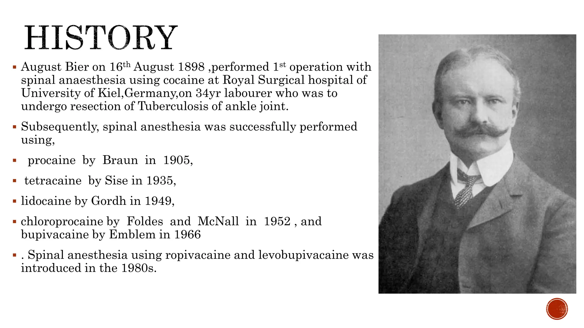
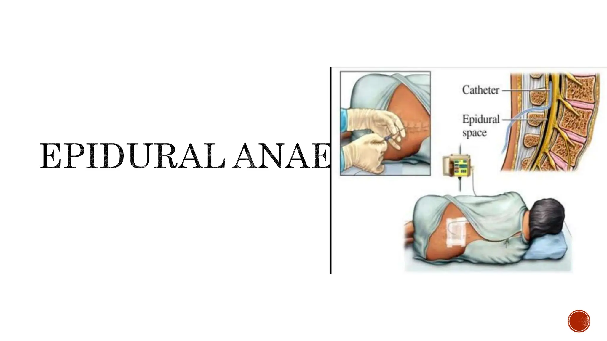
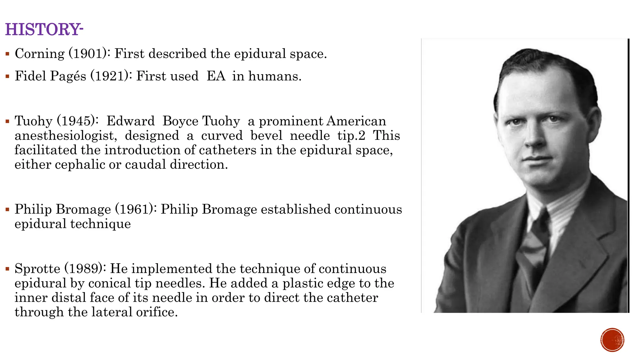
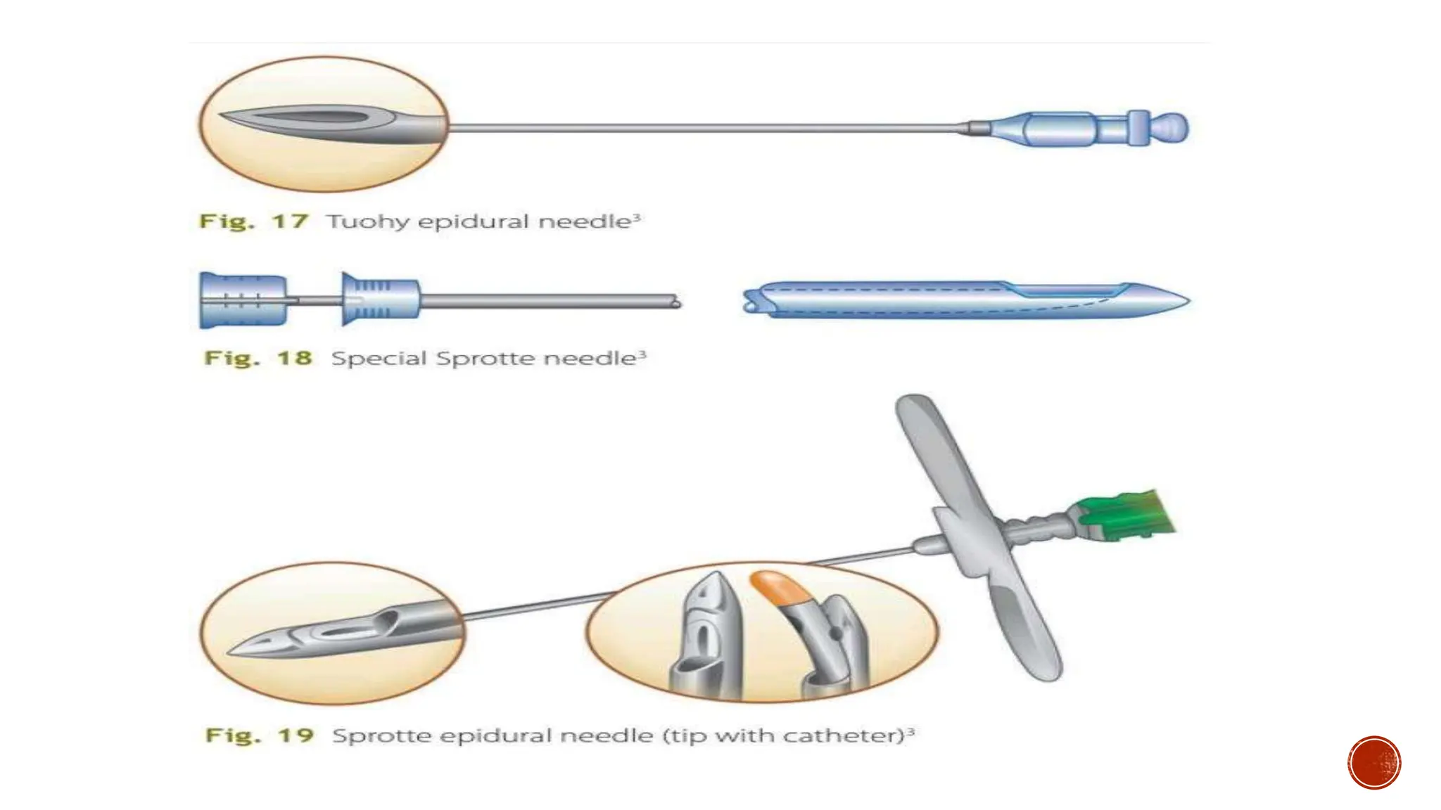
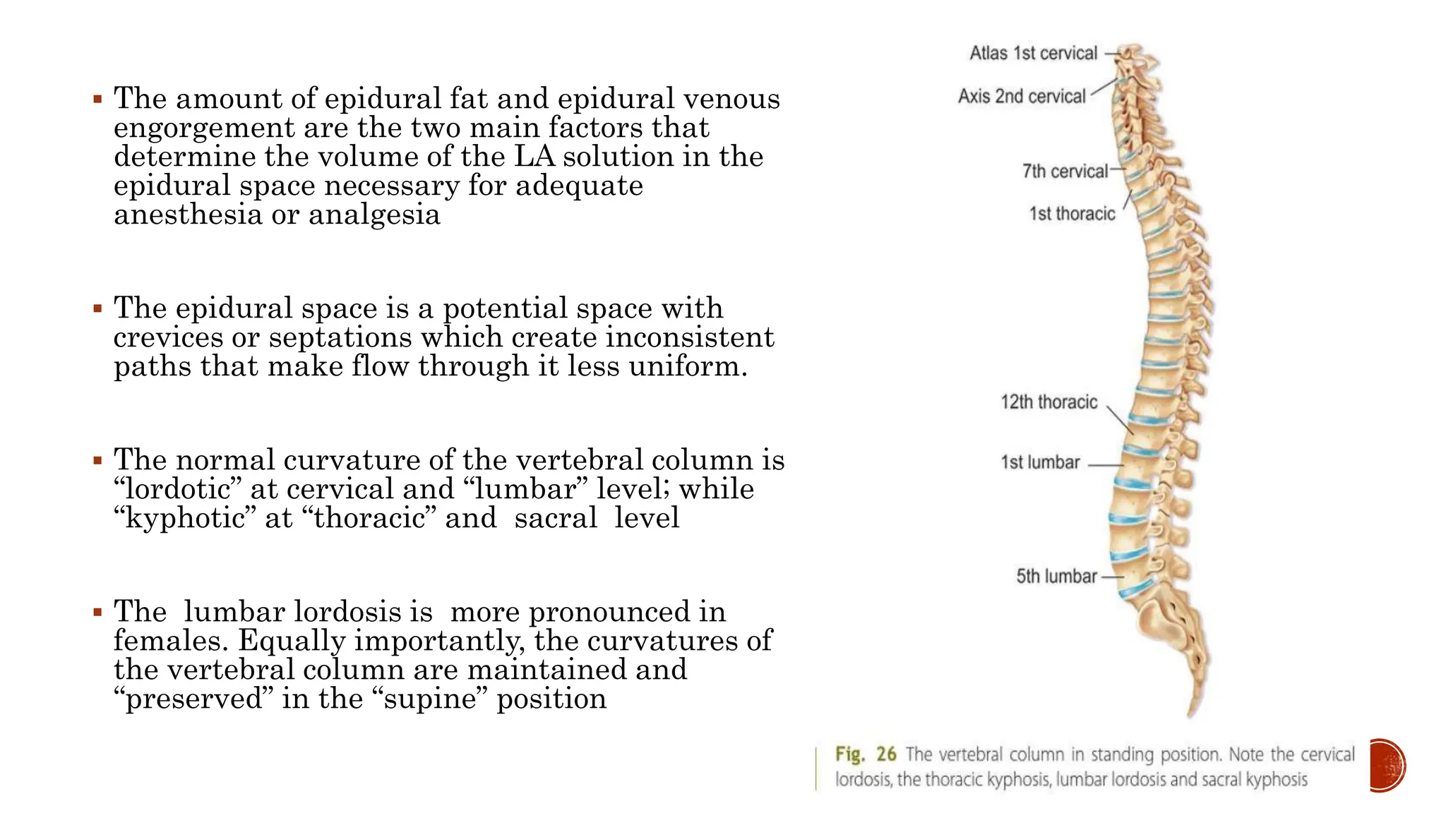
Presenter-Dr. Sindhu R V,
1st year postgraduate, Anesthesia
Moderator-Dr.Poornima madam
 History
 Anatomy
 Physiologic effects
 Indications
 Contraindications
 Factors affecting block height
 Technique
 Block monitoring
 Epidural Anaesthesia
 Combined spinal and epidural anaesthesia
 August Bier on 16th August 1898 ,performed 1st operation with
spinal anaesthesia using cocaine at Royal Surgical hospital of
University of Kiel,Germany,on 34yr labourer who was to
undergo resection of Tuberculosis of ankle joint.
 Subsequently, spinal anesthesia was successfully performed
using,
 procaine by Braun in 1905,
 tetracaine by Sise in 1935,
 lidocaine by Gordh in 1949,
 chloroprocaine by Foldes and McNall in 1952 , and
bupivacaine by Emblem in 1966
 . Spinal anesthesia using ropivacaine and levobupivacaine was
introduced in the 1980s.
 The spinal cord is continuous with the brainstem proximally and terminates
distally in the conus medullaris as the filum terminale (fibrous extension) and the
cauda equina (neural extension).
 The distal termination varies from L3 in infants to the lower border of L1 in
adults.
 Three membranes covering spinal cord (from innermost to outermost): the pia
mater, the arachnoid mater, and the dura mater
 The pia mater is a highly vascular membrane that closely invests the spinal cord
and brain.
 The arachnoid mater is a delicate, nonvascular membrane that functions as the
principal barrier to drugs crossing into (and out of) the CSF and is
estimated to account for 90% of the resistance to drug migration
 The cerebrospinal fluid (CSF) resides in the space between the pia mater and the
arachnoid mater, termed the subarachnoid (or intrathecal) space.
 The cerebrospinal fluid (CSF) resides in the space between the pia mater and the
arachnoid mater, termed the subarachnoid (or intrathecal) space.
 Approximately 500 mL of CSF is formed daily by the choroid plexuses of the
cerebral ventricles.
 Neuraxial anesthesia evokes blockade of the sympathetic and somatic (sensory
and motor) nervous systems, along with compensatory reflexes and unopposed
parasympathetic activity.
 CARDIOASCULAR EFFECTS-
 Decreased stroke volume and heart rate caused by blockade of the
peripheral (T1-L2) and cardiac (T1-T4) sympathetic fibers as well as
adrenal medullary secretion.
 Sympathectomy usually decreases stroke volume. Venous and
arterial vasodilation reduces preload (venous return) and afterload
(systemic vascular resistance), respectively.
 Heart rate may decrease during a high neuraxial block as a result of blockade of
the cardioaccelerator fibers arising from T1-T4
 Although hypotension will trigger a compensatory baroreceptor sympathetic
response (vasoconstriction and increased heart rate) above the level of blockade,
the reduction in venous return and right atrial filling causes a decrease in signal
output from intrinsic chronotropic stretch receptors located in the right atrium
and great veins,leading to a marked increase in parasympathetic activity (vagal
tone). The two opposing responses are usually in check with a minimal change in
heart rate (or a slight reduction)
 250- to 2000-mL preblock hydration regimens may temporarily increase preload
and cardiac output but do not consistently increase arterial blood pressure or
prevent hypotension
 Useful techniques to prevent hypotension include the repeated low-dose local
anesthetic boluses through a continuous spinal catheter, smalldose unilateral
spinal anesthesia, and selective small-dose spinal anesthesia.
 Spinal anesthesia–induced hypotension may decrease regional cerebral blood flow
(CBF) in elderly patients and those with preexisting hypertension.
 When the level of spinal anesthesia was purposely increased to produce higher
levels of block (T4) in normotensive and hypertensive patients, CBF was
unchanged in the normotensive group (45 to 46 mL/100 g per minute), whereas a
19% decrease occurred in the apparently untreated hypertensive patients (46.5 to
37.5 mL/100 g per minute).
 decrease in vital capacity follows a reduction in expiratory reserve volume related
to paralysis of the abdominal muscles necessary for forced exhalation rather than
a decrease in phrenic or diaphragmatic function
 The magnitude of decline in vital capacity is proportional to the BMI value
(vital capacity –19% for BMI 30 to 40 kg/m2 versus –33% for BMI >40 kg/m2).
 Neuraxial blockade from T6 to L1 disrupts splanchnic sympathetic innervation to
the gastrointestinal tract, resulting in a contracted gut and hyperperistalsis.
 Nausea and vomiting may be associated with neuraxial block in as much as 20%
of patients and they are primarily related to gastrointestinal hyperperistalsis
caused by unopposed parasympathetic (vagal) activity
 Renal good flow is maintained through autoregulation,and there is little effect of
neuraxial anesthesia on kidney function
 Neuraxial Anesthesia at lumbar and sacral levels block both sympathetic and
parasympathetic control of bladder function.
 Loss of autonomic bladder control results in urinary retention until the block
wears off.
 Spinal anesthesia is most commonly used for patients who require surgical
anesthesia for procedures of known duration that involve the lower extremities,
perineum, pelvic girdle, or lower abdomen
 Descriptions of spinal anesthesia as the primary surgical anesthetic have more
recently expanded to include lumbar spine surgery,61 as well as upper
abdominal procedures, such as laparoscopic cholecystectomy.
 ABSOLUTE-
 Lack of consent
 Severe hypovolemia
 Increased intracranial pressure
 Coagulopathy or other bleeding disorders
 Infection at the site of injection.
 RELATIVE-
 Sepsis
 Demyelinating lesions
 Severe spinal deformity
 Preexisting neurological deficits
 Stenotic valvular heart lesions
 Baricity-
 Baricity is the ratio of the density of a local anesthetic solution to the density of
CSF.
 Density is defined as the mass per unit volume of solution (g/mL) at a specific
temperature.
 density varies inversely with temperature, the baricity of a local anesthetic
solution is conventionally defined at 37° C.
The density of CSF is 1.00059 g/L.
Local anesthetic solutions that have the same density as CSF are termed isobaric,
those that have a higher density than CSF are termed hyperbaric, and those with a
lower density than CSF are termed hypobaric.
 The spread of hyperbaric solutions is more predictable, with less interpatient
variability
 Dextrose and sterile water are commonly added to render local anesthetic
solutions either hyperbaric or hypobaric, respectively.
 Dose,volume and concentration-
 Dose=volume×concentration
 Dose is the most reliable determinant of local anaesthetic spread and block height
 The CSF volume is an important patient-related factor that significantly
influences peak block height and regression of sensory and motor blockade
 In theory, the increased abdominal mass in obese patients, and possible increased
epidural fat, may decrease the CSF volume and therefore increase the spread of
local anesthetic and block height
 The density of CSF is lower in women compared with men, premenopausal
compared with postmenopausal women, and pregnant compared with
nonpregnant women
 Advanced age is associated with increased block height
 In older patients, CSF volume decreases, whereas its specific gravity increases.
Further, the nerve roots appear more sensitive to local anesthetic in the aged
population
 Gender can theoretically affect block height by several mechanisms. CSF density
is higher in males, thereby reducing the baricity of local anesthetic solution and
possibly limiting the extent of cephalad spread
 Scoliosis, although it possibly makes insertion of the needle more difficult, will
have little effect on local anesthetic spread if the patient is turned supine.
Kyphosis, however, in a supine patient may affect the spread of a hyperbaric
solution.
 Patient position, needle type and alignment, and the level of injection are each
procedure-related factors that can affect block height
 Intrathecal local anesthetic appears to stop spreading 20 to 25 minutes after
injection, thus positioning of the patient is most important during this time
period, but particularly in the initial few minutes.
 Although a 10-degree headup tilt can reduce the spread of hyperbaric solutions
without hemodynamic compromise
 head-down tilt does not always increase the spread of hyperbaric bupivacaine.
Flexion of the hips in combination with the Trendelenburg position flattens the
lumbar lordosis and has been shown to increase cephalad spread of hyperbaric
solutions.1
 The specific needle type and orientation of the orifice may affect block quality.
With hypobaric solutions, cephalad alignment of the orifice of Whitacre, but not
Sprotte, needles produces greater spread
 even when the difference is only one interspace more cephalad, the block height is
greater
 Injection rate and barbotage (repeated aspiration and reinjection of CSF) of
isobaric and hyperbaric solutions have not consistently been shown to affect block
height.
 PREPARATION
 Informed consent must be obtained, with adequate documentation of the
discussion of risk
 The patient should have adequate intravenous access and be monitored with
pulse oximetry, noninvasive arterial blood pressure, and electrocardiogram
 Spinal needle-
. Needle tip shapes fall into two main categories: those that cut the
dura and those with a conical, pencil-point tip
Dura cutting-pitkin ,Quincke-babcock needles
Pencil point-Whitacre and Sprotte needles
 The use of small needles reduces the incidence of post–dural puncture headache
from 40% with a 22-G needle to less than 2% with a 29-G needle
 Pencil-point needles provide better tactile sensation of the different layers
encountered during needle insertion but, more importantly, they reduce the
incidence of post–dural puncture headache.
 most common organisms responsible for postspinal bacterial meningitis is
Streptococcus viridans, which is an oral commensal, emphasizing the purpose of
wearing a mask as part of a full aseptic technique
 Hands and forearms must be washed and all jewelry removed. A variety of
solutions may be used to clean the back, such as chlorhexidine or alcohol (alone or
in combination), or iodine solutions. Chlorhexidine and alcohol together have been
concluded to be most effective.
 If chlorhexidine is used, it is important that the solution is allowed to dry
completely before skin puncture because chlorhexidine is neurotoxic.
 POSITION
 The three primary patient positions include the lateral decubitus, sitting,
and prone
 Lateral-Patients are placed with their back parallel to the edge of the
operating table nearest the anesthesiologist, thighs flexed onto the abdomen,
with the neck flexed to allow the forehead to be as close as possible to the
knees in an attempt to “open up” the vertebral spaces
 Sitting-Hypotension may also be more common for a person in the sitting
position.
 prone position- is rarely used but may be chosen when the patient is to be
maintained in that position during the surgical procedures likee rectal,
perineal, or lumbar procedures
PROJECTION AND PUNCTURE
The spinal cord ends at the level of L1-L2 and so needle insertion above this level
should be avoided. The intercristal line is the line drawn between the two iliac
crests and traditionally corresponds to the level of the L4 vertebral body or the L4-
L5 interspace,
Once the appropriate space has been selected, a subcutaneous skin wheal of local
anesthetic is developed over this space, and the introducer is inserted at a slight
cephalad angle of 10 to 15 degrees through skin, subcutaneous tissue, and
supraspinous ligament to reach the substance of the interspinous ligament.
until the characteristic change in resistance is noted as the needle passes
through the ligamentum flavum and dura. On passing through the dura, there is
often a slight “click” or “pop” sensation. The stylet is then removed, and CSF
should appear at the needle hub
 The paramedian approach may be especially useful in the setting of diffuse
calcification of the interspinous ligament
 In the paramedian approach, a skin wheal is raised 1 cm lateral and 1 cm caudad
to the corresponding spinous process.
 A longer needle (e.g., 3 to 5 cm) is then used to infiltrate deeper tissues in a
cephalomedial plane.
 After CSF is obtained, the block is carried out in a manner similar to that for the
midline approach
 Once the spinal anesthetic has been administered, the onset, extent, and quality
of the sensory and motor blocks must be assessed while heart rate and arterial
blood pressure are also being monitored for any resultant sympathetic blockade
 cold sensation and pinprick representing C- and A-delta fibers, respectively, are
used moreoften than mechanical stimuli such as touch, pressure, and von Frey
hairs, which reflect the A-beta nerves. Loss of sensation to cold usually occurs
first, verified using an ethyl chloride spray, ice, or alcohol, followed by the loss of
sensation to pinprick, verified using a needle that does not pierce the skin.20
Finally, loss of sensation to touch occurs.
 Motor block-
 The modified Bromage scale is most commonly used.
 In practice, the combination of sympathetic block with an adequate sensory level
and motor block (inability to straight-leg raise ensures at least that lumbar nerves
are blocked) are used to confirm spinal efficacy.
 Ensuring that the level of block using cold or pinprick is two to three segments
above the expected level of surgical stimulus is commonly considered adequate.
 CNS-
 Serious neurologic complications associated with neuraxial anesthesia are rare.
 Paraplegia
 Epidural hematoma
 Cauda Equina syndrome
 Nerve injury
 Post dural puncture headache
 Transient neurological symptoms
 CVS-
 Bradycardia
 Hypotension
 Cardiac arrest
 RESPIRATORY-
 Risk of respiratory depression with neuraxial opioids Is dose dependent.
 INFECTION-
 Bacterial meningitis- staphylococcus, streptococcus.
 Backache
 Nausea and Vomiting
 Urinary retention
 Pruritis,shivering.
HISTORY-
 Corning (1901): First described the epidural space.
 Fidel Pagés (1921): First used EA in humans.
 Tuohy (1945): Edward Boyce Tuohy a prominent American
anesthesiologist, designed a curved bevel needle tip.2 This
facilitated the introduction of catheters in the epidural space,
either cephalic or caudal direction.
 Philip Bromage (1961): Philip Bromage established continuous
epidural technique
 Sprotte (1989): He implemented the technique of continuous
epidural by conical tip needles. He added a plastic edge to the
inner distal face of its needle in order to direct the catheter
through the lateral orifice.
 TUOHY NEEDLE- 10 cm in length, 3½ inch marked with 1 cm
gradations. 16 G and 18 G are available. Tip is called Huber’ Tip
 Pediatric 19 G, 5 cm in length with 0.5 cm markings are available.
This allows the passage of a 21 G catheter.
 MODIFIEDTUOHY NEEDLE(TuohyHusteadneedle): Short length,
short rounded tip, and a bevel opening located 2.7 mm from the tip
to minimize accidental dural puncture.
 PORTEX NEEDLES: 16G (light blue), 18G (dark blue) sizes.
 The contents of the epidural space are fat, lymphatics and veins with nerve roots
that cross it.
 Veins in the epidural space form a plexus called Batson’s venous plexus. These
veins connect with the iliac and azygos veins and are significant because of a
lack of valves commonly found in veins.
 It is the lack of these valves in conjunction with a compressed inferior vena cava
from a gravid uterus, which results in the venous engorgement of epidural veins
found in the parturients
 The amount of epidural fat and epidural venous
engorgement are the two main factors that
determine the volume of the LA solution in the
epidural space necessary for adequate
anesthesia or analgesia
 The epidural space is a potential space with
crevices or septations which create inconsistent
paths that make flow through it less uniform.
 The normal curvature of the vertebral column is
“lordotic” at cervical and “lumbar” level; while
“kyphotic” at “thoracic” and sacral level
 The lumbar lordosis is more pronounced in
females. Equally importantly, the curvatures of
the vertebral column are maintained and
“preserved” in the “supine” position
 When epidural catheter is inserted in an interspace,
the tip of the epidural catheter reaches 1 to 1.5
interspace higher (cephalad) than the space of
insertion because about 5–6 cm of catheter are kept
inside the epidural space
 The taller the person, the greater is the height of the
vertebral body and therefore the lesser is the height
reached by the tip of the epidural catheter in the
epidural space cephalad
 . The shorter the person, the lesser is the height of
the vertebral body and therefore the greater is the
height reached by the tip of the epidural catheter in
the epidural space cephalad.
 This is because in both the scenarios, about 5–6 cm
(i.e. the constant length of the catheter) are kept
inside the epidural space.
 After proper positioning and sterile preparation, place a wheal with LA at a
predetermined site of injection.
 Identify the midline and introduce the epidural needle up to ligamentum flavum.
 In the lumbar area, the depth of the ligamentum flavum from the skin is about 4–
6 cm. The average thickness of ligamentum flavum is 5–6 mm.
 Once the epidural needle is on the ligamentum flavum, remove the stylet and
attach the special plastic syringe with a plunger that has a very low resistance.
 Once the needle enters the ligamentum flavum, there is a distinctive sensation of
increased resistance, as this is a dense ligament with a leathery consistency.
 Once the needle crosses the ligamentum flavum, and
enters the epidural space, there will be smooth
injection of saline or air into the epidural space.
 Normal saline as well as air can be used in the syringe
to identify LOR as the tip of the needle enters the
epidural space after crossing ligamentum flavum.
 Now remove the syringe and gently thread the catheter
into the epidural space. It should be advanced till 15–
18 cm appear at the hub of the needle. Now remove the
needle carefully
 The markings on the needle will show the depth of
the needle from the skin to the epidural space
 . This distance will determine the depth to which the
epidural catheter should be inserted from skin
 Test dose before injecting the full dose of LA through the catheter is a must.
 Test dose consists of 3 mL of 2% lignocaine (preservative-free) with 1:200,000
epinephrine.
 Lignocaine (60 mg) injected intrathecally will give rise to Spinal Anaesthesia and
epinephrine (15 µg) injected intravascularly will give rise to an increase in
heart rate (HR) by 20% or more. BP may be elevated or remain normal.
 If test dose is negative then incremental doses of 5 mL of LA every 5–7 minutes
should be injected via the catheter .
Volume of Local Anesthetic-
An epidural should be placed at an appropriate level that corresponds to the
dermatome level of the intended surgical procedure since epidural produces a
segmental block.
The rule of thumb for dosing an epidural is 1–2 mL of LA per dermatome segment.
if an epidural catheter is placed at L4-L5, and for surgical purpose a T4 sensory block
is required, then dose the patient with 12–24 mL of LA. There are 4 lumbar
dermatomes (L1-L4) and 8 thoracic dermatomes (T12-T4), so a total of 12
dermatomes
Therefore, 12 × 1–2 mL/ dermatome = 12–24 mL of LA solution is required.
Age-
 As the patients age, less LA is required to achieve the same level of block as their
younger counterparts. This is due to change in size and compliance of the epidural
space.
Height of the Patient-
 The shorter the patient, the lesser is the LA solution required to achieve the
same level of anesthesia as a tall patient
Gravity
 Positioning the patient after injection of LA into the epidural space impacts its
spread and height, but not to the degree that it does with SA
 INDICATIONS
 Orthopedics—especially hip and knee replacements, lower limbs surgeries.
 •Gynecological, urogenital, rectal, perianal, pelvic, upper and lower abdominal
surgeries.
 •Microvascular surgeries of lower limbs
 . •In labor analgesia (walking epidural) with intrathecal opioids and later
continued with epidural doses of bupivacaine if cesarean section required.
Combined spinal-epidural anesthesia can be given by three methods:
 1. Single-segment “needle-through-needle” method
 2. Single-segment/double-barrel method
 3. Double-segment method.
 T he present day anesthesia practice recommends the use of single-segment “needle-
through-needle” technique over double-segment technique because:
 •It causes less discomfort and trauma to the patient
 •With it, there is 50% decrease in morbidity associated with intervertebral space penetration
(backache, epidural vein puncture hematoma, infection) in contrast to two level penetration.
 Since in the double-segment method, the higher interspace is used for insertion of
epidural catheter and the lower interspace for spinal, it is either a high epidural or a
low spinal.
 The hemodynamic alterations will be certainly greater with a high epidural. With the
single level needlethrough-needle method, an ideal and lower interspace for both
spinal and epidural can be chosen to minimize the hemodynamic alterations.
Epidural and spinal anesthesia are types of regional anesthesia that block pain impulses from the lower body to the brain

Epidural and spinal anesthesia are types of regional anesthesia that block pain impulses from the lower body to the brain

  • 1.
    Presenter-Dr. Sindhu RV, 1st year postgraduate, Anesthesia Moderator-Dr.Poornima madam
  • 2.
     History  Anatomy Physiologic effects  Indications  Contraindications  Factors affecting block height  Technique  Block monitoring  Epidural Anaesthesia  Combined spinal and epidural anaesthesia
  • 3.
     August Bieron 16th August 1898 ,performed 1st operation with spinal anaesthesia using cocaine at Royal Surgical hospital of University of Kiel,Germany,on 34yr labourer who was to undergo resection of Tuberculosis of ankle joint.  Subsequently, spinal anesthesia was successfully performed using,  procaine by Braun in 1905,  tetracaine by Sise in 1935,  lidocaine by Gordh in 1949,  chloroprocaine by Foldes and McNall in 1952 , and bupivacaine by Emblem in 1966  . Spinal anesthesia using ropivacaine and levobupivacaine was introduced in the 1980s.
  • 4.
     The spinalcord is continuous with the brainstem proximally and terminates distally in the conus medullaris as the filum terminale (fibrous extension) and the cauda equina (neural extension).  The distal termination varies from L3 in infants to the lower border of L1 in adults.  Three membranes covering spinal cord (from innermost to outermost): the pia mater, the arachnoid mater, and the dura mater  The pia mater is a highly vascular membrane that closely invests the spinal cord and brain.  The arachnoid mater is a delicate, nonvascular membrane that functions as the principal barrier to drugs crossing into (and out of) the CSF and is estimated to account for 90% of the resistance to drug migration  The cerebrospinal fluid (CSF) resides in the space between the pia mater and the arachnoid mater, termed the subarachnoid (or intrathecal) space.
  • 5.
     The cerebrospinalfluid (CSF) resides in the space between the pia mater and the arachnoid mater, termed the subarachnoid (or intrathecal) space.  Approximately 500 mL of CSF is formed daily by the choroid plexuses of the cerebral ventricles.
  • 10.
     Neuraxial anesthesiaevokes blockade of the sympathetic and somatic (sensory and motor) nervous systems, along with compensatory reflexes and unopposed parasympathetic activity.  CARDIOASCULAR EFFECTS-  Decreased stroke volume and heart rate caused by blockade of the peripheral (T1-L2) and cardiac (T1-T4) sympathetic fibers as well as adrenal medullary secretion.  Sympathectomy usually decreases stroke volume. Venous and arterial vasodilation reduces preload (venous return) and afterload (systemic vascular resistance), respectively.
  • 11.
     Heart ratemay decrease during a high neuraxial block as a result of blockade of the cardioaccelerator fibers arising from T1-T4  Although hypotension will trigger a compensatory baroreceptor sympathetic response (vasoconstriction and increased heart rate) above the level of blockade, the reduction in venous return and right atrial filling causes a decrease in signal output from intrinsic chronotropic stretch receptors located in the right atrium and great veins,leading to a marked increase in parasympathetic activity (vagal tone). The two opposing responses are usually in check with a minimal change in heart rate (or a slight reduction)  250- to 2000-mL preblock hydration regimens may temporarily increase preload and cardiac output but do not consistently increase arterial blood pressure or prevent hypotension  Useful techniques to prevent hypotension include the repeated low-dose local anesthetic boluses through a continuous spinal catheter, smalldose unilateral spinal anesthesia, and selective small-dose spinal anesthesia.
  • 12.
     Spinal anesthesia–inducedhypotension may decrease regional cerebral blood flow (CBF) in elderly patients and those with preexisting hypertension.  When the level of spinal anesthesia was purposely increased to produce higher levels of block (T4) in normotensive and hypertensive patients, CBF was unchanged in the normotensive group (45 to 46 mL/100 g per minute), whereas a 19% decrease occurred in the apparently untreated hypertensive patients (46.5 to 37.5 mL/100 g per minute).
  • 13.
     decrease invital capacity follows a reduction in expiratory reserve volume related to paralysis of the abdominal muscles necessary for forced exhalation rather than a decrease in phrenic or diaphragmatic function  The magnitude of decline in vital capacity is proportional to the BMI value (vital capacity –19% for BMI 30 to 40 kg/m2 versus –33% for BMI >40 kg/m2).
  • 14.
     Neuraxial blockadefrom T6 to L1 disrupts splanchnic sympathetic innervation to the gastrointestinal tract, resulting in a contracted gut and hyperperistalsis.  Nausea and vomiting may be associated with neuraxial block in as much as 20% of patients and they are primarily related to gastrointestinal hyperperistalsis caused by unopposed parasympathetic (vagal) activity
  • 15.
     Renal goodflow is maintained through autoregulation,and there is little effect of neuraxial anesthesia on kidney function  Neuraxial Anesthesia at lumbar and sacral levels block both sympathetic and parasympathetic control of bladder function.  Loss of autonomic bladder control results in urinary retention until the block wears off.
  • 16.
     Spinal anesthesiais most commonly used for patients who require surgical anesthesia for procedures of known duration that involve the lower extremities, perineum, pelvic girdle, or lower abdomen  Descriptions of spinal anesthesia as the primary surgical anesthetic have more recently expanded to include lumbar spine surgery,61 as well as upper abdominal procedures, such as laparoscopic cholecystectomy.
  • 17.
     ABSOLUTE-  Lackof consent  Severe hypovolemia  Increased intracranial pressure  Coagulopathy or other bleeding disorders  Infection at the site of injection.
  • 18.
     RELATIVE-  Sepsis Demyelinating lesions  Severe spinal deformity  Preexisting neurological deficits  Stenotic valvular heart lesions
  • 20.
     Baricity-  Baricityis the ratio of the density of a local anesthetic solution to the density of CSF.  Density is defined as the mass per unit volume of solution (g/mL) at a specific temperature.  density varies inversely with temperature, the baricity of a local anesthetic solution is conventionally defined at 37° C. The density of CSF is 1.00059 g/L. Local anesthetic solutions that have the same density as CSF are termed isobaric, those that have a higher density than CSF are termed hyperbaric, and those with a lower density than CSF are termed hypobaric.
  • 21.
     The spreadof hyperbaric solutions is more predictable, with less interpatient variability  Dextrose and sterile water are commonly added to render local anesthetic solutions either hyperbaric or hypobaric, respectively.  Dose,volume and concentration-  Dose=volume×concentration  Dose is the most reliable determinant of local anaesthetic spread and block height
  • 22.
     The CSFvolume is an important patient-related factor that significantly influences peak block height and regression of sensory and motor blockade  In theory, the increased abdominal mass in obese patients, and possible increased epidural fat, may decrease the CSF volume and therefore increase the spread of local anesthetic and block height  The density of CSF is lower in women compared with men, premenopausal compared with postmenopausal women, and pregnant compared with nonpregnant women  Advanced age is associated with increased block height  In older patients, CSF volume decreases, whereas its specific gravity increases. Further, the nerve roots appear more sensitive to local anesthetic in the aged population
  • 23.
     Gender cantheoretically affect block height by several mechanisms. CSF density is higher in males, thereby reducing the baricity of local anesthetic solution and possibly limiting the extent of cephalad spread  Scoliosis, although it possibly makes insertion of the needle more difficult, will have little effect on local anesthetic spread if the patient is turned supine. Kyphosis, however, in a supine patient may affect the spread of a hyperbaric solution.
  • 24.
     Patient position,needle type and alignment, and the level of injection are each procedure-related factors that can affect block height  Intrathecal local anesthetic appears to stop spreading 20 to 25 minutes after injection, thus positioning of the patient is most important during this time period, but particularly in the initial few minutes.  Although a 10-degree headup tilt can reduce the spread of hyperbaric solutions without hemodynamic compromise  head-down tilt does not always increase the spread of hyperbaric bupivacaine. Flexion of the hips in combination with the Trendelenburg position flattens the lumbar lordosis and has been shown to increase cephalad spread of hyperbaric solutions.1
  • 25.
     The specificneedle type and orientation of the orifice may affect block quality. With hypobaric solutions, cephalad alignment of the orifice of Whitacre, but not Sprotte, needles produces greater spread  even when the difference is only one interspace more cephalad, the block height is greater  Injection rate and barbotage (repeated aspiration and reinjection of CSF) of isobaric and hyperbaric solutions have not consistently been shown to affect block height.
  • 26.
     PREPARATION  Informedconsent must be obtained, with adequate documentation of the discussion of risk  The patient should have adequate intravenous access and be monitored with pulse oximetry, noninvasive arterial blood pressure, and electrocardiogram  Spinal needle- . Needle tip shapes fall into two main categories: those that cut the dura and those with a conical, pencil-point tip Dura cutting-pitkin ,Quincke-babcock needles Pencil point-Whitacre and Sprotte needles
  • 28.
     The useof small needles reduces the incidence of post–dural puncture headache from 40% with a 22-G needle to less than 2% with a 29-G needle  Pencil-point needles provide better tactile sensation of the different layers encountered during needle insertion but, more importantly, they reduce the incidence of post–dural puncture headache.  most common organisms responsible for postspinal bacterial meningitis is Streptococcus viridans, which is an oral commensal, emphasizing the purpose of wearing a mask as part of a full aseptic technique  Hands and forearms must be washed and all jewelry removed. A variety of solutions may be used to clean the back, such as chlorhexidine or alcohol (alone or in combination), or iodine solutions. Chlorhexidine and alcohol together have been concluded to be most effective.  If chlorhexidine is used, it is important that the solution is allowed to dry completely before skin puncture because chlorhexidine is neurotoxic.
  • 29.
     POSITION  Thethree primary patient positions include the lateral decubitus, sitting, and prone  Lateral-Patients are placed with their back parallel to the edge of the operating table nearest the anesthesiologist, thighs flexed onto the abdomen, with the neck flexed to allow the forehead to be as close as possible to the knees in an attempt to “open up” the vertebral spaces  Sitting-Hypotension may also be more common for a person in the sitting position.  prone position- is rarely used but may be chosen when the patient is to be maintained in that position during the surgical procedures likee rectal, perineal, or lumbar procedures
  • 30.
    PROJECTION AND PUNCTURE Thespinal cord ends at the level of L1-L2 and so needle insertion above this level should be avoided. The intercristal line is the line drawn between the two iliac crests and traditionally corresponds to the level of the L4 vertebral body or the L4- L5 interspace, Once the appropriate space has been selected, a subcutaneous skin wheal of local anesthetic is developed over this space, and the introducer is inserted at a slight cephalad angle of 10 to 15 degrees through skin, subcutaneous tissue, and supraspinous ligament to reach the substance of the interspinous ligament. until the characteristic change in resistance is noted as the needle passes through the ligamentum flavum and dura. On passing through the dura, there is often a slight “click” or “pop” sensation. The stylet is then removed, and CSF should appear at the needle hub
  • 31.
     The paramedianapproach may be especially useful in the setting of diffuse calcification of the interspinous ligament  In the paramedian approach, a skin wheal is raised 1 cm lateral and 1 cm caudad to the corresponding spinous process.  A longer needle (e.g., 3 to 5 cm) is then used to infiltrate deeper tissues in a cephalomedial plane.  After CSF is obtained, the block is carried out in a manner similar to that for the midline approach
  • 32.
     Once thespinal anesthetic has been administered, the onset, extent, and quality of the sensory and motor blocks must be assessed while heart rate and arterial blood pressure are also being monitored for any resultant sympathetic blockade  cold sensation and pinprick representing C- and A-delta fibers, respectively, are used moreoften than mechanical stimuli such as touch, pressure, and von Frey hairs, which reflect the A-beta nerves. Loss of sensation to cold usually occurs first, verified using an ethyl chloride spray, ice, or alcohol, followed by the loss of sensation to pinprick, verified using a needle that does not pierce the skin.20 Finally, loss of sensation to touch occurs.
  • 33.
     Motor block- The modified Bromage scale is most commonly used.
  • 34.
     In practice,the combination of sympathetic block with an adequate sensory level and motor block (inability to straight-leg raise ensures at least that lumbar nerves are blocked) are used to confirm spinal efficacy.  Ensuring that the level of block using cold or pinprick is two to three segments above the expected level of surgical stimulus is commonly considered adequate.
  • 35.
     CNS-  Seriousneurologic complications associated with neuraxial anesthesia are rare.  Paraplegia  Epidural hematoma  Cauda Equina syndrome  Nerve injury  Post dural puncture headache  Transient neurological symptoms
  • 36.
     CVS-  Bradycardia Hypotension  Cardiac arrest  RESPIRATORY-  Risk of respiratory depression with neuraxial opioids Is dose dependent.  INFECTION-  Bacterial meningitis- staphylococcus, streptococcus.
  • 37.
     Backache  Nauseaand Vomiting  Urinary retention  Pruritis,shivering.
  • 39.
    HISTORY-  Corning (1901):First described the epidural space.  Fidel Pagés (1921): First used EA in humans.  Tuohy (1945): Edward Boyce Tuohy a prominent American anesthesiologist, designed a curved bevel needle tip.2 This facilitated the introduction of catheters in the epidural space, either cephalic or caudal direction.  Philip Bromage (1961): Philip Bromage established continuous epidural technique  Sprotte (1989): He implemented the technique of continuous epidural by conical tip needles. He added a plastic edge to the inner distal face of its needle in order to direct the catheter through the lateral orifice.
  • 44.
     TUOHY NEEDLE-10 cm in length, 3½ inch marked with 1 cm gradations. 16 G and 18 G are available. Tip is called Huber’ Tip  Pediatric 19 G, 5 cm in length with 0.5 cm markings are available. This allows the passage of a 21 G catheter.  MODIFIEDTUOHY NEEDLE(TuohyHusteadneedle): Short length, short rounded tip, and a bevel opening located 2.7 mm from the tip to minimize accidental dural puncture.  PORTEX NEEDLES: 16G (light blue), 18G (dark blue) sizes.
  • 45.
     The contentsof the epidural space are fat, lymphatics and veins with nerve roots that cross it.  Veins in the epidural space form a plexus called Batson’s venous plexus. These veins connect with the iliac and azygos veins and are significant because of a lack of valves commonly found in veins.  It is the lack of these valves in conjunction with a compressed inferior vena cava from a gravid uterus, which results in the venous engorgement of epidural veins found in the parturients
  • 46.
     The amountof epidural fat and epidural venous engorgement are the two main factors that determine the volume of the LA solution in the epidural space necessary for adequate anesthesia or analgesia  The epidural space is a potential space with crevices or septations which create inconsistent paths that make flow through it less uniform.  The normal curvature of the vertebral column is “lordotic” at cervical and “lumbar” level; while “kyphotic” at “thoracic” and sacral level  The lumbar lordosis is more pronounced in females. Equally importantly, the curvatures of the vertebral column are maintained and “preserved” in the “supine” position
  • 47.
     When epiduralcatheter is inserted in an interspace, the tip of the epidural catheter reaches 1 to 1.5 interspace higher (cephalad) than the space of insertion because about 5–6 cm of catheter are kept inside the epidural space  The taller the person, the greater is the height of the vertebral body and therefore the lesser is the height reached by the tip of the epidural catheter in the epidural space cephalad  . The shorter the person, the lesser is the height of the vertebral body and therefore the greater is the height reached by the tip of the epidural catheter in the epidural space cephalad.  This is because in both the scenarios, about 5–6 cm (i.e. the constant length of the catheter) are kept inside the epidural space.
  • 48.
     After properpositioning and sterile preparation, place a wheal with LA at a predetermined site of injection.  Identify the midline and introduce the epidural needle up to ligamentum flavum.  In the lumbar area, the depth of the ligamentum flavum from the skin is about 4– 6 cm. The average thickness of ligamentum flavum is 5–6 mm.  Once the epidural needle is on the ligamentum flavum, remove the stylet and attach the special plastic syringe with a plunger that has a very low resistance.  Once the needle enters the ligamentum flavum, there is a distinctive sensation of increased resistance, as this is a dense ligament with a leathery consistency.
  • 49.
     Once theneedle crosses the ligamentum flavum, and enters the epidural space, there will be smooth injection of saline or air into the epidural space.  Normal saline as well as air can be used in the syringe to identify LOR as the tip of the needle enters the epidural space after crossing ligamentum flavum.  Now remove the syringe and gently thread the catheter into the epidural space. It should be advanced till 15– 18 cm appear at the hub of the needle. Now remove the needle carefully  The markings on the needle will show the depth of the needle from the skin to the epidural space  . This distance will determine the depth to which the epidural catheter should be inserted from skin
  • 50.
     Test dosebefore injecting the full dose of LA through the catheter is a must.  Test dose consists of 3 mL of 2% lignocaine (preservative-free) with 1:200,000 epinephrine.  Lignocaine (60 mg) injected intrathecally will give rise to Spinal Anaesthesia and epinephrine (15 µg) injected intravascularly will give rise to an increase in heart rate (HR) by 20% or more. BP may be elevated or remain normal.  If test dose is negative then incremental doses of 5 mL of LA every 5–7 minutes should be injected via the catheter .
  • 52.
    Volume of LocalAnesthetic- An epidural should be placed at an appropriate level that corresponds to the dermatome level of the intended surgical procedure since epidural produces a segmental block. The rule of thumb for dosing an epidural is 1–2 mL of LA per dermatome segment. if an epidural catheter is placed at L4-L5, and for surgical purpose a T4 sensory block is required, then dose the patient with 12–24 mL of LA. There are 4 lumbar dermatomes (L1-L4) and 8 thoracic dermatomes (T12-T4), so a total of 12 dermatomes Therefore, 12 × 1–2 mL/ dermatome = 12–24 mL of LA solution is required.
  • 53.
    Age-  As thepatients age, less LA is required to achieve the same level of block as their younger counterparts. This is due to change in size and compliance of the epidural space. Height of the Patient-  The shorter the patient, the lesser is the LA solution required to achieve the same level of anesthesia as a tall patient Gravity  Positioning the patient after injection of LA into the epidural space impacts its spread and height, but not to the degree that it does with SA
  • 54.
     INDICATIONS  Orthopedics—especiallyhip and knee replacements, lower limbs surgeries.  •Gynecological, urogenital, rectal, perianal, pelvic, upper and lower abdominal surgeries.  •Microvascular surgeries of lower limbs  . •In labor analgesia (walking epidural) with intrathecal opioids and later continued with epidural doses of bupivacaine if cesarean section required.
  • 55.
    Combined spinal-epidural anesthesiacan be given by three methods:  1. Single-segment “needle-through-needle” method  2. Single-segment/double-barrel method  3. Double-segment method.  T he present day anesthesia practice recommends the use of single-segment “needle- through-needle” technique over double-segment technique because:  •It causes less discomfort and trauma to the patient  •With it, there is 50% decrease in morbidity associated with intervertebral space penetration (backache, epidural vein puncture hematoma, infection) in contrast to two level penetration.
  • 56.
     Since inthe double-segment method, the higher interspace is used for insertion of epidural catheter and the lower interspace for spinal, it is either a high epidural or a low spinal.  The hemodynamic alterations will be certainly greater with a high epidural. With the single level needlethrough-needle method, an ideal and lower interspace for both spinal and epidural can be chosen to minimize the hemodynamic alterations.