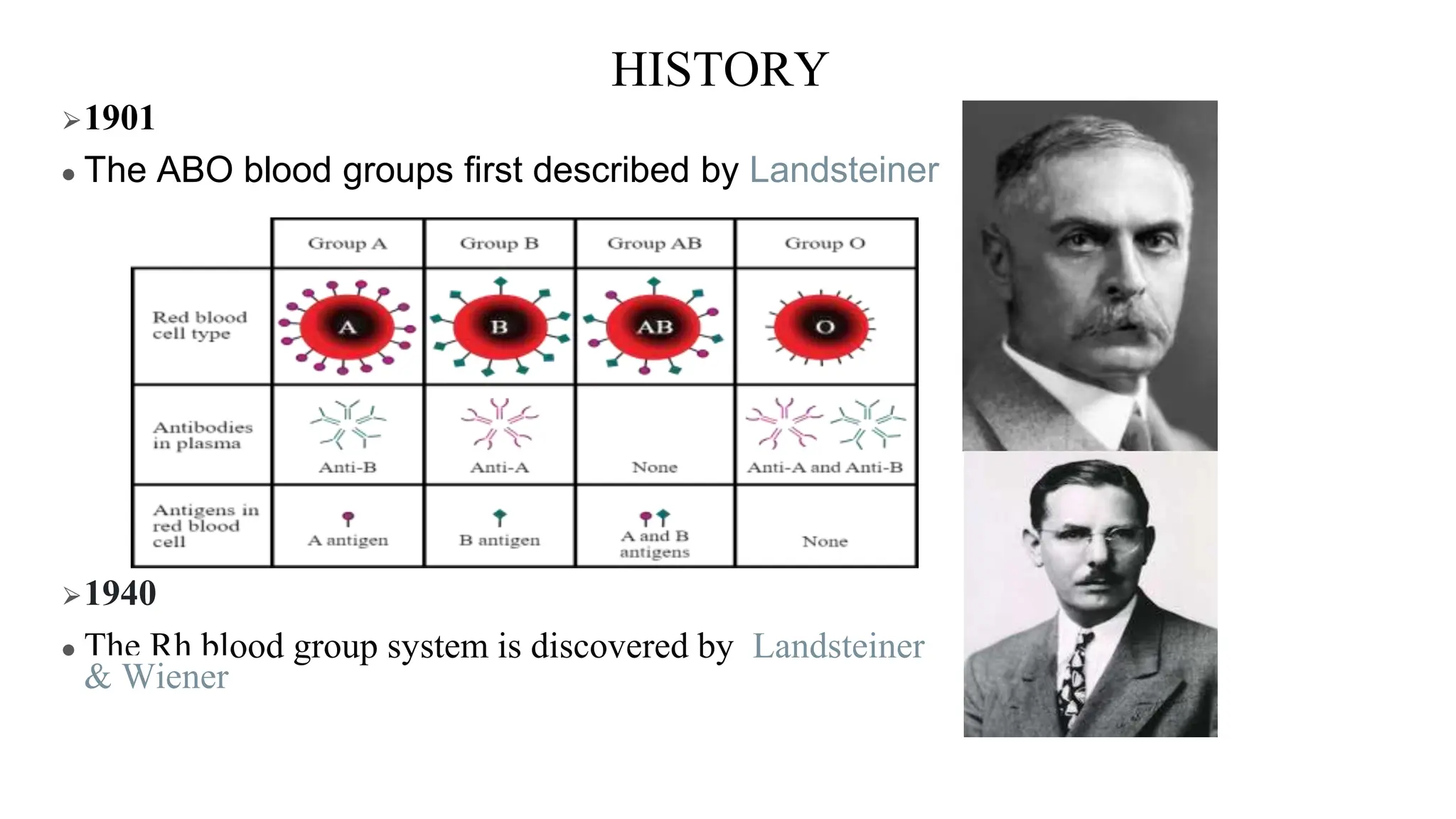
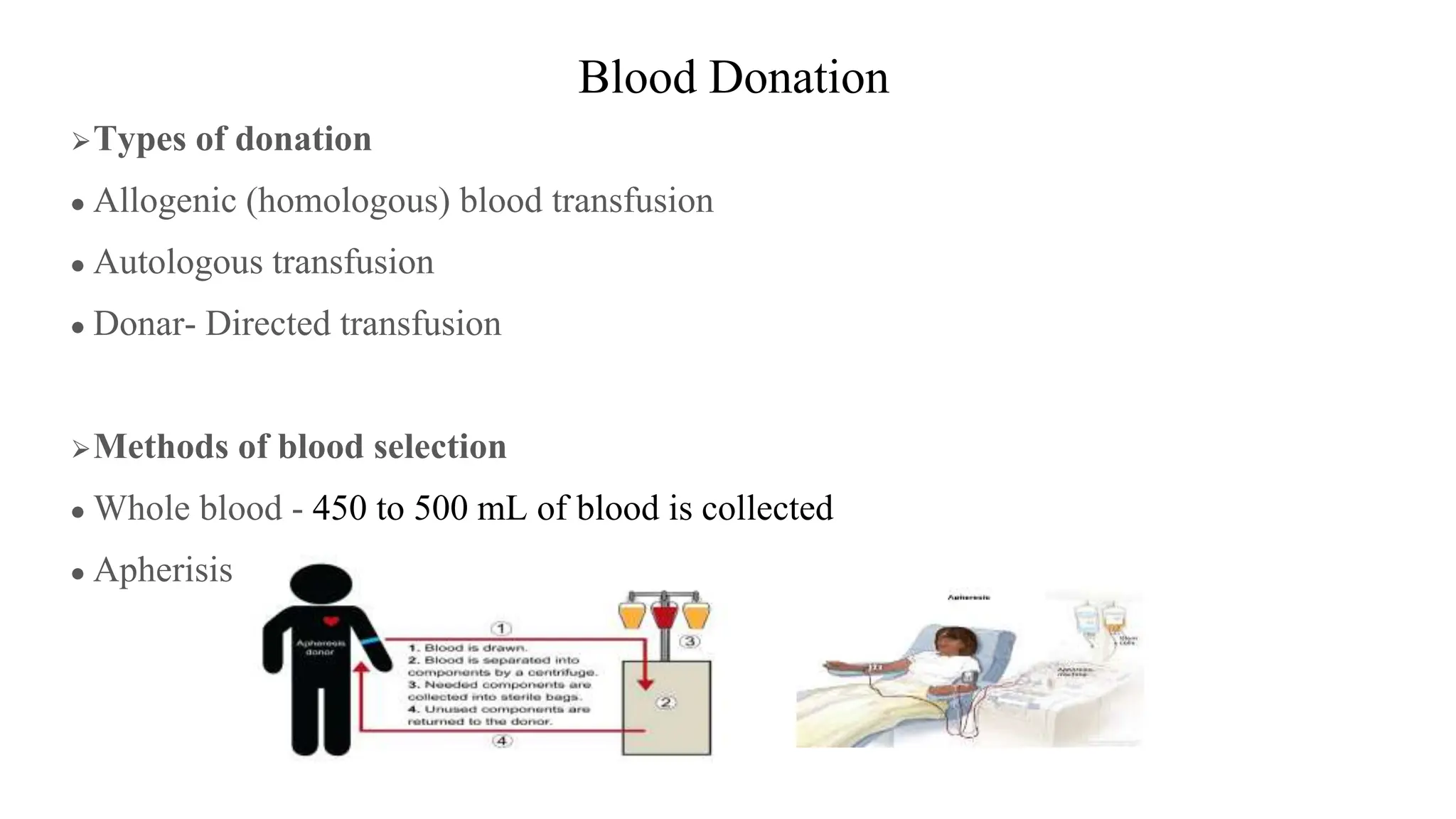
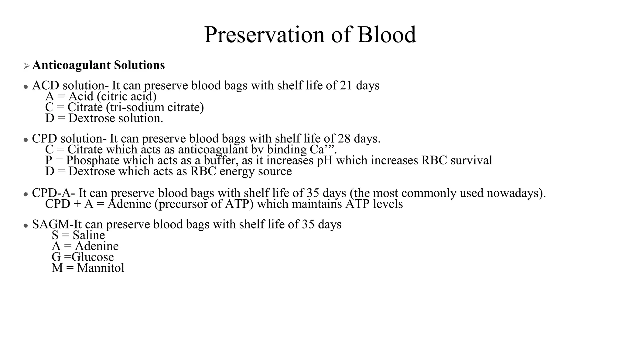
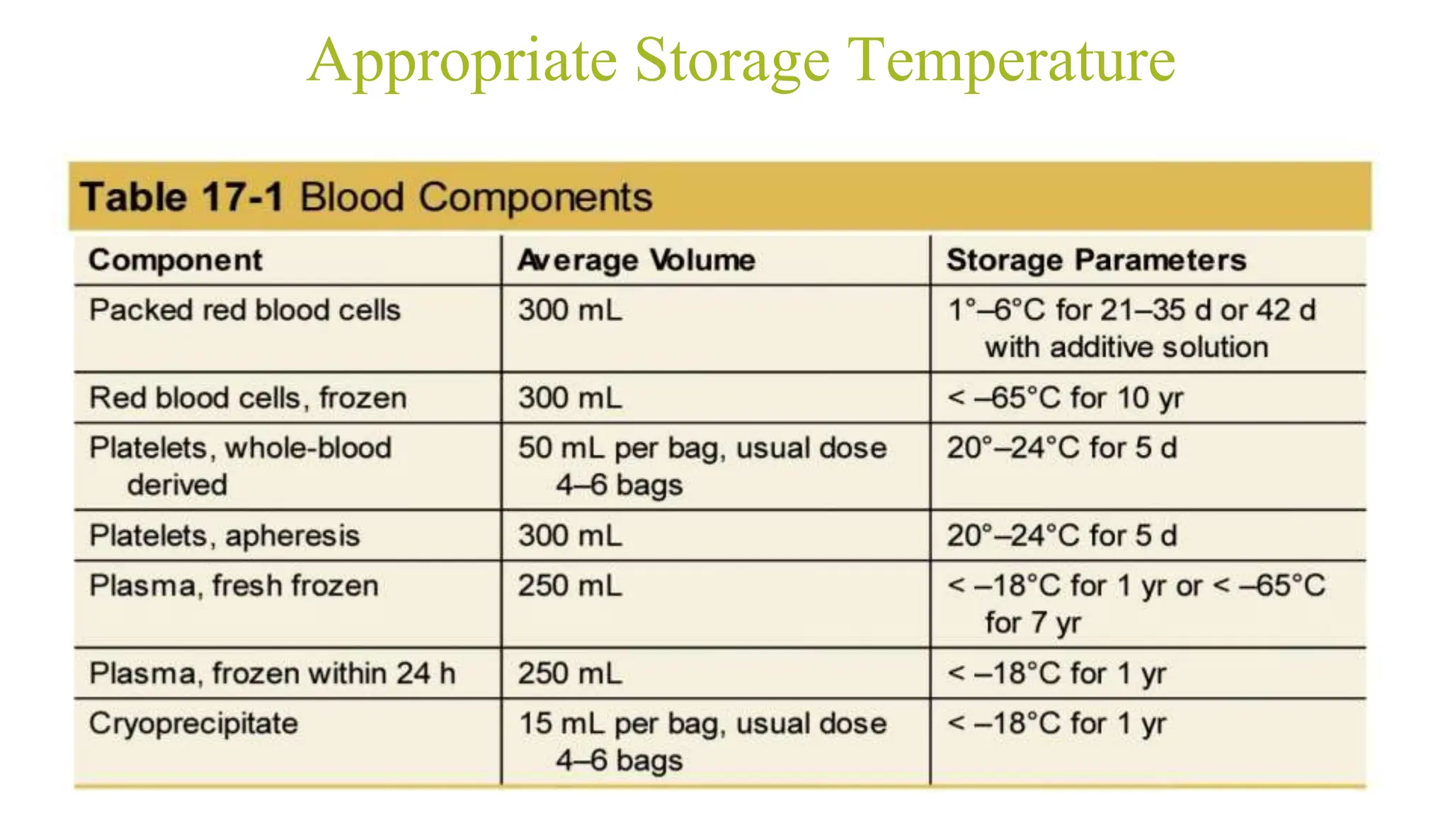
BLOOD COMPONENTS AND
BLOOD TRANSFUSION.
PRESENTER - DR VARUN T D
POST GRADUATE
MODERATOR-DR RAMYA
ASSISTANT PROFESSOR
DEPARTMENT OF ANAESTHESIOLOGY
HISTORY
⮚ 1492
● The first recorded attempt of a blood transfusion was
given to Pope Innocent VII in an attempt to save his life
by giving him the blood from the three young blood
donors
● The blood was transferred through the mouth, as the
concept of intravenous circulation had not yet been
discovered
HISTORY
⮚1628
● William Harvey discovered the circulation of blood.
⮚1665
● The first documented animal to animal (dog) blood transfusion was
performed by Richard Lower
⮚1667
● The first transfusion from animal (sheep) to human by Jean B
Denis
⮚1818
● An obstetrician James Blundell performed the first transfusion of
human blood for postpartum hemorrhage case where husband
donated blood to a lady.
HISTORY
⮚1901
● The ABO blood groups first described by Landsteiner
⮚1940
● The Rh blood group system is discovered by Landsteiner
& Wiener
BOMBAY PHENOTYPE
⮚ H system
• On group O’red cells there is no A or B antigen& the membrane
expresses ‘H’ antigen
⮚ BOMBAY PHENOTYPE
• Seen in persons with red cells which lack H, A&B antigens &
whose plasma contains antiH, antiA & antiB
• Pts with this blood group should be transfused only with this group
because hemolysis occurs if transfused with other groups which may
lead to death
Blood Donation
⮚Types of donation
● Allogenic (homologous) blood transfusion
● Autologous transfusion
● Donar- Directed transfusion
⮚Methods of blood selection
● Whole blood - 450 to 500 mL of blood is collected
● Apherisis
Preservation of Blood
⮚Anticoagulant Solutions
● ACD solution- It can preserve blood bags with shelf life of 21 days
A = Acid (citric acid)
C = Citrate (tri-sodium citrate)
D = Dextrose solution.
● CPD solution- It can preserve blood bags with shelf life of 28 days.
C = Citrate which acts as anticoagulant bv binding Ca’”.
P = Phosphate which acts as a buffer, as it increases pH which increases RBC survival
D = Dextrose which acts as RBC energy source
● CPD-A- It can preserve blood bags with shelf life of 35 days (the most commonly used nowadays).
CPD + A = Adenine (precursor of ATP) which maintains ATP levels
● SAGM-It can preserve blood bags with shelf life of 35 days
S = Saline
A = Adenine
G =Glucose
M = Mannitol
Appropriate Storage Temperature
Preparation of Blood components
BLOOD COMPONENTS.
BLOOD COMPONENT PRODUCTION.
● For Donors - Minimum hemoglobin/hematocrit level - 12.5 g/dL or 38% for
women and 13 g/dL or 39% for men.
● In whole-blood donations, 450 to 500 mL of blood is collected into citrate
anticoagulant.
● Donors are deferred for 8 weeks after a whole-blood donation to avoid iron
deficiency.
● WHOLE BLOOD.
● Volume-350-450ml.
● Anticoagulants - 65ml
● 200mg iron
● Hematocrit - 40-50%
● Plasma-150ml.
● Contains all blood elements and plasma.
● 1 unit will increase Hct by 3% Hb by 1g/dl.
● The use of whole blood is inefficient.
● Risk of volume overload
Packed RBCs
⮚Packed RBCs:Hct- 75 - 80%
● It should be the most commonly used blood transfusion because it allows optimal utilization of
blood bank resources.
● Indications:
1) For patients requiring RBC (but not volume) replacement e.g., a severely anemic patient in
compensated congestive heart failure or patients with active bleeding
2) Exchange blood transfusion during treatment of a patient with sickle cell disease
3) When volume replacement is required (e.g., most surgical patients)
● Add normal saline to the packed RBCs.
● Avoid lactated Ringer's injection because its calcium content may reverse the anticoagulant
effects of the citrate preservative
● Avoid 5% glucose because it is distilled water which may cause RBC hemolysis
.
OTHER FORMS OF RBC TRANSFUSION
⮚Leukocyte poor RBCs:
● They are prepared by centrifugation or filtration of blood units to remove leukocytes
● About 70% of leukocytes are removed, but the other 30% remain with the RBCs.
● Indication:
• To decrease febrile reactions by decreasing the possibility of HLA alloimmunization
• To decrease the incidence of cytomegalovirus infection
⮚Irradiated RBCs
● RBCs are irradiated by gamma irradiation to inactivate donor leukocytes and render them incapable of
participation in the immune response.
● Indication:
• To reduce graft-versus-host disease
⮚Washed RBCs
● RBCs are washed in saline to remove most plasma proteins, some leukocytes, and platelets.
● Indication:
• To decrease hypersensitivity allergic reaction in patients sensitive to a plasma component.
• To decrease incidence of anaphylactic reactions in IgA-deficient patients
. • To avoid transfusion of anti-A and anti-B antibodies when O-negative blood is used in patients who
are type A, B, or AB.
RBC.
The recommendations of the ASA practice guidelines
● Considerable morbidity and to a lesser extent mortality are associated with
blood transfusion therapy.
● Blood and blood products should be transfused only when there are clear
therapeutic indications
Recommendations:
Trigger-
Liberal strategy-HB< 10g/dl.
Restrictive strategy-HB< 7g/dl.
The ASA’s most recent publication on the Practice Guidelines for Perioperative
Blood Management defines restrictive practices as “hemoglobin criteria for
transfusion less than 8 g/dL and hematocrit values less than 25%
PLATELETS.
● most commonly stored at room temperature to preserve clotting function
● this increases the risk of bacterial growth.
.
Fresh Frozen Plasma
● It should be frozen within 6 hours after collection and can be stored frozen at -18°C to -
30°C for up to 1 year.
● Once thawed 30-37c, it must be transfused within 24 hours
● It contains all plasma proteins and lipids including all clotting factors
● Each unit of FFP generally increases the level of each clotting factor by 2-3% in adults.
● Indications:
1- Correction of abnormal coagulation tests
2- Treatment of isolated factor deficiency either prophylactic before surgery or with
bleeding.
3- Reversal of warfarin therapy.
4- Correction of coagulopathy associated with liver diseases.
5- In patients who have received massive blood transfusion.
6- In patients who continue to bleed after platelet transfusion.
● Dose:
• The amount of plasma must be individualized according to the level of coagulation
factor deficiency.
• The initial FFP dose is 10-15 ml/kg. FFP at that dose, achieves a minimum of 30% of
clotting factor concentration
CRYOPRECIPITATE.
● created by a controlled thaw of FFP
● containing concentrated doses of fibrinogen (at least 150 mg/unit), fibronectin,
vWF, FVIII, and FXIII.
● only indicated for critical bleeding with evidence of hypofibrinogenemia.
Plasma derivatives
● They include albumin, immunoglobulins (IGs), clotting factors, and other
proteins.
Cryoprecipitate:
only indicated for critical bleeding with evidence of hypofibrinogenemia
ALBUMIN.
● The derivative is available in 5% and 25% solutions.
● available for use as volume expanders.
● They can be given without regard to ABO blood type.
● Indications:
1. Hypoproteinemia
2. Burns
Intravenous immunoglobulins:
● given for immune support or for immunomodulation to suppress native antibody production
● antibody to specific antigens of interest, such as viruses (HBV, CMV, varicella zoster) or the
Rh blood group D antigen (RhIG to prevent anti-D formation in RhD-negative women).
Antithrombin III concentrates:
• Antithrombin III deficiency
• Shock and DIC
Factors VIII concentrates:
• Used in treatment of hemophilia A
Factor IX concentrates:
• Used in treatment of hemophilia B
Blood Products and Transfusion Practice.
COMPATABILITY TESTING.
Routine RBC compatibility testing includes
● ABO and RhD typing
● an antibody screen for IgG non-ABO RBC antibodies, and
● an RBC cross-match
RBCs must be ABO compatible to avoid intravascular hemolysis, and RhD-
negative patients should receive D-negative RBCs to avoid anti-D
alloimmunization
Rh, Kell, Kidd, Duffy, and some other nonABO antibodies can also hemolyze
transfused RBCs.
25
plasm
a
No
No
Yes
Yes
Yes
No
Yes
No
Yes
No Yes
Yes
Yes
Yes
No
No
The Risks of Blood Product Administration:
1.Infectious Risks of Blood Product Administration
2. Noninfectious Risks of Blood Product Administration.
A.Immune-Mediated Transfusion Reactions
● Febrile Nonhemolytic Transfusion Reactions
● Allergic Reactions
● Acute hemolytic transfusion reactions
● Delayed hemolytic transfusion reactions
● Transfusion-Related Immunomodulation
● Alloimmunization
● Transfusion-Related Acute Lung Injury
● Transfusion-Associated Graft-Versus-Host Disease.
● Posttransfusion Purpura
B. Nonimmune-Mediated Transfusion Reactions.
● Transfusion-Associated Circulatory Overload
● Metabolic Derangements
● Iron Overload
1. Infectious Risks of Blood Product Administration
Since the introduction of Nucleic Acid Amplification Testing (NAT) for major
transfusion-transmittable viral infections-the risk of infection from blood
product transfusion has decreased substantially
NAT increased the sensitivity of donor screening by testing for the presence
of specific viral DNA or RNA.
This significantly shortened the window period between when a donor gets
infected and when the viral load is detectable.
.
Bacterial Contamination.
Frequent pathogens implicated in bacterial contamination of blood products stem
from skin flora, including Staphylococcus and Bacillus species
Gram-negative species such as Escherichia coli, Enterobacter cloacae, and
especially Yersinia enterocolitica more frequently lead to sepsis given the higher
tolerance for cold storage
Platelet concentrates carry the highest risk of bacterial contamination, since they
are stored at 20° to 24°C, which provides a more suitable environment for the
exponential bacterial replication to occur over 4 to 5 days in storage.
.
.
ALLOIMMUNIZATION.
● refers to the induction of an immune response to allogenic antigen exposure
● Unlike classic ABO antigens, which consist of carbohydrate chains, most of
the non-ABO alloantigens (Kell, Kidd, Duffy, etc.) result from single amino
acid polymorphisms between the recipient and donor.
● alloimmunization prompts an amnestic IgG-mediated humoral immunity to
foreign proteins and does not result in RBC destruction until the second
antigen exposure
● RISK FACTOR-chronically transfused patients such as those with sickle cell
disease, hematologic malignancy, or thalassemia
Transfusion-Associated Graft-Versus-Host Disease
● It occurs when donor T-cell lymphocytes engraft in the recipient and attack
host cells they recognize as foreign.
● Patients at risk- immunocompromised
Immunocompetent patients may also be at risk when
transfused with directed donations from blood relations with similar HLA types
.
● classically presents 8 to 10 days after transfusion, but clinical suspicion
should exist for up to 6 weeks.
● Symptoms-fever, maculopapular rash, liver dysfunction, watery diarrhea,
and pancytopenia.
Nonimmune-Mediated Transfusion Reactions
1.Transfusion-Associated Circulatory Overload.
Three of the following finding
● respiratory distress within 6 hours of transfusion,
● increased central venous pressure, increased brain natriuretic peptide,
clinical signs of volume overload, and
● radiographic evidence of bilateral acute pulmonary edema.
RISK FACTORS - critically ill patients with a history of heart failure, chronic
diuretic dependence, and underlining renal insufficiency.
● The signs and symptoms of TACO can be confused with TAD and TRALI,-but
it differs from TRALI in that it is not immune mediated or associated with
increased capillary permeability, and it responds rapidly to diuretic therapy
and afterload reduction.
● Preventative measures
slowing the rate of transfusion when appropriate.
frequent assessment of vital signs, symptoms, and pulmonary status, and
rapid treatment of volume overload with diuretics
2.Metabolic Derangements.
● patients receiving large-volume transfusion or rapid infusion rates
● Hyperkalemia-storage time for blood products increases- leaking of potassium and metabolize
glucose into lactate, resulting in hyperkalemia and/or acidemia.
Hyperkalemia results from high-volume transfusion, especially when infusion rates exceed 100
to 150 mL/hr.
The acidosis is quickly cleared by physiologic buffers, as citrate preservative in blood products
is metabolized to bicarbonate.
● CITRATE TOXICITY :rapid infusion rates, massive transfusion, or in patients with liver dysfunction,
citrate accumulates in the plasma and chelates calcium, resulting in hypocalcemia.
● Hypothermia:especially during rapid infusion of previously cold or recently thawed blood products.
Coagulation factor activity decreases by 10% for every 1°C decrease in core body
temperature
IRON OVERLOAD.
● Each unit of RBCs contains an average of 200 to 250 mg of iron which
accumulates in patients who require frequent transfusions for chronic anemia
or hemoglobinopathies.
● Stored iron is not directly detrimental to organ systems; however, iron
metabolism produces harmful intracellular free radicals, which in turn cause
cellular dysfunction and organ failure.
● Chelation therapy with agents such as deferoxamine or deferasirox are the
first line of treatment.
● Plasma exchange transfusion therapy.
MASSIVE TRANSFUSION.
Definition:
Replacement of one entire blood volume within 24hrs.
Also defined as:
• Transfusion of >4 units within 1hr when ongoing need is foreseeable
• Transfusion of >10 units within 24hhrs,
• Replacement of 50% of total blood volume with in 3hrs.
*Transfusion of >20 units in 24hrs.
.
Blood Conservation Techniques
● Blood conservation techniques reduce the risk associated with blood
transfusion and improve patient outcomes.
● Strategies should be patient specific and include preoperative optimisation of
haemoglobin, management of comorbidities and medications, intraoperative
blood preservation techniques, and careful postoperative management.
● A multidisciplinary and multimodal approach is key to effective blood
conservation.
.
PREOPERATIVE STRATEGIES
● A patient-specific risk assessment for transfusion should be carried out for all
patients - - - -potential for high blood loss during surgery, for example,
cardiac, spinal, or major lower-limb procedures.
● Preoperative optimisation includes the identification and treatment of
preoperative anaemia with consideration of patient comorbidities such as
ischaemic heart disease and the appropriate management of perioperative
anticoagulation.
1.IDENTIFYING AND TREATING ANAEMIA
key to effective treatment and optimisation
● IRON-Increased requirement, limited supply, and blood loss can all lead to a
true deficiency–Early correction with iron replacement treatment is required.
● oral iron such as ferrous sulphate, ferrous fumarate, or ferrous gluconate.
● Parenteral iron therapy can be used if there is a functional iron deficiency or
if oral treatment is not tolerated or is ineffective or when time is limited for
adequate replacement.
● An increase in Hb is seen within 2 weeks of parenteral iron therapy and is
maximal at approximately 6 weeks.
● Erythropoiesis-stimulating agents (ESA) may also be of benefit to increase
RBC production.
● Other causes of anaemia once identified can be treated with replacement of
the relevant component, for example, vitamin B12 or folic acid, or involve
more specific management plans from the haematologist.
2.Anticoagulants.
● Many patients take medications that interfere with coagulation and can lead to
a higher risk of perioperative bleeding.
● It is important to balance the risks and benefits of stopping these medications
perioperatively depending on the thromboembolic versus intraoperative
bleeding risk
● this may require a multidisciplinary discussion between the anaesthetist,
surgeon, haematologist, and cardiologist to make a detailed plan.
● If the risk of stopping treatment is high, then bridging therapy can also be
considered using a shorter-acting heparin to cover the immediate operative
period.
Intraoperative
● Reducing blood loss intraoperatively involves a multidisciplinary approach.
● These include combined consideration of
patient positioning,
thermoregulation,
regional anaesthesia,
blood pressure management,
operative techniques, haemodilution
cell salvage,
and the use of antifibrinolytics.
Patient Positioning
● Circulatory changes induced by the perioperative positioning of patients can
be used to reduce or increase blood loss by promoting or opposing venous
drainage in a field.
Thermoregulation
● Hypothermia, can lead to altered enzyme function in the clotting cascade and
reduced platelet function.
● The combination of these factors may result in abnormal haemostasis with an
increased risk of intraoperative bleeding.
● Hypothermia also leads to an increase in Hb oxygen affinity, reduced cardiac
output, and ultimately a reduction in tissue oxygenation.
REGIONAL ANAESTHESIA.
Loss of sympathetic tone as a result of regional anaesthesia may help reduce the
arterial and peripheral venous blood pressure in the surgical field, resulting in less
intraoperative blood loss.
Blood Pressure Management
● Lowering the mean arterial pressure (MAP) in a controlled way reduces end-
organ blood flow and may help decrease blood loss (eg, in ear, nose, and
throat surgery)
● The risks of inducing hypotension must be tailored for an individual patient
and balanced to ensure appropriate vital organ perfusion.
● A reduction of 30% below the patient’s usual MAP is thought to be clinically
acceptable for ASA 1 Patients.
OPERATIVE TECHNIQUE.
● Preventative measures such as tourniquets can be considered, especially
with lower-limb surgery.
● Vessel ligation should be proactive rather than reactive.
● Other methods to stop bleeding early include pressure, ligation, diathermy,
and topical vasoconstrictors.
Antifibrinolytics.
Tranexamic acid is an antifibrinolytic used increasingly during surgery to decrease
blood loss by helping to prevent the breakdown of fibrin and promoting the
maintenance of blood clots.
Autologous Blood Transfusion:
encompasses three separate processes:
(1) preoperative autologous blood donation;
(2) acute normovolemic hemodilution (ANH); and
(3) perioperative blood cell salvage.
preoperative autologous blood donation.
● patients donating their own whole blood in the weeks preceding a planned
surgical procedure to ensure that they receive autologous blood.
● PAD eliminates the risk of transfusion-transmitted infection and
alloimmunization, and it may also decrease the risk of TRALI.
● protocol- early donation and use of iron supplementation and erythrocyte-
stimulating agents (ESAs) to allow for sufficient erythrogenesis.
● indicated for use in patients in whom it would be difficult to find compatible
blood products due to multiple antibodies or rare blood types, and those who
refuse to receive allogenic transfusion.
● Typically, each donation session collects a single or double unit of whole
blood and can technically be repeated weekly until 72 hours prior to the
scheduled procedure.
● Hemoglobin must remain above 11 g/dL before donation.. Q
ACUTE NORMOVOLEMIC HEMODILUTION.
● Normovolaemic haemodilution involves removing a patient’s blood and
replacing it with crystalloid to maintain intravascular volume.
● The haematocrit is low; tissue perfusion and blood flow are high due to low
plasma viscosity.
● Blood loss intraoperatively will contain fewer RBCs. Once the blood loss has
stopped, the patient’s own blood can then be returned.
● Volume to be removed = EBV × [(Hcti – Hctt)/Hctave]
● ANH is most effective in patients with high preoperative hemoglobin levels,
minimal cardiovascular comorbidities that allow intraoperative anemia, and a
surgical risk for largevolume blood loss.
PERIOPERATIVE BLOOD SALVAGE.
● cell salvage involves the collection of shed surgical blood, which is
filtered and washed prior to reinfusion.
● carried out intraoperatively or postoperatively.
Intra operative blood salvage.
● IOBS uses a double-lumen suction catheter with one port for aspiration from
the surgical field and the other for the addition of an anticoagulant solution,
usually heparin or citrate.
● Suctioned blood is collected in a reservoir, filtered to remove large debris, and
centrifuged to produce RBC concentrates. The final step of washing clears
the product of residual contaminants such as plasma, platelets, free
hemoglobin, cellular fragments, WBCs, and the remaining heparin or citrate9
● The resultant red cells are resuspended in saline and ready for reinfusion
● IOBS yields a hematocrit ranging from 55% to 70%.
● Evidence shows that salvaged blood has better oxygencarrying capacity and
tissue oxygenation than stored blood
● indicated for patients with low preoperative hemoglobin who cannot tolerate
PAD or ANH, those unwilling to consent to allogenic transfusion, and patients
with pre-existing bleeding risks or multiple alloantibodies.
● CONTRAINDICATION FOR CELLS SALVAGE.
POSTOPERATIVE BLOOD SALVAGE
● collection and reinfusion of blood shed into surgical wound drains in the
immediate postoperative period.
● Recovered blood product can be processed in two ways:
A. Washed.
B. Filtered before reinfusion.
Postoperative:
● Blood conservation postoperatively includes close observation and
maintaining a high index of suspicion if there are concerns about ongoing
blood loss
● Pressure dressings can be useful.
● Cell salvage, using the appropriate aseptic collection systems, can also be
considered for collections in drains.
THANK YOU.

BLOOD COMPONENTS AND BLOOD TRANSFUSION. .pptx

  • 1.
    BLOOD COMPONENTS AND BLOODTRANSFUSION. PRESENTER - DR VARUN T D POST GRADUATE MODERATOR-DR RAMYA ASSISTANT PROFESSOR DEPARTMENT OF ANAESTHESIOLOGY
  • 2.
    HISTORY ⮚ 1492 ● Thefirst recorded attempt of a blood transfusion was given to Pope Innocent VII in an attempt to save his life by giving him the blood from the three young blood donors ● The blood was transferred through the mouth, as the concept of intravenous circulation had not yet been discovered
  • 3.
    HISTORY ⮚1628 ● William Harveydiscovered the circulation of blood. ⮚1665 ● The first documented animal to animal (dog) blood transfusion was performed by Richard Lower ⮚1667 ● The first transfusion from animal (sheep) to human by Jean B Denis ⮚1818 ● An obstetrician James Blundell performed the first transfusion of human blood for postpartum hemorrhage case where husband donated blood to a lady.
  • 4.
    HISTORY ⮚1901 ● The ABOblood groups first described by Landsteiner ⮚1940 ● The Rh blood group system is discovered by Landsteiner & Wiener
  • 5.
    BOMBAY PHENOTYPE ⮚ Hsystem • On group O’red cells there is no A or B antigen& the membrane expresses ‘H’ antigen ⮚ BOMBAY PHENOTYPE • Seen in persons with red cells which lack H, A&B antigens & whose plasma contains antiH, antiA & antiB • Pts with this blood group should be transfused only with this group because hemolysis occurs if transfused with other groups which may lead to death
  • 6.
    Blood Donation ⮚Types ofdonation ● Allogenic (homologous) blood transfusion ● Autologous transfusion ● Donar- Directed transfusion ⮚Methods of blood selection ● Whole blood - 450 to 500 mL of blood is collected ● Apherisis
  • 7.
    Preservation of Blood ⮚AnticoagulantSolutions ● ACD solution- It can preserve blood bags with shelf life of 21 days A = Acid (citric acid) C = Citrate (tri-sodium citrate) D = Dextrose solution. ● CPD solution- It can preserve blood bags with shelf life of 28 days. C = Citrate which acts as anticoagulant bv binding Ca’”. P = Phosphate which acts as a buffer, as it increases pH which increases RBC survival D = Dextrose which acts as RBC energy source ● CPD-A- It can preserve blood bags with shelf life of 35 days (the most commonly used nowadays). CPD + A = Adenine (precursor of ATP) which maintains ATP levels ● SAGM-It can preserve blood bags with shelf life of 35 days S = Saline A = Adenine G =Glucose M = Mannitol
  • 8.
  • 9.
  • 10.
    BLOOD COMPONENTS. BLOOD COMPONENTPRODUCTION. ● For Donors - Minimum hemoglobin/hematocrit level - 12.5 g/dL or 38% for women and 13 g/dL or 39% for men. ● In whole-blood donations, 450 to 500 mL of blood is collected into citrate anticoagulant. ● Donors are deferred for 8 weeks after a whole-blood donation to avoid iron deficiency.
  • 11.
    ● WHOLE BLOOD. ●Volume-350-450ml. ● Anticoagulants - 65ml ● 200mg iron ● Hematocrit - 40-50% ● Plasma-150ml. ● Contains all blood elements and plasma. ● 1 unit will increase Hct by 3% Hb by 1g/dl. ● The use of whole blood is inefficient. ● Risk of volume overload
  • 12.
    Packed RBCs ⮚Packed RBCs:Hct-75 - 80% ● It should be the most commonly used blood transfusion because it allows optimal utilization of blood bank resources. ● Indications: 1) For patients requiring RBC (but not volume) replacement e.g., a severely anemic patient in compensated congestive heart failure or patients with active bleeding 2) Exchange blood transfusion during treatment of a patient with sickle cell disease 3) When volume replacement is required (e.g., most surgical patients) ● Add normal saline to the packed RBCs. ● Avoid lactated Ringer's injection because its calcium content may reverse the anticoagulant effects of the citrate preservative ● Avoid 5% glucose because it is distilled water which may cause RBC hemolysis
  • 13.
  • 14.
    OTHER FORMS OFRBC TRANSFUSION ⮚Leukocyte poor RBCs: ● They are prepared by centrifugation or filtration of blood units to remove leukocytes ● About 70% of leukocytes are removed, but the other 30% remain with the RBCs. ● Indication: • To decrease febrile reactions by decreasing the possibility of HLA alloimmunization • To decrease the incidence of cytomegalovirus infection ⮚Irradiated RBCs ● RBCs are irradiated by gamma irradiation to inactivate donor leukocytes and render them incapable of participation in the immune response. ● Indication: • To reduce graft-versus-host disease ⮚Washed RBCs ● RBCs are washed in saline to remove most plasma proteins, some leukocytes, and platelets. ● Indication: • To decrease hypersensitivity allergic reaction in patients sensitive to a plasma component. • To decrease incidence of anaphylactic reactions in IgA-deficient patients . • To avoid transfusion of anti-A and anti-B antibodies when O-negative blood is used in patients who are type A, B, or AB.
  • 15.
    RBC. The recommendations ofthe ASA practice guidelines ● Considerable morbidity and to a lesser extent mortality are associated with blood transfusion therapy. ● Blood and blood products should be transfused only when there are clear therapeutic indications Recommendations: Trigger- Liberal strategy-HB< 10g/dl. Restrictive strategy-HB< 7g/dl.
  • 16.
    The ASA’s mostrecent publication on the Practice Guidelines for Perioperative Blood Management defines restrictive practices as “hemoglobin criteria for transfusion less than 8 g/dL and hematocrit values less than 25%
  • 17.
    PLATELETS. ● most commonlystored at room temperature to preserve clotting function ● this increases the risk of bacterial growth.
  • 18.
  • 19.
    Fresh Frozen Plasma ●It should be frozen within 6 hours after collection and can be stored frozen at -18°C to - 30°C for up to 1 year. ● Once thawed 30-37c, it must be transfused within 24 hours ● It contains all plasma proteins and lipids including all clotting factors ● Each unit of FFP generally increases the level of each clotting factor by 2-3% in adults. ● Indications: 1- Correction of abnormal coagulation tests 2- Treatment of isolated factor deficiency either prophylactic before surgery or with bleeding. 3- Reversal of warfarin therapy. 4- Correction of coagulopathy associated with liver diseases. 5- In patients who have received massive blood transfusion. 6- In patients who continue to bleed after platelet transfusion. ● Dose: • The amount of plasma must be individualized according to the level of coagulation factor deficiency. • The initial FFP dose is 10-15 ml/kg. FFP at that dose, achieves a minimum of 30% of clotting factor concentration
  • 20.
    CRYOPRECIPITATE. ● created bya controlled thaw of FFP ● containing concentrated doses of fibrinogen (at least 150 mg/unit), fibronectin, vWF, FVIII, and FXIII. ● only indicated for critical bleeding with evidence of hypofibrinogenemia. Plasma derivatives ● They include albumin, immunoglobulins (IGs), clotting factors, and other proteins.
  • 21.
    Cryoprecipitate: only indicated forcritical bleeding with evidence of hypofibrinogenemia
  • 22.
    ALBUMIN. ● The derivativeis available in 5% and 25% solutions. ● available for use as volume expanders. ● They can be given without regard to ABO blood type. ● Indications: 1. Hypoproteinemia 2. Burns
  • 23.
    Intravenous immunoglobulins: ● givenfor immune support or for immunomodulation to suppress native antibody production ● antibody to specific antigens of interest, such as viruses (HBV, CMV, varicella zoster) or the Rh blood group D antigen (RhIG to prevent anti-D formation in RhD-negative women). Antithrombin III concentrates: • Antithrombin III deficiency • Shock and DIC Factors VIII concentrates: • Used in treatment of hemophilia A Factor IX concentrates: • Used in treatment of hemophilia B
  • 24.
    Blood Products andTransfusion Practice. COMPATABILITY TESTING. Routine RBC compatibility testing includes ● ABO and RhD typing ● an antibody screen for IgG non-ABO RBC antibodies, and ● an RBC cross-match RBCs must be ABO compatible to avoid intravascular hemolysis, and RhD- negative patients should receive D-negative RBCs to avoid anti-D alloimmunization Rh, Kell, Kidd, Duffy, and some other nonABO antibodies can also hemolyze transfused RBCs.
  • 25.
  • 26.
  • 27.
    The Risks ofBlood Product Administration: 1.Infectious Risks of Blood Product Administration 2. Noninfectious Risks of Blood Product Administration. A.Immune-Mediated Transfusion Reactions ● Febrile Nonhemolytic Transfusion Reactions ● Allergic Reactions ● Acute hemolytic transfusion reactions ● Delayed hemolytic transfusion reactions ● Transfusion-Related Immunomodulation ● Alloimmunization ● Transfusion-Related Acute Lung Injury
  • 28.
    ● Transfusion-Associated Graft-Versus-HostDisease. ● Posttransfusion Purpura B. Nonimmune-Mediated Transfusion Reactions. ● Transfusion-Associated Circulatory Overload ● Metabolic Derangements ● Iron Overload
  • 29.
    1. Infectious Risksof Blood Product Administration Since the introduction of Nucleic Acid Amplification Testing (NAT) for major transfusion-transmittable viral infections-the risk of infection from blood product transfusion has decreased substantially NAT increased the sensitivity of donor screening by testing for the presence of specific viral DNA or RNA. This significantly shortened the window period between when a donor gets infected and when the viral load is detectable.
  • 30.
  • 31.
    Bacterial Contamination. Frequent pathogensimplicated in bacterial contamination of blood products stem from skin flora, including Staphylococcus and Bacillus species Gram-negative species such as Escherichia coli, Enterobacter cloacae, and especially Yersinia enterocolitica more frequently lead to sepsis given the higher tolerance for cold storage Platelet concentrates carry the highest risk of bacterial contamination, since they are stored at 20° to 24°C, which provides a more suitable environment for the exponential bacterial replication to occur over 4 to 5 days in storage.
  • 32.
  • 33.
  • 34.
    ALLOIMMUNIZATION. ● refers tothe induction of an immune response to allogenic antigen exposure ● Unlike classic ABO antigens, which consist of carbohydrate chains, most of the non-ABO alloantigens (Kell, Kidd, Duffy, etc.) result from single amino acid polymorphisms between the recipient and donor. ● alloimmunization prompts an amnestic IgG-mediated humoral immunity to foreign proteins and does not result in RBC destruction until the second antigen exposure ● RISK FACTOR-chronically transfused patients such as those with sickle cell disease, hematologic malignancy, or thalassemia
  • 35.
    Transfusion-Associated Graft-Versus-Host Disease ●It occurs when donor T-cell lymphocytes engraft in the recipient and attack host cells they recognize as foreign. ● Patients at risk- immunocompromised Immunocompetent patients may also be at risk when transfused with directed donations from blood relations with similar HLA types . ● classically presents 8 to 10 days after transfusion, but clinical suspicion should exist for up to 6 weeks. ● Symptoms-fever, maculopapular rash, liver dysfunction, watery diarrhea, and pancytopenia.
  • 36.
    Nonimmune-Mediated Transfusion Reactions 1.Transfusion-AssociatedCirculatory Overload. Three of the following finding ● respiratory distress within 6 hours of transfusion, ● increased central venous pressure, increased brain natriuretic peptide, clinical signs of volume overload, and ● radiographic evidence of bilateral acute pulmonary edema. RISK FACTORS - critically ill patients with a history of heart failure, chronic diuretic dependence, and underlining renal insufficiency.
  • 37.
    ● The signsand symptoms of TACO can be confused with TAD and TRALI,-but it differs from TRALI in that it is not immune mediated or associated with increased capillary permeability, and it responds rapidly to diuretic therapy and afterload reduction. ● Preventative measures slowing the rate of transfusion when appropriate. frequent assessment of vital signs, symptoms, and pulmonary status, and rapid treatment of volume overload with diuretics
  • 38.
    2.Metabolic Derangements. ● patientsreceiving large-volume transfusion or rapid infusion rates ● Hyperkalemia-storage time for blood products increases- leaking of potassium and metabolize glucose into lactate, resulting in hyperkalemia and/or acidemia. Hyperkalemia results from high-volume transfusion, especially when infusion rates exceed 100 to 150 mL/hr. The acidosis is quickly cleared by physiologic buffers, as citrate preservative in blood products is metabolized to bicarbonate. ● CITRATE TOXICITY :rapid infusion rates, massive transfusion, or in patients with liver dysfunction, citrate accumulates in the plasma and chelates calcium, resulting in hypocalcemia. ● Hypothermia:especially during rapid infusion of previously cold or recently thawed blood products. Coagulation factor activity decreases by 10% for every 1°C decrease in core body temperature
  • 39.
    IRON OVERLOAD. ● Eachunit of RBCs contains an average of 200 to 250 mg of iron which accumulates in patients who require frequent transfusions for chronic anemia or hemoglobinopathies. ● Stored iron is not directly detrimental to organ systems; however, iron metabolism produces harmful intracellular free radicals, which in turn cause cellular dysfunction and organ failure. ● Chelation therapy with agents such as deferoxamine or deferasirox are the first line of treatment. ● Plasma exchange transfusion therapy.
  • 40.
    MASSIVE TRANSFUSION. Definition: Replacement ofone entire blood volume within 24hrs. Also defined as: • Transfusion of >4 units within 1hr when ongoing need is foreseeable • Transfusion of >10 units within 24hhrs, • Replacement of 50% of total blood volume with in 3hrs. *Transfusion of >20 units in 24hrs.
  • 41.
  • 43.
    Blood Conservation Techniques ●Blood conservation techniques reduce the risk associated with blood transfusion and improve patient outcomes. ● Strategies should be patient specific and include preoperative optimisation of haemoglobin, management of comorbidities and medications, intraoperative blood preservation techniques, and careful postoperative management. ● A multidisciplinary and multimodal approach is key to effective blood conservation.
  • 44.
  • 45.
    PREOPERATIVE STRATEGIES ● Apatient-specific risk assessment for transfusion should be carried out for all patients - - - -potential for high blood loss during surgery, for example, cardiac, spinal, or major lower-limb procedures. ● Preoperative optimisation includes the identification and treatment of preoperative anaemia with consideration of patient comorbidities such as ischaemic heart disease and the appropriate management of perioperative anticoagulation.
  • 46.
    1.IDENTIFYING AND TREATINGANAEMIA key to effective treatment and optimisation
  • 47.
    ● IRON-Increased requirement,limited supply, and blood loss can all lead to a true deficiency–Early correction with iron replacement treatment is required. ● oral iron such as ferrous sulphate, ferrous fumarate, or ferrous gluconate. ● Parenteral iron therapy can be used if there is a functional iron deficiency or if oral treatment is not tolerated or is ineffective or when time is limited for adequate replacement. ● An increase in Hb is seen within 2 weeks of parenteral iron therapy and is maximal at approximately 6 weeks. ● Erythropoiesis-stimulating agents (ESA) may also be of benefit to increase RBC production. ● Other causes of anaemia once identified can be treated with replacement of the relevant component, for example, vitamin B12 or folic acid, or involve more specific management plans from the haematologist.
  • 48.
    2.Anticoagulants. ● Many patientstake medications that interfere with coagulation and can lead to a higher risk of perioperative bleeding. ● It is important to balance the risks and benefits of stopping these medications perioperatively depending on the thromboembolic versus intraoperative bleeding risk ● this may require a multidisciplinary discussion between the anaesthetist, surgeon, haematologist, and cardiologist to make a detailed plan. ● If the risk of stopping treatment is high, then bridging therapy can also be considered using a shorter-acting heparin to cover the immediate operative period.
  • 49.
    Intraoperative ● Reducing bloodloss intraoperatively involves a multidisciplinary approach. ● These include combined consideration of patient positioning, thermoregulation, regional anaesthesia, blood pressure management, operative techniques, haemodilution cell salvage, and the use of antifibrinolytics.
  • 50.
    Patient Positioning ● Circulatorychanges induced by the perioperative positioning of patients can be used to reduce or increase blood loss by promoting or opposing venous drainage in a field. Thermoregulation ● Hypothermia, can lead to altered enzyme function in the clotting cascade and reduced platelet function. ● The combination of these factors may result in abnormal haemostasis with an increased risk of intraoperative bleeding. ● Hypothermia also leads to an increase in Hb oxygen affinity, reduced cardiac output, and ultimately a reduction in tissue oxygenation.
  • 51.
    REGIONAL ANAESTHESIA. Loss ofsympathetic tone as a result of regional anaesthesia may help reduce the arterial and peripheral venous blood pressure in the surgical field, resulting in less intraoperative blood loss. Blood Pressure Management ● Lowering the mean arterial pressure (MAP) in a controlled way reduces end- organ blood flow and may help decrease blood loss (eg, in ear, nose, and throat surgery) ● The risks of inducing hypotension must be tailored for an individual patient and balanced to ensure appropriate vital organ perfusion. ● A reduction of 30% below the patient’s usual MAP is thought to be clinically acceptable for ASA 1 Patients.
  • 52.
    OPERATIVE TECHNIQUE. ● Preventativemeasures such as tourniquets can be considered, especially with lower-limb surgery. ● Vessel ligation should be proactive rather than reactive. ● Other methods to stop bleeding early include pressure, ligation, diathermy, and topical vasoconstrictors. Antifibrinolytics. Tranexamic acid is an antifibrinolytic used increasingly during surgery to decrease blood loss by helping to prevent the breakdown of fibrin and promoting the maintenance of blood clots.
  • 53.
    Autologous Blood Transfusion: encompassesthree separate processes: (1) preoperative autologous blood donation; (2) acute normovolemic hemodilution (ANH); and (3) perioperative blood cell salvage.
  • 54.
    preoperative autologous blooddonation. ● patients donating their own whole blood in the weeks preceding a planned surgical procedure to ensure that they receive autologous blood. ● PAD eliminates the risk of transfusion-transmitted infection and alloimmunization, and it may also decrease the risk of TRALI. ● protocol- early donation and use of iron supplementation and erythrocyte- stimulating agents (ESAs) to allow for sufficient erythrogenesis. ● indicated for use in patients in whom it would be difficult to find compatible blood products due to multiple antibodies or rare blood types, and those who refuse to receive allogenic transfusion. ● Typically, each donation session collects a single or double unit of whole blood and can technically be repeated weekly until 72 hours prior to the scheduled procedure. ● Hemoglobin must remain above 11 g/dL before donation.. Q
  • 55.
    ACUTE NORMOVOLEMIC HEMODILUTION. ●Normovolaemic haemodilution involves removing a patient’s blood and replacing it with crystalloid to maintain intravascular volume. ● The haematocrit is low; tissue perfusion and blood flow are high due to low plasma viscosity. ● Blood loss intraoperatively will contain fewer RBCs. Once the blood loss has stopped, the patient’s own blood can then be returned. ● Volume to be removed = EBV × [(Hcti – Hctt)/Hctave] ● ANH is most effective in patients with high preoperative hemoglobin levels, minimal cardiovascular comorbidities that allow intraoperative anemia, and a surgical risk for largevolume blood loss.
  • 56.
    PERIOPERATIVE BLOOD SALVAGE. ●cell salvage involves the collection of shed surgical blood, which is filtered and washed prior to reinfusion. ● carried out intraoperatively or postoperatively. Intra operative blood salvage.
  • 57.
    ● IOBS usesa double-lumen suction catheter with one port for aspiration from the surgical field and the other for the addition of an anticoagulant solution, usually heparin or citrate. ● Suctioned blood is collected in a reservoir, filtered to remove large debris, and centrifuged to produce RBC concentrates. The final step of washing clears the product of residual contaminants such as plasma, platelets, free hemoglobin, cellular fragments, WBCs, and the remaining heparin or citrate9 ● The resultant red cells are resuspended in saline and ready for reinfusion ● IOBS yields a hematocrit ranging from 55% to 70%. ● Evidence shows that salvaged blood has better oxygencarrying capacity and tissue oxygenation than stored blood ● indicated for patients with low preoperative hemoglobin who cannot tolerate PAD or ANH, those unwilling to consent to allogenic transfusion, and patients with pre-existing bleeding risks or multiple alloantibodies.
  • 58.
  • 59.
    POSTOPERATIVE BLOOD SALVAGE ●collection and reinfusion of blood shed into surgical wound drains in the immediate postoperative period. ● Recovered blood product can be processed in two ways: A. Washed. B. Filtered before reinfusion.
  • 60.
    Postoperative: ● Blood conservationpostoperatively includes close observation and maintaining a high index of suspicion if there are concerns about ongoing blood loss ● Pressure dressings can be useful. ● Cell salvage, using the appropriate aseptic collection systems, can also be considered for collections in drains.
  • 61.