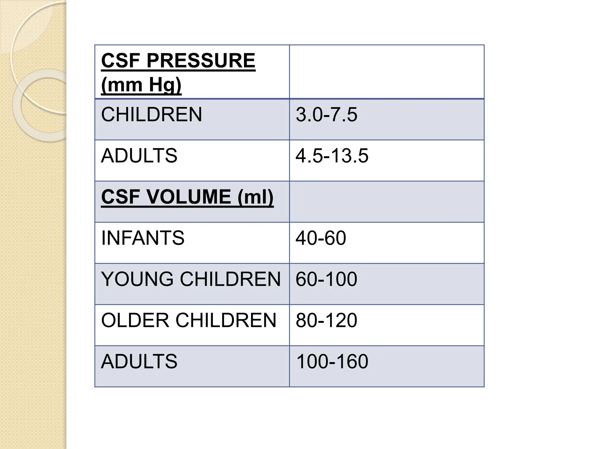
CSF PHYSIOLOGY AND
CIRCULATION
Presenter : Dr Rakshitha R
Moderator : Dr Lakshmi
INTRODUCTION
 Cerebrospinal fluid is a clear fluid
present in the ventricles of the brain,
the central canal of the spinal cord
and the subarachnoid space.
Function of CSF
 Protects, lubricates the brain.
 Provides nutrients
 Removes waste.
 Transport medium for nutrients and
metabolites, endocrine substances and
neurotransmitters.
 Biomarker for disease diagnosis.
 Contains growth factor and micro RNA
for neurogenesis.
 Blood-CSF barrier
FORMATION OF CSF
 3 sites :
Choroid plexus (50-70%)
Ependymal surfaces of ventricles
Perivascular spaces.
 Formed at rate of : 500-600ml/day OR
: 0.35-0.40ml/min
• Turn over time : 5-7hours = 4
times/day
• CSF volume : 150ml (125ml in cranial
and subarachnoid spaces+ 25ml in
ventricle)
CSF PRESSURE
(mm Hg)
CHILDREN 3.0-7.5
ADULTS 4.5-13.5
CSF VOLUME (ml)
INFANTS 40-60
YOUNG CHILDREN 60-100
OLDER CHILDREN 80-120
ADULTS 100-160
Choroid plexus
 It is a cauliflower like growth of blood
vessels covered by a thin layer of
epithelial cells.
 It projects into temporal horn of lateral
ventricle, posterior part of 3rd ventricle
and roof of 4th ventricle.
Nerve supply: branches of vagus and
glossopharyngeal nerve and sympathetic
nerves.
Blood supply:
Body of lateral ventricle Posterior choroidal artery
Body of third ventricle Anterior choroidal artery
Temporal horns Superior cerebellar artery
Fourth ventricle Posterior inferior cerebellar
artery.
Formation of CSF at choroid
plexus
 Filteration of plasma across choroidal
capillary endothelium into interstitial
compartment according to pressure
gradient.
 Secretion of ions and water across
epithelial cells.
CIRCULATION OF CSF
ABSORPTION OF CSF
• All CSF formed is reabsorbed into the
venous circulation through arachnoid
villi or granulations.
• These villi project the subarachnoid
spaces into the venous sinuses of the
brain and occasionally into the venous
of the spinal cord.
EFFECT OF DRUGS ON CSF
FORMATION
Desfluran
e
Enflurane Halothane Isoflurane Sevofluran
e
Nitrous
Oxide
secretion + + -- 0 -- 0
absorptio
n
0 -- -- + -- 0
Ketamine Etomidate Propofol Pentobarbital Thiopental
secretion 0 -- 0 0 0
absorption -- + 0 0 --
Midazolam Flumzenil Fentanyl Sufentanyl Lidocaine
secretion 0 0 0 0 --
absorption -- 0 + + 0
INHALED ANESTHETICS
IV ANESTHETICS
OTHER IV ANESTHETICS
OTHER DRUGS :
 Acetazolamide inhibit carbonic
anhydrase enzyme,constricts arterioles
=> decrease CSF formation.
 Furosemide inhibit sodium chloride
transport => decrease CSF formation.
 Theophyllin is a phosphodiesterase
inhibitor -increase CAMP- stimulate
sodium potassium pump => increase
CSF production.
 Digoxin inhibit sodium potassium
ATPase pump=> decrease CSF
formation.
 Vasopressin constrict choroid plexus
blood vessel => decrease CSF
production.
 Muscle relaxant has no effect on CSF
formation.
 Dexamethasone inhibit sodium
potassium ATPase => decrease CSF
production.
 Alpha agonist causes vasoconstriction
=> decrease CSF formation.
 Cholinergic drugs acts on muscarinic
receptor on choroid epithelium =>
decrease CSF formation.
 Hypothermia (1˚C)decrease cerebral
blood flow => decrease CSF
formation. (11%)
 Hypocapnia decrease cerebral blood
flow => decrease CSF production
 Metabolic alkalosis decrease
formation of CSF.
Composition of CSF
Intracranial Pressure(ICP)
 The intracranial and spinal vault
contains neural tissue, blood and CSF
and is enclosed by duramater and
bone.
 The pressure within this space –
intracranial pressure.
 Normal ICP is <15mm Hg.
 This pressure is regulated by rate of
formation of CSF and resistance to
CSF reabsorption by arachnoid villi.
Monro Kellie Doctrine
 The sum of volumes of brain, CSF and
intracranial blood is constant.
 An increase in volume of one component will
be compensated by decrease in one or both
of the remaining two.
Causes of increase ICP
Physiological causes:
 Coughing
 Sneezing
 Straining
Signs and symptoms
 Headache
 Nausea
 Vomiting
 Papilledema
Methods to measure ICP
 Pressure transducers can be placed
under aseptic conditions into subdural
space (subdural bolt), brain
parenchyma or ventricle
(ventriculostomy)
 Lumbar arachnoid catheter.
Methods to decrease ICP
 Elevation of the head.
 Hyperventilation.
 CSF drainage.
 Hyperosmotic drugs.
 Diuretics.
 Corticosteroids.
 Vasoconstricting anesthetics –
barbiturates, propofol.
 Surgical decompression.
Hydrocephalus
 One of the most common pediatric and
adult neurosurgical disorder,
 An abnormal increase in amount of CSF
resulting from a disturbance of formation,
flow or absorption of CSF thus resulting in
enlarged cerebral ventricles.
Clinical presentation :
In infants and newborn :
 Cranium enlarges>facial growth
 Irritable, lethargy
 Delayed milestones
 Vomiting
 Decrease oral intake
 Macewen’s sign- cracked pot sound
on percussion over dilated ventricle
 Hyperactive reflexes
Older child/adults:
Symptoms of increased ICP-
 Headache
 Papilledema
 Nausea, vomiting
 Upward gaze
 Abducens palsy
Treatment :
Medical therapy –
 Acetazolamide :25mg/kg/day PO
divided TID increase by 25mg/kg/day
until 100mg/kg/day.
 Furosemide : 1mg/kg/day PO divided
TID
 Correct electrolyte abnormalities
Neurosurgical procedures –
 Ventriculostomy
 Choroid plexectomy
 Shunting – ventriculoperitoneal shunt
- ventriculoatrial shunt
- ventriculopleural shunt
- lumboperitoneal shunt
- torkildsen shunt
Anesthesia management
 History taking and physical examination
– severity of ICP and neurological status.
 Premedication : avoid sedation.If
required titrated dose of oral/intranasal
midazolam may be used..
 Prophylactic antibiotics should be given
to lower shunt infection rate.
 Monitoring : pulse oximetry, ECG, blood
pressure, capnometry, temperature
 Patient head is placed 30˚head up
position.
 Induction : high ICP and vomiting increase
risk of aspiration. So rapid sequence
induction with thiopentone (3-5mg/kg) or
propofol (2-4mg/kg) followed by
succinylcholine or rocuronium is used.
 Institute hyperventilation.(PaCO2 of 25-30
mm Hg).
 Avoid spontaneous ventilation in
ventriculopleural shunt ( to prevent
pneumothorax) and ventriculoatrial shunt (
to prevent air embolism).
 Maintainance: air, oxygen, isoflurane/
sevoflurane, fentanyl, paracetamol,
intermediate acting non depolarising
muscle relaxants.
 Replace loss of intravascular volume
due to emesis or diuresis with saline.
 Maintain normothermia.
 Extubate when fully awake.
 Post operatively patient should be
nursed flat so as to avoid subdural
hemorrhage which may occur
because of rapid collpse of ventricles
in head high position. Monitor mental
status as reobstruction can occur.
References
 Guyton and Hall Textbook Of Medical
Physiology.
 Stoelting’s Pharmacology and
Physiology in Anesthetic Practice.
 Stoelting’s Anesthesia Co-Existing
Diseaase.
THANK YOU

C S F PHYSIOLOGY AND CIRCULATION.pptx

  • 1.
    CSF PHYSIOLOGY AND CIRCULATION Presenter: Dr Rakshitha R Moderator : Dr Lakshmi
  • 2.
    INTRODUCTION  Cerebrospinal fluidis a clear fluid present in the ventricles of the brain, the central canal of the spinal cord and the subarachnoid space.
  • 3.
    Function of CSF Protects, lubricates the brain.  Provides nutrients  Removes waste.  Transport medium for nutrients and metabolites, endocrine substances and neurotransmitters.  Biomarker for disease diagnosis.  Contains growth factor and micro RNA for neurogenesis.  Blood-CSF barrier
  • 5.
    FORMATION OF CSF 3 sites : Choroid plexus (50-70%) Ependymal surfaces of ventricles Perivascular spaces.
  • 6.
     Formed atrate of : 500-600ml/day OR : 0.35-0.40ml/min • Turn over time : 5-7hours = 4 times/day • CSF volume : 150ml (125ml in cranial and subarachnoid spaces+ 25ml in ventricle)
  • 7.
    CSF PRESSURE (mm Hg) CHILDREN3.0-7.5 ADULTS 4.5-13.5 CSF VOLUME (ml) INFANTS 40-60 YOUNG CHILDREN 60-100 OLDER CHILDREN 80-120 ADULTS 100-160
  • 8.
    Choroid plexus  Itis a cauliflower like growth of blood vessels covered by a thin layer of epithelial cells.  It projects into temporal horn of lateral ventricle, posterior part of 3rd ventricle and roof of 4th ventricle.
  • 9.
    Nerve supply: branchesof vagus and glossopharyngeal nerve and sympathetic nerves. Blood supply: Body of lateral ventricle Posterior choroidal artery Body of third ventricle Anterior choroidal artery Temporal horns Superior cerebellar artery Fourth ventricle Posterior inferior cerebellar artery.
  • 11.
    Formation of CSFat choroid plexus  Filteration of plasma across choroidal capillary endothelium into interstitial compartment according to pressure gradient.  Secretion of ions and water across epithelial cells.
  • 13.
  • 15.
    ABSORPTION OF CSF •All CSF formed is reabsorbed into the venous circulation through arachnoid villi or granulations. • These villi project the subarachnoid spaces into the venous sinuses of the brain and occasionally into the venous of the spinal cord.
  • 17.
    EFFECT OF DRUGSON CSF FORMATION
  • 18.
    Desfluran e Enflurane Halothane IsofluraneSevofluran e Nitrous Oxide secretion + + -- 0 -- 0 absorptio n 0 -- -- + -- 0 Ketamine Etomidate Propofol Pentobarbital Thiopental secretion 0 -- 0 0 0 absorption -- + 0 0 -- Midazolam Flumzenil Fentanyl Sufentanyl Lidocaine secretion 0 0 0 0 -- absorption -- 0 + + 0 INHALED ANESTHETICS IV ANESTHETICS OTHER IV ANESTHETICS
  • 19.
    OTHER DRUGS : Acetazolamide inhibit carbonic anhydrase enzyme,constricts arterioles => decrease CSF formation.  Furosemide inhibit sodium chloride transport => decrease CSF formation.  Theophyllin is a phosphodiesterase inhibitor -increase CAMP- stimulate sodium potassium pump => increase CSF production.
  • 20.
     Digoxin inhibitsodium potassium ATPase pump=> decrease CSF formation.  Vasopressin constrict choroid plexus blood vessel => decrease CSF production.  Muscle relaxant has no effect on CSF formation.  Dexamethasone inhibit sodium potassium ATPase => decrease CSF production.
  • 21.
     Alpha agonistcauses vasoconstriction => decrease CSF formation.  Cholinergic drugs acts on muscarinic receptor on choroid epithelium => decrease CSF formation.  Hypothermia (1˚C)decrease cerebral blood flow => decrease CSF formation. (11%)  Hypocapnia decrease cerebral blood flow => decrease CSF production  Metabolic alkalosis decrease formation of CSF.
  • 22.
  • 25.
    Intracranial Pressure(ICP)  Theintracranial and spinal vault contains neural tissue, blood and CSF and is enclosed by duramater and bone.  The pressure within this space – intracranial pressure.  Normal ICP is <15mm Hg.  This pressure is regulated by rate of formation of CSF and resistance to CSF reabsorption by arachnoid villi.
  • 26.
    Monro Kellie Doctrine The sum of volumes of brain, CSF and intracranial blood is constant.  An increase in volume of one component will be compensated by decrease in one or both of the remaining two.
  • 30.
    Causes of increaseICP Physiological causes:  Coughing  Sneezing  Straining
  • 33.
    Signs and symptoms Headache  Nausea  Vomiting  Papilledema
  • 34.
    Methods to measureICP  Pressure transducers can be placed under aseptic conditions into subdural space (subdural bolt), brain parenchyma or ventricle (ventriculostomy)  Lumbar arachnoid catheter.
  • 35.
    Methods to decreaseICP  Elevation of the head.  Hyperventilation.  CSF drainage.  Hyperosmotic drugs.  Diuretics.  Corticosteroids.  Vasoconstricting anesthetics – barbiturates, propofol.  Surgical decompression.
  • 36.
    Hydrocephalus  One ofthe most common pediatric and adult neurosurgical disorder,  An abnormal increase in amount of CSF resulting from a disturbance of formation, flow or absorption of CSF thus resulting in enlarged cerebral ventricles.
  • 38.
    Clinical presentation : Ininfants and newborn :  Cranium enlarges>facial growth  Irritable, lethargy  Delayed milestones  Vomiting  Decrease oral intake  Macewen’s sign- cracked pot sound on percussion over dilated ventricle  Hyperactive reflexes
  • 40.
    Older child/adults: Symptoms ofincreased ICP-  Headache  Papilledema  Nausea, vomiting  Upward gaze  Abducens palsy
  • 41.
    Treatment : Medical therapy–  Acetazolamide :25mg/kg/day PO divided TID increase by 25mg/kg/day until 100mg/kg/day.  Furosemide : 1mg/kg/day PO divided TID  Correct electrolyte abnormalities
  • 42.
    Neurosurgical procedures – Ventriculostomy  Choroid plexectomy  Shunting – ventriculoperitoneal shunt - ventriculoatrial shunt - ventriculopleural shunt - lumboperitoneal shunt - torkildsen shunt
  • 43.
    Anesthesia management  Historytaking and physical examination – severity of ICP and neurological status.  Premedication : avoid sedation.If required titrated dose of oral/intranasal midazolam may be used..  Prophylactic antibiotics should be given to lower shunt infection rate.  Monitoring : pulse oximetry, ECG, blood pressure, capnometry, temperature  Patient head is placed 30˚head up position.
  • 44.
     Induction :high ICP and vomiting increase risk of aspiration. So rapid sequence induction with thiopentone (3-5mg/kg) or propofol (2-4mg/kg) followed by succinylcholine or rocuronium is used.  Institute hyperventilation.(PaCO2 of 25-30 mm Hg).  Avoid spontaneous ventilation in ventriculopleural shunt ( to prevent pneumothorax) and ventriculoatrial shunt ( to prevent air embolism).  Maintainance: air, oxygen, isoflurane/ sevoflurane, fentanyl, paracetamol, intermediate acting non depolarising muscle relaxants.
  • 45.
     Replace lossof intravascular volume due to emesis or diuresis with saline.  Maintain normothermia.  Extubate when fully awake.  Post operatively patient should be nursed flat so as to avoid subdural hemorrhage which may occur because of rapid collpse of ventricles in head high position. Monitor mental status as reobstruction can occur.
  • 46.
    References  Guyton andHall Textbook Of Medical Physiology.  Stoelting’s Pharmacology and Physiology in Anesthetic Practice.  Stoelting’s Anesthesia Co-Existing Diseaase.
  • 47.