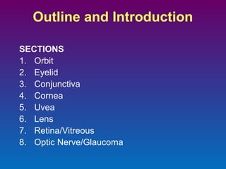
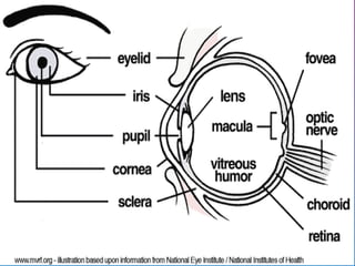
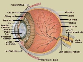
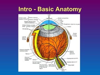
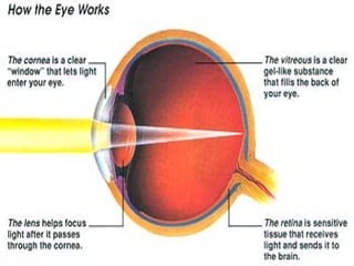
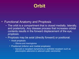
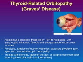
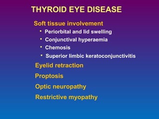
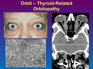
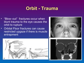
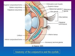
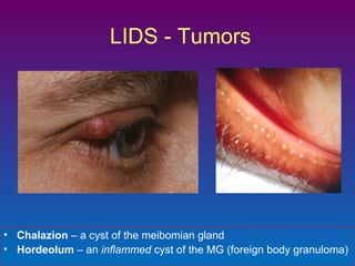
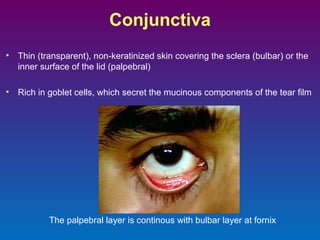
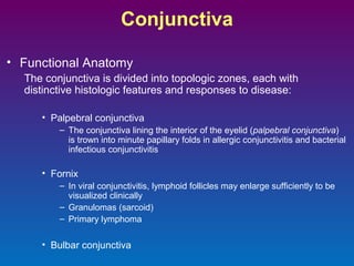
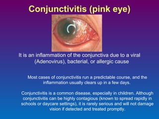
The document provides an outline and introduction for a lecture on eye pathology. It begins with an intro on basic eye anatomy and then outlines the main sections to be covered: Orbit, Eyelid, Conjunctiva, Cornea, Uvea, Lens, Retina/Vitreous, Optic Nerve/Glaucoma. Under each section, some key pathological conditions are briefly described, such as thyroid-related orbitopathy under Orbit, chalazion vs hordeolum under Eyelid, conjunctivitis types under Conjunctiva, and refractive surgery techniques under Cornea.
1-IT IS A MIDDLE VASCULAR COAT OF EYEBALL.
2-IT MAINLY CONSIST OF THREE PARTS IRIS, CHOROID, CILIARY BODY.
3- CILIARY BODY CAN HOLD THE LENS AND PLAY IMPORTANT ROLE IN ACCOMODATION.
Aphakia and its causes. Correction of Aphakia. Advantages and disadvantages of different corrections. Surgeries and related signs and symptoms of aphakia. Complications related to Aphakia.
The aqueous humour is a transparent, watery fluid similar to plasma, but containing low protein concentrations. It is secreted from the ciliary epithelium, a structure supporting the lens
The lacrimal apparatus is the physiological system containing the orbital structures for tear production and drainage. It consists of: The lacrimal gland, which secretes the tears, and its excretory ducts, which convey the fluid to the surface of the human eye;it is a serous gland located in lacrimal fossa.
The tear film is a complex mixture of substances secreted from multiple sources on the ocular surface, including the lacrimal gland, the accessory lacrimal glands, the meibomian glands, and the goblet cells.
The retina is the sensory membrane that lines the inner surface of the back of the eyeball. It's composed of several layers, including one that contains specialized cells called photoreceptors.
Photoreceptor cells take light focused by the cornea and lens and convert it into chemical and nervous signals which are transported to visual centers in the brain by way of the optic nerve.
In the visual cortex of the brain (which, ironically, is located in the back of the brain), these signals are converted into images and visual perceptions.
Eyes are an important part of the body. In this Presentation we are introducing some common Eye disorders & some natural remedies to treat them.
For more information & any help write directly to us: support@nirogam.com
Nirogam India Pvt. Ltd.
F-32 & 33, 1st Floor
DLF Centre Point, Sector – 11,
Bata Mor, Main Mathura Road,
Faridabad, Haryana (India) - 121006
Mob: +91-9958171405 # 9015525552
Ph: 0129-4076777, 4006805
Email: info@nirogam.com
Facebook: www.facebook.com/nirogam
Web: www.nirogam.com
OCT-Angiography is a dye-less method, unlike traditional Fluorescein Angiography and ICG. OCT-Angiography is three dimensional, which allows to scroll through depths and layers, showing superficial and deeper capillaries.
Systemic Diseases and the Eye
Various systemic diseases affect the eye and it's functioning. Here we present those common systemic diseases that are responsible to cause effects in the eye.
What are the diseases that affect eye?
Eye is affected by the following diseases:
Systemic Hypertension (Increased blood pressure of the body)
Diabetes Melitus (Increased blood sugar level)
Systemic Lupus Erythromatosus (SLE)
AIDS and other Venereal Diseases like Syphilis
Sickle Cell Anemia,
Eales Disease and many more.
Look at the slides.
presentation on intraolcular tumors including detailed explaination on their pathology diagnosis and treatment including details of retinoblastoma. enucleation
Dermoid cysts, capillary hemangiomas, and rhabdomyosarcoma are the most common paediatric orbital tumours.
Retinoblastoma is the most common malignant ocular tumour in children.
Neuroblastoma can involve the orbit via metastases and is the most common metastatic tumor to the orbit in children.
Lymphoid tumors, cavernous hemangiomas, and meningiomas are the most common orbital tumours in adults.
Other tumors include those of the lacrimal gland, tumors from the surrounding sinuses, metastatic tumors such as breast cancer in women, and neural-based tumors
Knee anatomy and clinical tests 2024.pdfvimalpl1234
This includes all relevant anatomy and clinical tests compiled from standard textbooks, Campbell,netter etc..It is comprehensive and best suited for orthopaedicians and orthopaedic residents.
Adv. biopharm. APPLICATION OF PHARMACOKINETICS : TARGETED DRUG DELIVERY SYSTEMSAkankshaAshtankar
MIP 201T & MPH 202T
ADVANCED BIOPHARMACEUTICS & PHARMACOKINETICS : UNIT 5
APPLICATION OF PHARMACOKINETICS : TARGETED DRUG DELIVERY SYSTEMS By - AKANKSHA ASHTANKAR
Tom Selleck Health: A Comprehensive Look at the Iconic Actor’s Wellness Journeygreendigital
Tom Selleck, an enduring figure in Hollywood. has captivated audiences for decades with his rugged charm, iconic moustache. and memorable roles in television and film. From his breakout role as Thomas Magnum in Magnum P.I. to his current portrayal of Frank Reagan in Blue Bloods. Selleck's career has spanned over 50 years. But beyond his professional achievements. fans have often been curious about Tom Selleck Health. especially as he has aged in the public eye.
Follow us on: Pinterest
Introduction
Many have been interested in Tom Selleck health. not only because of his enduring presence on screen but also because of the challenges. and lifestyle choices he has faced and made over the years. This article delves into the various aspects of Tom Selleck health. exploring his fitness regimen, diet, mental health. and the challenges he has encountered as he ages. We'll look at how he maintains his well-being. the health issues he has faced, and his approach to ageing .
Early Life and Career
Childhood and Athletic Beginnings
Tom Selleck was born on January 29, 1945, in Detroit, Michigan, and grew up in Sherman Oaks, California. From an early age, he was involved in sports, particularly basketball. which played a significant role in his physical development. His athletic pursuits continued into college. where he attended the University of Southern California (USC) on a basketball scholarship. This early involvement in sports laid a strong foundation for his physical health and disciplined lifestyle.
Transition to Acting
Selleck's transition from an athlete to an actor came with its physical demands. His first significant role in "Magnum P.I." required him to perform various stunts and maintain a fit appearance. This role, which he played from 1980 to 1988. necessitated a rigorous fitness routine to meet the show's demands. setting the stage for his long-term commitment to health and wellness.
Fitness Regimen
Workout Routine
Tom Selleck health and fitness regimen has evolved. adapting to his changing roles and age. During his "Magnum, P.I." days. Selleck's workouts were intense and focused on building and maintaining muscle mass. His routine included weightlifting, cardiovascular exercises. and specific training for the stunts he performed on the show.
Selleck adjusted his fitness routine as he aged to suit his body's needs. Today, his workouts focus on maintaining flexibility, strength, and cardiovascular health. He incorporates low-impact exercises such as swimming, walking, and light weightlifting. This balanced approach helps him stay fit without putting undue strain on his joints and muscles.
Importance of Flexibility and Mobility
In recent years, Selleck has emphasized the importance of flexibility and mobility in his fitness regimen. Understanding the natural decline in muscle mass and joint flexibility with age. he includes stretching and yoga in his routine. These practices help prevent injuries, improve posture, and maintain mobilit
micro teaching on communication m.sc nursing.pdfAnurag Sharma
Microteaching is a unique model of practice teaching. It is a viable instrument for the. desired change in the teaching behavior or the behavior potential which, in specified types of real. classroom situations, tends to facilitate the achievement of specified types of objectives.
Ozempic: Preoperative Management of Patients on GLP-1 Receptor Agonists Saeid Safari
Preoperative Management of Patients on GLP-1 Receptor Agonists like Ozempic and Semiglutide
ASA GUIDELINE
NYSORA Guideline
2 Case Reports of Gastric Ultrasound
Title: Sense of Smell
Presenter: Dr. Faiza, Assistant Professor of Physiology
Qualifications:
MBBS (Best Graduate, AIMC Lahore)
FCPS Physiology
ICMT, CHPE, DHPE (STMU)
MPH (GC University, Faisalabad)
MBA (Virtual University of Pakistan)
Learning Objectives:
Describe the primary categories of smells and the concept of odor blindness.
Explain the structure and location of the olfactory membrane and mucosa, including the types and roles of cells involved in olfaction.
Describe the pathway and mechanisms of olfactory signal transmission from the olfactory receptors to the brain.
Illustrate the biochemical cascade triggered by odorant binding to olfactory receptors, including the role of G-proteins and second messengers in generating an action potential.
Identify different types of olfactory disorders such as anosmia, hyposmia, hyperosmia, and dysosmia, including their potential causes.
Key Topics:
Olfactory Genes:
3% of the human genome accounts for olfactory genes.
400 genes for odorant receptors.
Olfactory Membrane:
Located in the superior part of the nasal cavity.
Medially: Folds downward along the superior septum.
Laterally: Folds over the superior turbinate and upper surface of the middle turbinate.
Total surface area: 5-10 square centimeters.
Olfactory Mucosa:
Olfactory Cells: Bipolar nerve cells derived from the CNS (100 million), with 4-25 olfactory cilia per cell.
Sustentacular Cells: Produce mucus and maintain ionic and molecular environment.
Basal Cells: Replace worn-out olfactory cells with an average lifespan of 1-2 months.
Bowman’s Gland: Secretes mucus.
Stimulation of Olfactory Cells:
Odorant dissolves in mucus and attaches to receptors on olfactory cilia.
Involves a cascade effect through G-proteins and second messengers, leading to depolarization and action potential generation in the olfactory nerve.
Quality of a Good Odorant:
Small (3-20 Carbon atoms), volatile, water-soluble, and lipid-soluble.
Facilitated by odorant-binding proteins in mucus.
Membrane Potential and Action Potential:
Resting membrane potential: -55mV.
Action potential frequency in the olfactory nerve increases with odorant strength.
Adaptation Towards the Sense of Smell:
Rapid adaptation within the first second, with further slow adaptation.
Psychological adaptation greater than receptor adaptation, involving feedback inhibition from the central nervous system.
Primary Sensations of Smell:
Camphoraceous, Musky, Floral, Pepperminty, Ethereal, Pungent, Putrid.
Odor Detection Threshold:
Examples: Hydrogen sulfide (0.0005 ppm), Methyl-mercaptan (0.002 ppm).
Some toxic substances are odorless at lethal concentrations.
Characteristics of Smell:
Odor blindness for single substances due to lack of appropriate receptor protein.
Behavioral and emotional influences of smell.
Transmission of Olfactory Signals:
From olfactory cells to glomeruli in the olfactory bulb, involving lateral inhibition.
Primitive, less old, and new olfactory systems with different path
263778731218 Abortion Clinic /Pills In Harare ,sisternakatoto
263778731218 Abortion Clinic /Pills In Harare ,ABORTION WOMEN’S CLINIC +27730423979 IN women clinic we believe that every woman should be able to make choices in her pregnancy. Our job is to provide compassionate care, safety,affordable and confidential services. That’s why we have won the trust from all generations of women all over the world. we use non surgical method(Abortion pills) to terminate…Dr.LISA +27730423979women Clinic is committed to providing the highest quality of obstetrical and gynecological care to women of all ages. Our dedicated staff aim to treat each patient and her health concerns with compassion and respect.Our dedicated group ABORTION WOMEN’S CLINIC +27730423979 IN women clinic we believe that every woman should be able to make choices in her pregnancy. Our job is to provide compassionate care, safety,affordable and confidential services. That’s why we have won the trust from all generations of women all over the world. we use non surgical method(Abortion pills) to terminate…Dr.LISA +27730423979women Clinic is committed to providing the highest quality of obstetrical and gynecological care to women of all ages. Our dedicated staff aim to treat each patient and her health concerns with compassion and respect.Our dedicated group of receptionists, nurses, and physicians have worked together as a teamof receptionists, nurses, and physicians have worked together as a team wwww.lisywomensclinic.co.za/
Basavarajeeyam is an important text for ayurvedic physician belonging to andhra pradehs. It is a popular compendium in various parts of our country as well as in andhra pradesh. The content of the text was presented in sanskrit and telugu language (Bilingual). One of the most famous book in ayurvedic pharmaceutics and therapeutics. This book contains 25 chapters called as prakaranas. Many rasaoushadis were explained, pioneer of dhatu druti, nadi pareeksha, mutra pareeksha etc. Belongs to the period of 15-16 century. New diseases like upadamsha, phiranga rogas are explained.
Recomendações da OMS sobre cuidados maternos e neonatais para uma experiência pós-natal positiva.
Em consonância com os ODS – Objetivos do Desenvolvimento Sustentável e a Estratégia Global para a Saúde das Mulheres, Crianças e Adolescentes, e aplicando uma abordagem baseada nos direitos humanos, os esforços de cuidados pós-natais devem expandir-se para além da cobertura e da simples sobrevivência, de modo a incluir cuidados de qualidade.
Estas diretrizes visam melhorar a qualidade dos cuidados pós-natais essenciais e de rotina prestados às mulheres e aos recém-nascidos, com o objetivo final de melhorar a saúde e o bem-estar materno e neonatal.
Uma “experiência pós-natal positiva” é um resultado importante para todas as mulheres que dão à luz e para os seus recém-nascidos, estabelecendo as bases para a melhoria da saúde e do bem-estar a curto e longo prazo. Uma experiência pós-natal positiva é definida como aquela em que as mulheres, pessoas que gestam, os recém-nascidos, os casais, os pais, os cuidadores e as famílias recebem informação consistente, garantia e apoio de profissionais de saúde motivados; e onde um sistema de saúde flexível e com recursos reconheça as necessidades das mulheres e dos bebês e respeite o seu contexto cultural.
Estas diretrizes consolidadas apresentam algumas recomendações novas e já bem fundamentadas sobre cuidados pós-natais de rotina para mulheres e neonatos que recebem cuidados no pós-parto em unidades de saúde ou na comunidade, independentemente dos recursos disponíveis.
É fornecido um conjunto abrangente de recomendações para cuidados durante o período puerperal, com ênfase nos cuidados essenciais que todas as mulheres e recém-nascidos devem receber, e com a devida atenção à qualidade dos cuidados; isto é, a entrega e a experiência do cuidado recebido. Estas diretrizes atualizam e ampliam as recomendações da OMS de 2014 sobre cuidados pós-natais da mãe e do recém-nascido e complementam as atuais diretrizes da OMS sobre a gestão de complicações pós-natais.
O estabelecimento da amamentação e o manejo das principais intercorrências é contemplada.
Recomendamos muito.
Vamos discutir essas recomendações no nosso curso de pós-graduação em Aleitamento no Instituto Ciclos.
Esta publicação só está disponível em inglês até o momento.
Prof. Marcus Renato de Carvalho
www.agostodourado.com
The Gram stain is a fundamental technique in microbiology used to classify bacteria based on their cell wall structure. It provides a quick and simple method to distinguish between Gram-positive and Gram-negative bacteria, which have different susceptibilities to antibiotics
1. Today’s Quranic verse
‘Turn not your cheek away from people in scorn and pride, and
not on earth haughtily; for God does not love anyone who acts
proudly and boastfully. Be modest in your bearing and lower yo
voice; for the ugliest sound is the donkey’s braying.’
(Luqman 31: 18-19
2.
3. Pathology of the
Eye - I
Dr. Khurshid Anwar
https://www.facebook.com/pages/Interactive-Neuroscience/160305064167505
10. Orbit
• Functional Anatomy and Proptosis
– The orbit is a compartment that is closed medially, laterally,
and posteriorly. Any disease process that increases orbital
contents results in the forward displacement of the eye,
proptosis.
– Proptosis may be axial (directly forward) or positional.
• Axial proptosis
– Glioma and meningioma
• Positional (inferior and medial proptosis)
– Sarcoid or neoplasm (lymphoma or epithelial neoplasm such as
pleomorphic adenoma or adenoid cystic carcinoma)
11. Thyroid-Related Orbitopathy
(Graves’ Disease)
• Autoimmune condition, triggered by TSH-R Antibodies, with
lymphocytic infiltration, fibrosis and enlargement of extra-ocular
muscles.
• Proptosis, strabismus/muscle-restriction, exposure problems (dryeye), and compressive optic neuropathy.
• Treated with steroids, radiation therapy, or surgical decompression
(opening the orbital walls into the sinuses)
12. THYROID EYE DISEASE
Soft tissue involvement
• Periorbital and lid swelling
• Conjunctival hyperaemia
• Chemosis
• Superior limbic keratoconjunctivitis
Eyelid retraction
Proptosis
Optic neuropathy
Restrictive myopathy
14. Orbit - Trauma
• “Blow–out” fractures occur when
blunt trauma to the eye causes the
orbit to rupture
• Orbital Floor fractures can cause
restricted upgaze if there is muscle
entrapment
15. Orbit - Inflammation
• Orbital Cellulits frequently
extends from adjacent sinus
infections, or periocular trauma.
• A life and sight threatening
emergency! Can extend into the
cavernous sinus, and brain.
• “Pre-Septal” vs. “Post-Septal” can
be distinguished by involvement
of intraorbital structures
17. Orbit
• Other Orbital Inflammatory Conditions
– Orbital cellulitis
• Caused by fungal infection (Mucormycosis)
• Appears in systemic conditions such as Wegener granulomatosis
– Idiopathic orbital inflammation (orbital inflammatory
pseudotumor)
•
•
•
•
•
May be unilateral or bilateral
May be confined to the lacrimal gland (sclerosing dacryoadenitis)
Extraocular muscles (orbital myositis)
Tenon’s capsule (posterior scleritis)
In the long-term, patients may show evidence of systemic vasculitis
or other forms of connective tissue diseases
18. Orbital Neoplasms
– Most frequent primary neoplasms are vascular in origin
• capillary hemangioma (infancy and early childhood)
• lymphangioma
• encapsulated cavernous hemangioma (adults)
– Only a handful of orbital masses are encapsulated
• pleomorphic adenoma
• dermoid cyst
• neurilemmoma
– Malignant lymphoma
– In Children
• rhabdomyosarcoma is the most common primary malignancy of orbit.
• neuroblastomaa is the most common metastatic tumor.
– Metástasis: may produce characteristic periocular echymoses
• prostatic carcinoma
• metastatic neuroblastoma
• Wilms tumor
21. Eyelid
• Functional Anatomy
– The eyelid covers and protects the eye, and generates critical
components of the tear film
– The eyelid is a composite of skin externally and a mucosa (the
conjunctiva) on the surface apposed to the eye
– Eccrine and apocrine glands (glands of Moll) populate the eyelid.
– Blepharitis:
• Obstruction of the drainage system of the sebaceous glands by
chronic inflammation or by neoplasm
– Lipid extravasating into surrounding tissue and provoking a
granulomatose response is called a lipogranuloma or chalazion
23. LIDS - Tumors
• Benign
–
–
–
–
Chalazion vs. Hordeolum
Papillomas/Verrucae
Epidermal inclusion cysts
Many others
• Malignant
– Basal cell carcinoma (most common, located in lower eyelid &
medial canthus)
–
–
–
–
Sebaceous cell carcinoma (D/D chalazion, belephritis)
Squamous cell carcinoma
Melanoma (rare)
Kaposi sarcoma in AIDS
24. LIDS - Tumors
• Chalazion – a cyst of the meibomian gland
• Hordeolum – an inflammed cyst of the MG (foreign body granuloma)
25. Conjunctiva
•
Thin (transparent), non-keratinized skin covering the sclera (bulbar) or the
inner surface of the lid (palpebral)
•
Rich in goblet cells, which secret the mucinous components of the tear film
The palpebral layer is continous with bulbar layer at fornix
26. Conjunctiva
• Functional Anatomy
The conjunctiva is divided into topologic zones, each with
distinctive histologic features and responses to disease:
• Palpebral conjunctiva
– The conjunctiva lining the interior of the eyelid (palpebral conjunctiva)
is trown into minute papillary folds in allergic conjunctivitis and bacterial
infectious conjunctivitis
• Fornix
– In viral conjunctivitis, lymphoid follicles may enlarge sufficiently to be
visualized clinically
– Granulomas (sarcoid)
– Primary lymphoma
• Bulbar conjunctiva
27. Conjunctivitis (pink eye)
It is an inflammation of the conjunctiva due to a viral
(Adenovirus), bacterial, or allergic cause
Most cases of conjunctivitis run a predictable course, and the
inflammation usually clears up in a few days.
Conjunctivitis is a common disease, especially in children. Although
conjunctivitis can be highly contagious (known to spread rapidly in
schools or daycare settings), it is rarely serious and will not damage
vision if detected and treated promptly.
28. Trachoma
Trachoma is the world’s leading cause of preventable blindness of infectious
origin.
Caused by the bacterium Chlamydia trachomatis.
It spreads through direct personal contact, shared towels and cloths, and flies
that have come in contact with the eyes or nose of an infected person.
If left untreated, repeated trachoma infections can cause severe scarring of the
inside of the eyelid and can cause the eyelashes to scratch the cornea
(trichiasis). In addition to causing pain, trichiasis permanently damages the
cornea and can lead to irreversible blindness.
It spreads in areas that lack adequate access to water and sanitation, affects the
most marginalized communities in the world. Globally, almost 8 million people
are visually impaired by trachoma; 500 million are at risk of blindness from the
disease throughout 57 endemic countries
http://www.cdc.gov/healthywater/hygiene/disease/trachoma.html
30. Conjunctiva
• Conjunctival Scarring
– Caused by:
•
•
•
•
Chlamydia trachomatis (trachoma)
Caustic alkalis
A sequela to ocular cicatricial pemphigoid
Iatrogenically through reaction to drugs or as a
concequence of surgery
– Painful, dry eye
31. Conjunctiva
Degenerative conditions
• Pinguecula – on the conjunctiva only
• Pterygium – encroaching onto cornea
• Histologically identical
• Both involve “elastotic degeneration” of the conjunctiva,
usually due to chronic ultraviolet exposure.
32. Conjunctiva
• Pterygium and Pinguecula
– Appear as submucosal elevations on the conjunctiva
– Result from actinic damage
Pterygium
•
•
•
•
•
In the conjunctiva astride the limbus
Formed by fibrovascular connective tissue that migrates onto the cornea
May possibly induce mild astigmatism
Commonly excised
Occasionally precursors of actinic-induced neoplasms (SCC, Melanoma)
Pinguecula
• Does not invade the cornea as pterygium does
• Focal dehydration (dellen: a saucer-like depression in the corneal tissue)
• Solar elastosis: sun-damaged collagen with elastic-like properties, that are
the reason why the pinguecula is yellow in color
• Actinic granuloma may develop secondary to a foreign body
granulomatous reaction against the elastotic collagen
34. Conjunctival Neoplasm
– Squamous neoplasms (CIN – Conjunctival intraepithelial neoplasia & SCC)
• May be associated with the presence of human papillomavirus types 16 & 18
– Mucoepidermoid carcinoma (more aggressive course)
– Conjunctival nevi
– Conjunctival melanoma
• Tend to develop in the limbus
• Most cases develop through a phase of intraepithelial growth termed primary
acquired melanosis with atypia, which is analogous to melanoma in situ
• Spread first to the parotid or submandibular lymph nodes
• Mortality rate: 25%
– Lymphoma; Spread first to the parotid or submandibular lymph nodes
36. Sclera
• May appear “blue” in a variety of conditions:
– high intraocular pressure (staphyloma)
– osteogenesis imperfecta
– congenital melanosis oculi
• Heavily pigmented congenital nevus of the underlying uvea
• Together with periocular cutaneous pigmentation
– Nevus of Ota
37. Cornea
• Functional Anatomy
– The cornea and its overlying tear film – and not the lens –
make up the major refractive surface of the eye
– Corneal stroma lacks blood vessels and lymphatics (is very
transparent; corneal transplantation is usually successful)
– Scars can exist due to inflammation or trauma
38. Cornea - Histology
5 Layers:
1. Epithelium – Continuous with
conj, richly innervated by CN-V1
2. Bowman’s Membrane
3. Stroma – The thickest central
portion (90%). This is where
LASIK/Refractive surgery
happens! Primarily made up of
Type 1 Collagen in uniformlyspaced lamellar bundles.
4. Descemet’s membrane
5. Endothelium – pumps the water
out of the cornea and keeps it
clear
39. Cornea
The uniform spacing of the stromal collagen bundles at a
distance of approx ¼ wavelength light allows transparency .
41. Cornea - Refractive Surgery
•
•
Excimer Laser is applied to the stromal bed, underneath a reflected
corneal flap (LASIK= Laser-Assisted Stromal In-situ Keratomileusis).
The tissue is ablated precisely to counteract the refractive error of the eye.
43. Cornea-Pathology
• Functional Anatomy
– Descemet membrane increases in thickness with age
• It is the site of copper deposition in the Kayser-Fleischer ring of
Wilson disease
• Keratitis (inflammation of cornea) and Ulcers
– Bacterial (frequent in contact lens users, pseudomonas most common)
– Fungal
– Viral (especially herpes simplex and herpes zoster)
• Chronic herpes simplex keratitis may be associated with a
granulomatous reaction to Descemet’s membrane
– Protozoal (Acanthoamoeba)
– Autoimmune, Syphilis etc
44. Cornea - Bacterial Ulcer
Epithelial defect, infiltrate of white cells into the cornea, and a layered
leukocyte collection in the AC (Hypopyon)
45. Cornea - HSV Keratitis
Epithelial “dendritic” Keratitis
Stromal Keratits (note the
vessels and clouding)
chronic herpes simplex keratitis may be associated with a granulomatous reaction involving the
Descemet membrane
46. Corneal degenerations
May be either unilateral or bilateral & typically non-familial.
Calcific band keratopathy is characterized by deposition of calcium in the Bowman layer. This
condition may complicate chronic uveitis, especially in individuals with chronic juvenile
rheumatoid arthritis.
Actinic band keratopathy develops in individuals who are exposed chronically to high levels of
ultraviolet light. In this condition, extensive solar elastosis develops in the superficial layers of
corneal collagen in the sun-exposed interpalpebral fissure, hence the horizontally distributed
band of pathology. ("oil-droplet keratopathy.“)
Keratoconus is fairly common disorder characterized by progressive thinning and ectasia of the
cornea without evidence of inflammation or vascularization and leads to irregular astigmatism
that is difficult to correct with spectacles. Activation of collagenases, gelatinases, and matrix
metalloproteinases has been implicated in the pathogenesis of this condition
47. Cornea - Ectasia
Progressive deformation of cornea is an ectasia. Keratoconus is the most
common ectatic degenerative change. Ectasia can also be a complication
of refractive surgery.
48. Cornea – Dystrophy
Group of progressive, usually bilateral mostly genetically determined, noninflammatory opacifying disorders characterized by abnormal tissue
morphology, function, or abnormal depositions of material into the cornea.
MANY types, affecting each specific layer (epithelial, Bowmans membrane,
stroma or endothelium).
Fuchs Dystrophy
Affecting the endothelium, is one of the principal indications for corneal transplantation in the
United States. The two major clinical manifestations of Fuchs endothelial dystrophy-stromal
edema and bullous keratopathy-are both related to a primary loss of endothelial cells.
Macular Dystrophy
Least common, autosomal recessive, most severe, characterized by multiple, gray-white
opacities that are present in the corneal stroma and that extend out into the peripheral cornea)
Crystalline Dystrophy
Granular Dystrophy
Lattice Dystrophy
Map-Dot-Fingerprint Dystrophy
49. Cornea – Stromal Dystrophy
Granular Dystrophy
Hyaline material deposited in stroma
Diverse mutations in TGFB1 disrupt the folding of keratoepithelin, and depending on the exact
mutation, lead to the deposition of various types of proteinaceous deposits in the cornea.
50. Cornea – Stromal Dystrophy
Lattice Dystrophy
Amyloid deposition with “applegreen” birefringence. Stains with
Congo Red
52. The Uvea
“The uvea” is:
1.
2.
3.
The Iris
The Ciliary body
The Choroid
Each has a function
1.
2.
3.
Iris is a diaphragm for light
Ciliary body suspends & flexes the lens, & makes the aqueous
humor
The choroid helps nourish the outer retina
The choroid is among the most richly vascularized sites in the body
As in the retina, there are no lymphatics within the uvea
53. The Uvea - Angle
• The “angle” is a special region of the uvea where the iris
meets the cornea
– Regulates the outflow of Aqueous humor through the Canal of Schlem
– Determines the Intraocular pressure (Important in Glaucoma)
54. The Uvea - Inflammation
•
“Uveitis” is inflamation of any combination of the iris, ciliary body, or choroid.
•
Many etiologies (autoimmune, syphilis, sacrcoid, TB, HLA-B27, infectious,
idiopathic, etc…)
•
Many names (iritis, anterior uveitis, iridocylitis, choroiditis, posterior uveitis
etc.) depending on the location
•
Sometimes associated with SERIOUS systemic inflamatory diseases (eg.
arthritic diseases), inflamatory bowel disease, and vasculitis.
55. Uveitis
Causes:
• Infectious agents
– e.g.: pneumocystis carinii
• Idiopathic
– e.g.: sarcoidosis
• Autoimmune
Sympathetic ophthalmia
Sympathetic ophthalmia, an example of noninfectious autoimmune uveitis
Characterized by bilateral granulomatous inflammation typically affecting all components of the
uvea: a panuveitis.
May complicate a penetrating injury of the eye. In the injured eye, retinal antigens sequestered
from the immune system may gain access to lymphatics in the conjunctiva and thus set up a
delayed hypersensitivity reaction that affects not only the injured eye but also the contralateral,
noninjured eye.
Condition may develop from 2 weeks to many years after injury. Enucleation of a blind eye
(which can be the sympathizing eye rather than the directly injured eye) may yield diagnostic
findings.
Treated by the administration of systemic immunosuppressive agents.
56. The Uvea – Anterior Uveitis
• Anterior uveitis/iritis
• WBCs floating in the aqueous
57. Uvea – Posterior Uveitis
• Active Toxoplasmosis Choroiditis, and old scar (above)
58. The cherry-red spot in Tay-Sachs disease. A, Fundus photograph of the cherry-red
spot in Tay-Sachs disease. B, Photomicrograph of the macula in a patient with TaySachs disease, stained with periodic acid-Schiff to highlight the accumulation of
ganglioside material in the retinal ganglion cells. The presence of ganglion cells filled
with gangliosides outside the fovea blocks the transmission of the normal orange-red
color of the choroid, but absence of ganglion cells within the fovea (to the right of the
vertical bar) permits the normal orange-red color to be visualized, accounting for the
so-called cherry-red spot.
59. Uveal Neoplasm
– Most common intraocular malignancy of adults:
metastasis, typically to the choroid from breast and lung
• Suggestive of extremely short survival
– Nevi and Melanomas
• Uveal melanoma
–
–
–
–
–
Most common primary intraocular malignancy of adults (7-20/million)
Histologically spindle & epitheloid type
SIZE OF TUMOR/CELL TYPE/PROLIFERATIVE INDEX
Melanomas of the ciliary body and choroid are more aggressive
Most spread first to the liver; metastases appear many years after
treatment (dormancy).
– Mortality: 40% at ten years
• Uveal nevi
– Especially choroidal nevi, are common, affecting 10% of the
Caucasian population.
60. The Uvea - Tumors
• The uvea (especially choroid) is also richly pigmented, and primary
melanocytic tumors are common.
• Nevi and malignant melanomas are both relatively common, and can be
difficult to distinguish, clinically.
• Tumors with “spindle-B” or epithelioid histologic patterns are malignant
61. Anterior Segment
Functional Anatomy
The anterior chamber is bounded anteriorly
by the cornea, laterally by the trabecular
meshwork, and posteriorly by the iris.
Aqueous humor, formed by the pars plicata
of the ciliary body, enters the posterior
chamber, bathes the lens, and circulates
through the pupil to gain access to the
anterior chamber.
The lens is a closed epithelial system; the basement membrane of the lens
epitheliuim (known as the lens capsule) totally envelops the lens.
With aging, the size of the lens increases.
Neoplasms of the lens have not been described.
62. The Lens
• A transparent, avascular structure consisting of concentric
cellular fibers
• Highest protein content of the body (Crystallins), which
account for a high refractive index
• Interaction of the ciliary body muscle, through the zonular
fibers, cause dynamic shape changes.
• In concert with the cornea, helps to focus light on the retina.
63.
64. CATARACT
– The term cataract describes lenticular opacities that may be
congenital or acquired.
– Causes:
• Systemic diseases:
–
–
–
–
Galactosemia
Diabetes mellitus
Wilson disease
Atopic dermatitis
• Drugs:
– Corticosteroids
•
•
•
•
•
Radiation
Trauma
Many intraocular disorders
Age-related cataract (nuclear sclerosis)
Smoking
65. The Lens
•
•
•
•
•
The deepest fibers are the
oldest ones
The lens continues to fatten
throughout life
Central fibers become sclerotic
and opaque with time
Entire structure
encapsulated
Lens cells migrate and
elongate into fibers
66. The Lens - Cataract
Opacities of the lens develop with time, or insult
• UV light, steroids, and inflammation are pathogenic factors
67.
68. The Lens – Cataract surgery
• A opening into the lens
capsule is made
• The cataract is emulsified
with ultrasound energy,
and aspirated out of the
eye
69. The Lens – Cataract surgery
The dense, cloudy crystalline lens is removed, and replaced with an optical implant.
Inflammatory reactions to lens material may develop following exposure of the intact
lens cortex caused by rupture of the capsule due to trauma or as a result of cataract
extraction. It has been suggested that antigen-antibody complexes containing lens
cortical material develop especially in the presence of Propionibacterium acnes (which
acts as an adjuvant), generating a lens-induced uveitis
70. Rubella syndrome, or congenital rubella, is a group of physical abnormalities that
have developed in an infant as a result of maternal infection and subsequent fetal
infection with rubella virus. It is characterized by rash at birth, low birth weight, small
head size, heart abnormalities, visual problems and bulging fontanelle.
71. Hypermature Cataract & Glaucoma
Occasionally, the lens cortex may liquefy nearly entirely, a condition known
as hypermature or morgagnian cataract.
High-molecular-weight proteins from liquefied lens cortex may leak through
the lens capsule(phacolysis). This phacolytic protein-either free or
contained within macrophages-may clog the trabecular meshwork and
contribute to elevation in intra-ocular pressure and optic nerve damage;
phacolytic glaucoma is an example of secondary open-angle glaucoma.
72. GLAUCOMA
• The Anterior Segment and Glaucoma
The term glaucoma refers to a collection of diseases characterized by
distinctive changes in the visual field and in the cup of the optic nerve. Most
of the glaucomas are associated with elevated intraocular pressure, although
some patients with normal intraocular pressure may develop characteristic
optic nerve and visual field changes (normal or low-tension glaucoma).
It is the third most common cause of blindness in world
Risk Factors
Diagnosis of Glaucoma
Intraocular pressure ( IOP ) and its measurement. (tonometry)
Optic disc examination.
Visual Field examination ( perimetry )
Age
Race
Family
Diabetese
Eye injuries
Drugs
73.
74. Classification of Glaucoma
– Glaucoma may be classified as:
• Open angle glaucoma
– Increased resistance to aqueous outflow in the open angle
– Primary
– Secondary
• Angle closure glaucoma
– The peripheral zone of the iris adheres to the trabecular meshwork and
impedes the egress of aqueous from the eye.
– Primary
– Secondary
75. Open angle glaucoma
• Primary
– The most common form of glaucoma
– Few changes are apparent
– Mutations in the GLC1A gene which encodes the protein myocilin
located on chromosome 1 have been associated with a subset of
individuals with juvenile and adult primary open-angle glaucoma.
• Secondary
–
–
–
–
–
–
Phacolysis (liquefied lens cortex, leaking through the lens capsule)
Senescent red blood cells after trauma (ghost cell glaucoma)
iris epithelial pigment granules (pigmentary glaucoma),
fragments of oxytalan fibers (exfoliation glaucoma), and
necrotic tumors (melanomalytic glaucoma) .
Anything that contributes to elevate the pressure on the surface of the
eye (episcleral venous pressure) in the presence of an open angle can
contribute to other types of secondary open angle glaucoma
76. Angle closure glaucoma
Primary
– Typically develops in eyes with shallow anterior chambers
– Often found in patients with hyperopia (hypermetropia)
– Consequences:
» Pupillary block
» Iris bombé apposing it to the trabecular mesh work
» Corneal edema
» Bullous keratopathy
Secondary (causes)
– Contraction of various types of pathologic membranes that form over
the surface of the iris (chronic retinal ischemia is associated with the
up-regulation of VEGF)
– Necrotic tumors
– Iridocorneal endothelial syndrome
– Following intraocular surgery or penetrating trauma
– Tumors in the ciliary body
77.
78.
79. ENDOPHTHALAMITIS & PANOPHTHALAMITIS
Causes:
• Blunt trauma
• Corneal infections
• Uveitis
The term endophthalmitis is not applied clinically unless there is
suppurative inflammation within the vitreous humor is poorly tolerated by
the retina; after only a few hours of exposure to acute inflammation, the
retina may be irreversibly damaged
Exogenous (originating in the environment and gaining access to the
interior of the eye through a wound)
Endogenous (delivered to the eye hematogenously).
Panophthalmitis: inflammation within the eye that involves the
retina, choroid, and sclera and extends into the orbit may produce
proptosis