SlideShare a Scribd company logo
Eye Histo lo gy


                  Assoc. Prof. Dr. Karim Al-Jashamy
-The superior and inferior tarsal
plates are dense fibrous plates
that give support and form to the
eyelids. Tarsal glands produce oily
secretions to prevent tears from
leaking out, while the eyelids stay
dry.
- The obicularis oculi muscle
contains both palpebral and orbital
parts and acts to close the eyelids.
- The levator palpebrae superioris
muscle elevates the upper eyelid.
It is a skeletal muscle under
voluntary control.
The eye
•   A complex and highly developed
    photosensitive organ that permits an
    accurate analysis of the form, light
    intensity, and color reflected from
    objects.

•    Each eye is composed of three
    concentric layers: an external layer
    that consists of the sclera and the
    cornea; a middle layer also called the
    vascular layer consisting of the
    choroid, ciliary body, and iris; and an
    inner layer of nerve tissue, the retina,
    which consists of an outer pigment
    epithelium and an inner retina
    proper.

•   The photosensitive retina proper is
    part of the central nervous system
    and      communicates    with   the
    cerebrum through the optic nerve
    and extends forward to the ora
    serrata.
Diagram, showing the structure of the eye, retina, fovea, and ciliary body.
An enlarged diagram of the fovea is shown at the lower right: (1) axons of
ganglion cells; (2) bipolar cells; (3) rods; (4) cones. Enlarged diagrams of the
ciliary body (upper right) and retina (lower left)
•The sclera         consists of tough,
dense connective tissue made up
mainly of flat collagen bundles
intersecting in various directions while
remaining parallel to the surface of the
organ, a moderate amount of ground
substance, and a few fibroblasts.

•The external surface of the sclera the
episclera is connected by a loose
system of thin collagen fibers to a dense
layer of connective tissue called
Tenon's capsule. Tenon's capsule
comes into contact with the loose
conjunctival stroma at the junction of the
cornea with the sclera.
• Between the sclera and the choroid is      Section of choroid and sclera. The choroid is a
                                             highly vascular layer (arrowheads) of
the suprachoroidal lamina, a thin layer
                                             connective tissue containing melanocytes that
of loose connective tissue rich in           prevent the reflection of incident light.
melanocytes, fibroblasts, and elastic        The sclera is a dense layer of connective
fibers.                                      tissue rich in fibers of collagen type I,
                                             arranged in parallel bundles.
• The outer layer, or sclera,
  consists of dense fibrous
  connective tissue.
– The sclera is "white" of the eye.
– The sclera is continuous with the
  transparent substantia propria of the
  cornea.
– The exposed front surface of the eye,
  including the cornea, is also covered
  by a thin, non-keratinized stratified
  squamous epithelium.

1 - cornea
2 - iris
3 - posterior chamber of the eye
4 - lens
5 - vitreous body
6 - ciliary body
7 - retina
8 - choroid
9 - sclera
10 - canal of Sclemm
11 - growth area of the lens
12 - anterior epithelium of the lens
RETINA, CHOROID
• 1 - 8 - retina
  1 - optic nerve fibers
  2 - ganglion cell layer
  3 - inner plexiform
  layer
  4 - inner nuclear layer
  5 - outer plexiform
  layer
  6 - outer nuclear layer
  7 - outer processes of
  rods and cones
  8 - pigmented
  epithelium
  9 - choroid
  10 - sclera
• Cornea
• colorless and transparent.
• A transverse section of the cornea
  shows that it consists of five layers:
  epithelium,     Bowman's   membrane,
  stroma, Descemet's membrane, and
  endothelium.

•   The corneal epithelium is stratified,
    squamous, and nonkeratinized and
    consists of five or six layers of cells.

•    In the basal part of the epithelium are
    numerous mitotic figures that are
    responsible for the cornea's remarkable
    regenerative capacity

•  The surface corneal cells show
  microvilli protruding into the space
  filled by the precorneal tear film.
• This epithelial tissue is covered by a
  protective     layer    of     lipid and
  glycoprotein, about 7 m thick. The
  cornea has one of the richest sensory
  nerve supplies of any eye tissue.
CORNEA
•1 - anterior epithelium
 (stratified squamous
 epithelium)
 2 - anterior basement
 (Bowman's) membrane
 3 - substantia propria
 4 - posterior basement
 (Descemet's)
 membrane
 5 - posterior epithelium
 (simple squamous or
 endothelium)
•   This layer, Bowman's membrane, consists of collagen fibers crossing at random,
    a condensation of the intercellular substance, and no cells . Bowman's membrane
    contributes greatly to the stability and strength of the cornea.
•   The stroma is formed of many layers of parallel collagen bundles that cross at
    approximately right angles to each other. The collagen fibrils within each lamella
    are parallel to each other and run the full width of the cornea. Between the several
    layers, the cytoplasmic extensions of fibroblasts are flattened like the wings of a
    butterfly.

•   Descemet's membrane is a thick homogeneous structure composed of fine
    collagenous filaments organized in a three-dimensional network.

•   The endothelium of the cornea is a simple squamous epithelium. These cells
    possess organelles for secretion that are characteristic of cells engaged in active
    transport and protein synthesis and that may be related to the synthesis and
    maintenance of Descemet's membrane.

•   The corneal endothelium and epithelium are responsible for maintaining the
    transparency of the cornea. Both layers are capable of transporting sodium ions
    toward their apical surfaces. Chloride ions and water follow passively, maintaining
    the corneal stroma in a relatively dehydrated state. This state, along with the
    regular orientation of the very thin collagen fibrils of the stroma, accounts for the
    transparency of the cornea.
• The corneoscleral junction, or limbus, is an area of transition from
  the transparent collagen bundles of the cornea to the white opaque
  fibers of the sclera. It is highly vascularized, and its blood vessels
  assume an important role in corneal inflammatory processes.

• The cornea, an avascular structure, receives its metabolites by
  diffusion from adjacent vessels and from the fluid of the anterior
  chamber of the eye.

• In the region of the limbus in the stromal layer, irregular endothelium-
  lined channels, the trabecular meshwork, merge to form Schlemm's
  canal, which drains fluid from the anterior chamber of the eye.
  Schlemm's canal communicates externally with the venous system.
• Middle, or Vascular, Layer
• The middle (vascular) layer of the eye consists of three parts: choroid,
  ciliary body, and iris, known collectively as the uveal tract.
- The ciliary body contains ciliary muscle that is composed of smooth muscle. Contraction
and relaxation of the ciliary muscles change the tension of the zonular fibers, or suspensory
ligaments, of the lens. This allows the lens to change shape, a process known as
accommodation.
- The ciliary processes are folds of connective tissue that are covered by two layers of
epithelium. There is also a complex vasculature that cannot be seen easily. Fluid from these
vessels is processed and transported by the epithelial cells to the posterior chamber as
aqueous humor. The epithelial cells constitute the blood-aqueous barrier.
-
-   The aqueous humor enters the anterior
    chamber through the pupil as it flows
    between the lens and the iris.
-    Aqueous humor leaves the anterior
    chamber   through  the  trabecular
    meshwork and into the canal of
    Schlemm.
-   This    is    an     endothelial lines,
    circumferentially arranged vessel that
    communicates with veins in the sclera
    and returns the aqueous humor back to
    the general circulation.
-    Obstruction of the trabecular meshwork
    and canals of Schlemm are thought to
    be the major cause of elevated
    intraocular pressure, which could then
    lead to glaucoma.
- The iris is detailed here in higher magnification. Note
the anterior and posterior chambers to help orient
yourself.
- The anterior surface of the iris contains loose, variably
pigmented stroma. It is open to the circulating aqueous
humor within the anterior chamber.
- Two layers of heavily pigmented epithelium cover the
posterior surface of the iris.
- Note that the sphincter pupillae muscle can be easily
seen near the pupil margin. It is smooth muscle
controlled by parasympathetics. The dilator pupillae
muscle is more difficult to identify, but it dilates the pupil
upon sympathetic innervation.
Lens
• The lens consists of a
  lens capsule, the
  subcapsular epithelium
  and lens fibres. It does
  not contain blood
  vessels or nerves.

More Related Content

What's hot

Histology of the Lymphatic System
Histology of the Lymphatic System Histology of the Lymphatic System
Histology of the Lymphatic System
Garry D. Lasaga
 
Kidney histology
Kidney histologyKidney histology
Kidney histology
anamika gupta
 
Histology of endocrine system
Histology of  endocrine systemHistology of  endocrine system
Histology of endocrine systemMBBS IMS MSU
 
Histology of nerve system
Histology of nerve systemHistology of nerve system
Histology of nerve systemMBBS IMS MSU
 
Visual pathway
Visual pathway Visual pathway
Visual pathway
Laxmi Eye Institute
 
Histology of trachea and lung
Histology of trachea and lungHistology of trachea and lung
Histology of trachea and lung
mgmcri1234
 
Histology of respiratory system larynx trache and lung
Histology of respiratory system larynx trache and lungHistology of respiratory system larynx trache and lung
Histology of respiratory system larynx trache and lungkohlschuetter
 
Histology of small intestine
Histology of small intestine Histology of small intestine
Histology of small intestine
Shizra Imtiaz
 
PHYSIOLOGY OF VISION
PHYSIOLOGY OF VISIONPHYSIOLOGY OF VISION
PHYSIOLOGY OF VISION
Priyanka Raj
 
Anatomy of lacrimal apparatus
Anatomy of lacrimal apparatusAnatomy of lacrimal apparatus
Anatomy of lacrimal apparatus
Dr.Siddharth Gautam
 
Anatomy OF ORBIT
Anatomy OF ORBITAnatomy OF ORBIT
Anatomy OF ORBIT
SSSIHMS-PG
 
Histology of thyroid gland
Histology of thyroid glandHistology of thyroid gland
Histology of thyroid gland
chet08
 
RESPIRATORY SYSTEM HISTOLOGY
RESPIRATORY  SYSTEM HISTOLOGY RESPIRATORY  SYSTEM HISTOLOGY
RESPIRATORY SYSTEM HISTOLOGY
Pankaj Gaonkar
 
Bony orbit and its contents
Bony orbit and its contentsBony orbit and its contents
Bony orbit and its contents
mgmcri1234
 
Muscles of the eye
Muscles of the eyeMuscles of the eye
Muscles of the eye
Ashish Chaudhari
 
The orbit of eye
The orbit of eyeThe orbit of eye
The orbit of eye
Idris Siddiqui
 
03.18.09(d): Ear Histology
03.18.09(d): Ear Histology03.18.09(d): Ear Histology
03.18.09(d): Ear Histology
Open.Michigan
 
Histology of endocrine glands
Histology of endocrine glands Histology of endocrine glands
Histology of endocrine glands
Mehul Tandel
 
Accomodation reflex
Accomodation reflexAccomodation reflex
Accomodation reflex
Dr.Nusrat Tariq
 
Histology of large intestine
Histology of large intestineHistology of large intestine
Histology of large intestine
Hassan Tariq
 

What's hot (20)

Histology of the Lymphatic System
Histology of the Lymphatic System Histology of the Lymphatic System
Histology of the Lymphatic System
 
Kidney histology
Kidney histologyKidney histology
Kidney histology
 
Histology of endocrine system
Histology of  endocrine systemHistology of  endocrine system
Histology of endocrine system
 
Histology of nerve system
Histology of nerve systemHistology of nerve system
Histology of nerve system
 
Visual pathway
Visual pathway Visual pathway
Visual pathway
 
Histology of trachea and lung
Histology of trachea and lungHistology of trachea and lung
Histology of trachea and lung
 
Histology of respiratory system larynx trache and lung
Histology of respiratory system larynx trache and lungHistology of respiratory system larynx trache and lung
Histology of respiratory system larynx trache and lung
 
Histology of small intestine
Histology of small intestine Histology of small intestine
Histology of small intestine
 
PHYSIOLOGY OF VISION
PHYSIOLOGY OF VISIONPHYSIOLOGY OF VISION
PHYSIOLOGY OF VISION
 
Anatomy of lacrimal apparatus
Anatomy of lacrimal apparatusAnatomy of lacrimal apparatus
Anatomy of lacrimal apparatus
 
Anatomy OF ORBIT
Anatomy OF ORBITAnatomy OF ORBIT
Anatomy OF ORBIT
 
Histology of thyroid gland
Histology of thyroid glandHistology of thyroid gland
Histology of thyroid gland
 
RESPIRATORY SYSTEM HISTOLOGY
RESPIRATORY  SYSTEM HISTOLOGY RESPIRATORY  SYSTEM HISTOLOGY
RESPIRATORY SYSTEM HISTOLOGY
 
Bony orbit and its contents
Bony orbit and its contentsBony orbit and its contents
Bony orbit and its contents
 
Muscles of the eye
Muscles of the eyeMuscles of the eye
Muscles of the eye
 
The orbit of eye
The orbit of eyeThe orbit of eye
The orbit of eye
 
03.18.09(d): Ear Histology
03.18.09(d): Ear Histology03.18.09(d): Ear Histology
03.18.09(d): Ear Histology
 
Histology of endocrine glands
Histology of endocrine glands Histology of endocrine glands
Histology of endocrine glands
 
Accomodation reflex
Accomodation reflexAccomodation reflex
Accomodation reflex
 
Histology of large intestine
Histology of large intestineHistology of large intestine
Histology of large intestine
 

Viewers also liked

Taste Buds
Taste  BudsTaste  Buds
Taste Buds
Loretta Kreis
 
Respiratory System Anatomy
Respiratory System AnatomyRespiratory System Anatomy
Respiratory System Anatomy
bkwittmann
 
Taste ppt
Taste pptTaste ppt
Taste ppt
Aftab Badshah
 
Histology of special senses
Histology of special sensesHistology of special senses
Histology of special senses
mgmcri1234
 
Practical oral histology ii
Practical oral histology iiPractical oral histology ii
Practical oral histology ii
Titey
 
Lens and cataract
Lens and cataractLens and cataract
Lens and cataract
Bahaa Halwany
 

Viewers also liked (6)

Taste Buds
Taste  BudsTaste  Buds
Taste Buds
 
Respiratory System Anatomy
Respiratory System AnatomyRespiratory System Anatomy
Respiratory System Anatomy
 
Taste ppt
Taste pptTaste ppt
Taste ppt
 
Histology of special senses
Histology of special sensesHistology of special senses
Histology of special senses
 
Practical oral histology ii
Practical oral histology iiPractical oral histology ii
Practical oral histology ii
 
Lens and cataract
Lens and cataractLens and cataract
Lens and cataract
 

Similar to Histology of eye

د احمد اسامه هاشم عيون ophthalmology
د احمد اسامه هاشم عيون ophthalmologyد احمد اسامه هاشم عيون ophthalmology
د احمد اسامه هاشم عيون ophthalmology
Ahmed Osama Hashem
 
Eye anatomy
Eye anatomyEye anatomy
Eye anatomy
mohammed Qazzaz
 
Histology of the eye by a very good docotor in iraqi uni collage of med
Histology of the eye by a very good docotor in iraqi uni collage of medHistology of the eye by a very good docotor in iraqi uni collage of med
Histology of the eye by a very good docotor in iraqi uni collage of med
ZaraVvv
 
5. eye histology.pptx
5. eye histology.pptx5. eye histology.pptx
5. eye histology.pptx
Lahdks
 
Eye shalakya 2.pptx
Eye shalakya 2.pptxEye shalakya 2.pptx
Eye shalakya 2.pptx
RajuDsai
 
Uveal tract anatomy
Uveal tract anatomyUveal tract anatomy
Uveal tract anatomy
Othman Al-Abbadi
 
The eye atlas
The eye atlas The eye atlas
The eye atlas
sosojammoly
 
anatomyofeye-180217183456.pdf
anatomyofeye-180217183456.pdfanatomyofeye-180217183456.pdf
anatomyofeye-180217183456.pdf
EidTahir
 
Anatomy of eye
Anatomy of eyeAnatomy of eye
Anatomy of eye
sneha chavan
 
1-Anatomy-and embryology lecture of-the-EYE.pdf
1-Anatomy-and embryology lecture of-the-EYE.pdf1-Anatomy-and embryology lecture of-the-EYE.pdf
1-Anatomy-and embryology lecture of-the-EYE.pdf
RahulKumar4960
 
uveal part1 +2.pptx
uveal part1 +2.pptxuveal part1 +2.pptx
uveal part1 +2.pptx
LujainAlhunaiti
 
Anatomy of sclera
Anatomy of scleraAnatomy of sclera
Anatomy of sclera
Kanwal Perveen
 
Anatomy of cornea by alamin
Anatomy of cornea by alaminAnatomy of cornea by alamin
Anatomy of cornea by alamin
Al Amin
 
LACRIMAL APPARATUS.pptx
LACRIMAL APPARATUS.pptxLACRIMAL APPARATUS.pptx
LACRIMAL APPARATUS.pptx
Nasrinsultanarmc
 
Anatomy of cornea
Anatomy of corneaAnatomy of cornea
Anatomy of cornea
Satish Hadimani
 
Anatomy of eyeball
Anatomy of eyeballAnatomy of eyeball
Anatomy of eyeball
Rajit Adhikari
 
rajt-180629141431 (1).pptx
rajt-180629141431 (1).pptxrajt-180629141431 (1).pptx
rajt-180629141431 (1).pptx
SandipPradhan23
 
rajt-180629141431 (1).pptxuvealtractanat
rajt-180629141431 (1).pptxuvealtractanatrajt-180629141431 (1).pptxuvealtractanat
rajt-180629141431 (1).pptxuvealtractanat
MeenaSheokand2
 
rajt-180629141431 (1).pptx
rajt-180629141431 (1).pptxrajt-180629141431 (1).pptx
rajt-180629141431 (1).pptx
SANDIPPRADHANBWU
 
rajt-180629141431 (1).pptx
rajt-180629141431 (1).pptxrajt-180629141431 (1).pptx
rajt-180629141431 (1).pptx
SANDIPPRADHANBWU
 

Similar to Histology of eye (20)

د احمد اسامه هاشم عيون ophthalmology
د احمد اسامه هاشم عيون ophthalmologyد احمد اسامه هاشم عيون ophthalmology
د احمد اسامه هاشم عيون ophthalmology
 
Eye anatomy
Eye anatomyEye anatomy
Eye anatomy
 
Histology of the eye by a very good docotor in iraqi uni collage of med
Histology of the eye by a very good docotor in iraqi uni collage of medHistology of the eye by a very good docotor in iraqi uni collage of med
Histology of the eye by a very good docotor in iraqi uni collage of med
 
5. eye histology.pptx
5. eye histology.pptx5. eye histology.pptx
5. eye histology.pptx
 
Eye shalakya 2.pptx
Eye shalakya 2.pptxEye shalakya 2.pptx
Eye shalakya 2.pptx
 
Uveal tract anatomy
Uveal tract anatomyUveal tract anatomy
Uveal tract anatomy
 
The eye atlas
The eye atlas The eye atlas
The eye atlas
 
anatomyofeye-180217183456.pdf
anatomyofeye-180217183456.pdfanatomyofeye-180217183456.pdf
anatomyofeye-180217183456.pdf
 
Anatomy of eye
Anatomy of eyeAnatomy of eye
Anatomy of eye
 
1-Anatomy-and embryology lecture of-the-EYE.pdf
1-Anatomy-and embryology lecture of-the-EYE.pdf1-Anatomy-and embryology lecture of-the-EYE.pdf
1-Anatomy-and embryology lecture of-the-EYE.pdf
 
uveal part1 +2.pptx
uveal part1 +2.pptxuveal part1 +2.pptx
uveal part1 +2.pptx
 
Anatomy of sclera
Anatomy of scleraAnatomy of sclera
Anatomy of sclera
 
Anatomy of cornea by alamin
Anatomy of cornea by alaminAnatomy of cornea by alamin
Anatomy of cornea by alamin
 
LACRIMAL APPARATUS.pptx
LACRIMAL APPARATUS.pptxLACRIMAL APPARATUS.pptx
LACRIMAL APPARATUS.pptx
 
Anatomy of cornea
Anatomy of corneaAnatomy of cornea
Anatomy of cornea
 
Anatomy of eyeball
Anatomy of eyeballAnatomy of eyeball
Anatomy of eyeball
 
rajt-180629141431 (1).pptx
rajt-180629141431 (1).pptxrajt-180629141431 (1).pptx
rajt-180629141431 (1).pptx
 
rajt-180629141431 (1).pptxuvealtractanat
rajt-180629141431 (1).pptxuvealtractanatrajt-180629141431 (1).pptxuvealtractanat
rajt-180629141431 (1).pptxuvealtractanat
 
rajt-180629141431 (1).pptx
rajt-180629141431 (1).pptxrajt-180629141431 (1).pptx
rajt-180629141431 (1).pptx
 
rajt-180629141431 (1).pptx
rajt-180629141431 (1).pptxrajt-180629141431 (1).pptx
rajt-180629141431 (1).pptx
 

More from MBBS IMS MSU

Hema practical 05 hema staining
Hema practical 05 hema stainingHema practical 05 hema staining
Hema practical 05 hema stainingMBBS IMS MSU
 
Hema practical 03 coagulation
Hema practical 03 coagulationHema practical 03 coagulation
Hema practical 03 coagulationMBBS IMS MSU
 
Hema practical 02 hematology
Hema practical 02 hematologyHema practical 02 hematology
Hema practical 02 hematologyMBBS IMS MSU
 
Pharmacology anticoagulation
Pharmacology   anticoagulationPharmacology   anticoagulation
Pharmacology anticoagulationMBBS IMS MSU
 
Microbiology hiv-yf
Microbiology   hiv-yfMicrobiology   hiv-yf
Microbiology hiv-yfMBBS IMS MSU
 
Forensic medicine firearms and firearm injuries
Forensic medicine   firearms and firearm injuriesForensic medicine   firearms and firearm injuries
Forensic medicine firearms and firearm injuriesMBBS IMS MSU
 
Forensic medicine firearms and firearm injuries
Forensic medicine   firearms and firearm injuriesForensic medicine   firearms and firearm injuries
Forensic medicine firearms and firearm injuriesMBBS IMS MSU
 
Forensic medicine firearms and firearm injuries
Forensic medicine   firearms and firearm injuriesForensic medicine   firearms and firearm injuries
Forensic medicine firearms and firearm injuriesMBBS IMS MSU
 
Forensic medicine changes after death
Forensic medicine   changes after deathForensic medicine   changes after death
Forensic medicine changes after deathMBBS IMS MSU
 
Pharmacology cvs medicine
Pharmacology   cvs medicinePharmacology   cvs medicine
Pharmacology cvs medicineMBBS IMS MSU
 
Pharmacology antiarrhythmias
Pharmacology   antiarrhythmiasPharmacology   antiarrhythmias
Pharmacology antiarrhythmiasMBBS IMS MSU
 
Pharmacology angina
Pharmacology   anginaPharmacology   angina
Pharmacology anginaMBBS IMS MSU
 
Pathology hematology 3
Pathology   hematology 3Pathology   hematology 3
Pathology hematology 3MBBS IMS MSU
 
Forensic medicine the medico-legal autopsy
Forensic medicine   the medico-legal autopsyForensic medicine   the medico-legal autopsy
Forensic medicine the medico-legal autopsyMBBS IMS MSU
 
Forensic medicine post mortem artefact
Forensic medicine   post mortem artefactForensic medicine   post mortem artefact
Forensic medicine post mortem artefactMBBS IMS MSU
 
Pharmacology anemia and its treatment
Pharmacology   anemia and its treatmentPharmacology   anemia and its treatment
Pharmacology anemia and its treatmentMBBS IMS MSU
 
Pharmacology neuromuscular blockers & anemia
Pharmacology   neuromuscular blockers & anemiaPharmacology   neuromuscular blockers & anemia
Pharmacology neuromuscular blockers & anemiaMBBS IMS MSU
 
Pharmacology - Parkinsonism
Pharmacology - ParkinsonismPharmacology - Parkinsonism
Pharmacology - ParkinsonismMBBS IMS MSU
 
Forensic medicine medical negligence
Forensic medicine   medical negligenceForensic medicine   medical negligence
Forensic medicine medical negligenceMBBS IMS MSU
 
Forensic medicine medical negligence 2-bolam principle
Forensic medicine   medical negligence 2-bolam principleForensic medicine   medical negligence 2-bolam principle
Forensic medicine medical negligence 2-bolam principleMBBS IMS MSU
 

More from MBBS IMS MSU (20)

Hema practical 05 hema staining
Hema practical 05 hema stainingHema practical 05 hema staining
Hema practical 05 hema staining
 
Hema practical 03 coagulation
Hema practical 03 coagulationHema practical 03 coagulation
Hema practical 03 coagulation
 
Hema practical 02 hematology
Hema practical 02 hematologyHema practical 02 hematology
Hema practical 02 hematology
 
Pharmacology anticoagulation
Pharmacology   anticoagulationPharmacology   anticoagulation
Pharmacology anticoagulation
 
Microbiology hiv-yf
Microbiology   hiv-yfMicrobiology   hiv-yf
Microbiology hiv-yf
 
Forensic medicine firearms and firearm injuries
Forensic medicine   firearms and firearm injuriesForensic medicine   firearms and firearm injuries
Forensic medicine firearms and firearm injuries
 
Forensic medicine firearms and firearm injuries
Forensic medicine   firearms and firearm injuriesForensic medicine   firearms and firearm injuries
Forensic medicine firearms and firearm injuries
 
Forensic medicine firearms and firearm injuries
Forensic medicine   firearms and firearm injuriesForensic medicine   firearms and firearm injuries
Forensic medicine firearms and firearm injuries
 
Forensic medicine changes after death
Forensic medicine   changes after deathForensic medicine   changes after death
Forensic medicine changes after death
 
Pharmacology cvs medicine
Pharmacology   cvs medicinePharmacology   cvs medicine
Pharmacology cvs medicine
 
Pharmacology antiarrhythmias
Pharmacology   antiarrhythmiasPharmacology   antiarrhythmias
Pharmacology antiarrhythmias
 
Pharmacology angina
Pharmacology   anginaPharmacology   angina
Pharmacology angina
 
Pathology hematology 3
Pathology   hematology 3Pathology   hematology 3
Pathology hematology 3
 
Forensic medicine the medico-legal autopsy
Forensic medicine   the medico-legal autopsyForensic medicine   the medico-legal autopsy
Forensic medicine the medico-legal autopsy
 
Forensic medicine post mortem artefact
Forensic medicine   post mortem artefactForensic medicine   post mortem artefact
Forensic medicine post mortem artefact
 
Pharmacology anemia and its treatment
Pharmacology   anemia and its treatmentPharmacology   anemia and its treatment
Pharmacology anemia and its treatment
 
Pharmacology neuromuscular blockers & anemia
Pharmacology   neuromuscular blockers & anemiaPharmacology   neuromuscular blockers & anemia
Pharmacology neuromuscular blockers & anemia
 
Pharmacology - Parkinsonism
Pharmacology - ParkinsonismPharmacology - Parkinsonism
Pharmacology - Parkinsonism
 
Forensic medicine medical negligence
Forensic medicine   medical negligenceForensic medicine   medical negligence
Forensic medicine medical negligence
 
Forensic medicine medical negligence 2-bolam principle
Forensic medicine   medical negligence 2-bolam principleForensic medicine   medical negligence 2-bolam principle
Forensic medicine medical negligence 2-bolam principle
 

Recently uploaded

Knee anatomy and clinical tests 2024.pdf
Knee anatomy and clinical tests 2024.pdfKnee anatomy and clinical tests 2024.pdf
Knee anatomy and clinical tests 2024.pdf
vimalpl1234
 
Ophthalmology Clinical Tests for OSCE exam
Ophthalmology Clinical Tests for OSCE examOphthalmology Clinical Tests for OSCE exam
Ophthalmology Clinical Tests for OSCE exam
KafrELShiekh University
 
Physiology of Chemical Sensation of smell.pdf
Physiology of Chemical Sensation of smell.pdfPhysiology of Chemical Sensation of smell.pdf
Physiology of Chemical Sensation of smell.pdf
MedicoseAcademics
 
Superficial & Deep Fascia of the NECK.pptx
Superficial & Deep Fascia of the NECK.pptxSuperficial & Deep Fascia of the NECK.pptx
Superficial & Deep Fascia of the NECK.pptx
Dr. Rabia Inam Gandapore
 
263778731218 Abortion Clinic /Pills In Harare ,
263778731218 Abortion Clinic /Pills In Harare ,263778731218 Abortion Clinic /Pills In Harare ,
263778731218 Abortion Clinic /Pills In Harare ,
sisternakatoto
 
Hemodialysis: Chapter 3, Dialysis Water Unit - Dr.Gawad
Hemodialysis: Chapter 3, Dialysis Water Unit - Dr.GawadHemodialysis: Chapter 3, Dialysis Water Unit - Dr.Gawad
Hemodialysis: Chapter 3, Dialysis Water Unit - Dr.Gawad
NephroTube - Dr.Gawad
 
ARTHROLOGY PPT NCISM SYLLABUS AYURVEDA STUDENTS
ARTHROLOGY PPT NCISM SYLLABUS AYURVEDA STUDENTSARTHROLOGY PPT NCISM SYLLABUS AYURVEDA STUDENTS
ARTHROLOGY PPT NCISM SYLLABUS AYURVEDA STUDENTS
Dr. Vinay Pareek
 
ANATOMY AND PHYSIOLOGY OF URINARY SYSTEM.pptx
ANATOMY AND PHYSIOLOGY OF URINARY SYSTEM.pptxANATOMY AND PHYSIOLOGY OF URINARY SYSTEM.pptx
ANATOMY AND PHYSIOLOGY OF URINARY SYSTEM.pptx
Swetaba Besh
 
KDIGO 2024 guidelines for diabetologists
KDIGO 2024 guidelines for diabetologistsKDIGO 2024 guidelines for diabetologists
KDIGO 2024 guidelines for diabetologists
د.محمود نجيب
 
Pulmonary Thromboembolism - etilogy, types, medical- Surgical and nursing man...
Pulmonary Thromboembolism - etilogy, types, medical- Surgical and nursing man...Pulmonary Thromboembolism - etilogy, types, medical- Surgical and nursing man...
Pulmonary Thromboembolism - etilogy, types, medical- Surgical and nursing man...
VarunMahajani
 
NVBDCP.pptx Nation vector borne disease control program
NVBDCP.pptx Nation vector borne disease control programNVBDCP.pptx Nation vector borne disease control program
NVBDCP.pptx Nation vector borne disease control program
Sapna Thakur
 
Cervical & Brachial Plexus By Dr. RIG.pptx
Cervical & Brachial Plexus By Dr. RIG.pptxCervical & Brachial Plexus By Dr. RIG.pptx
Cervical & Brachial Plexus By Dr. RIG.pptx
Dr. Rabia Inam Gandapore
 
Flu Vaccine Alert in Bangalore Karnataka
Flu Vaccine Alert in Bangalore KarnatakaFlu Vaccine Alert in Bangalore Karnataka
Flu Vaccine Alert in Bangalore Karnataka
addon Scans
 
New Drug Discovery and Development .....
New Drug Discovery and Development .....New Drug Discovery and Development .....
New Drug Discovery and Development .....
NEHA GUPTA
 
Are There Any Natural Remedies To Treat Syphilis.pdf
Are There Any Natural Remedies To Treat Syphilis.pdfAre There Any Natural Remedies To Treat Syphilis.pdf
Are There Any Natural Remedies To Treat Syphilis.pdf
Little Cross Family Clinic
 
Novas diretrizes da OMS para os cuidados perinatais de mais qualidade
Novas diretrizes da OMS para os cuidados perinatais de mais qualidadeNovas diretrizes da OMS para os cuidados perinatais de mais qualidade
Novas diretrizes da OMS para os cuidados perinatais de mais qualidade
Prof. Marcus Renato de Carvalho
 
BENIGN PROSTATIC HYPERPLASIA.BPH. BPHpdf
BENIGN PROSTATIC HYPERPLASIA.BPH. BPHpdfBENIGN PROSTATIC HYPERPLASIA.BPH. BPHpdf
BENIGN PROSTATIC HYPERPLASIA.BPH. BPHpdf
DR SETH JOTHAM
 
Surat @ℂall @Girls ꧁❤8527049040❤꧂@ℂall @Girls Service Vip Top Model Safe
Surat @ℂall @Girls ꧁❤8527049040❤꧂@ℂall @Girls Service Vip Top Model SafeSurat @ℂall @Girls ꧁❤8527049040❤꧂@ℂall @Girls Service Vip Top Model Safe
Surat @ℂall @Girls ꧁❤8527049040❤꧂@ℂall @Girls Service Vip Top Model Safe
Savita Shen $i11
 
New Directions in Targeted Therapeutic Approaches for Older Adults With Mantl...
New Directions in Targeted Therapeutic Approaches for Older Adults With Mantl...New Directions in Targeted Therapeutic Approaches for Older Adults With Mantl...
New Directions in Targeted Therapeutic Approaches for Older Adults With Mantl...
i3 Health
 
micro teaching on communication m.sc nursing.pdf
micro teaching on communication m.sc nursing.pdfmicro teaching on communication m.sc nursing.pdf
micro teaching on communication m.sc nursing.pdf
Anurag Sharma
 

Recently uploaded (20)

Knee anatomy and clinical tests 2024.pdf
Knee anatomy and clinical tests 2024.pdfKnee anatomy and clinical tests 2024.pdf
Knee anatomy and clinical tests 2024.pdf
 
Ophthalmology Clinical Tests for OSCE exam
Ophthalmology Clinical Tests for OSCE examOphthalmology Clinical Tests for OSCE exam
Ophthalmology Clinical Tests for OSCE exam
 
Physiology of Chemical Sensation of smell.pdf
Physiology of Chemical Sensation of smell.pdfPhysiology of Chemical Sensation of smell.pdf
Physiology of Chemical Sensation of smell.pdf
 
Superficial & Deep Fascia of the NECK.pptx
Superficial & Deep Fascia of the NECK.pptxSuperficial & Deep Fascia of the NECK.pptx
Superficial & Deep Fascia of the NECK.pptx
 
263778731218 Abortion Clinic /Pills In Harare ,
263778731218 Abortion Clinic /Pills In Harare ,263778731218 Abortion Clinic /Pills In Harare ,
263778731218 Abortion Clinic /Pills In Harare ,
 
Hemodialysis: Chapter 3, Dialysis Water Unit - Dr.Gawad
Hemodialysis: Chapter 3, Dialysis Water Unit - Dr.GawadHemodialysis: Chapter 3, Dialysis Water Unit - Dr.Gawad
Hemodialysis: Chapter 3, Dialysis Water Unit - Dr.Gawad
 
ARTHROLOGY PPT NCISM SYLLABUS AYURVEDA STUDENTS
ARTHROLOGY PPT NCISM SYLLABUS AYURVEDA STUDENTSARTHROLOGY PPT NCISM SYLLABUS AYURVEDA STUDENTS
ARTHROLOGY PPT NCISM SYLLABUS AYURVEDA STUDENTS
 
ANATOMY AND PHYSIOLOGY OF URINARY SYSTEM.pptx
ANATOMY AND PHYSIOLOGY OF URINARY SYSTEM.pptxANATOMY AND PHYSIOLOGY OF URINARY SYSTEM.pptx
ANATOMY AND PHYSIOLOGY OF URINARY SYSTEM.pptx
 
KDIGO 2024 guidelines for diabetologists
KDIGO 2024 guidelines for diabetologistsKDIGO 2024 guidelines for diabetologists
KDIGO 2024 guidelines for diabetologists
 
Pulmonary Thromboembolism - etilogy, types, medical- Surgical and nursing man...
Pulmonary Thromboembolism - etilogy, types, medical- Surgical and nursing man...Pulmonary Thromboembolism - etilogy, types, medical- Surgical and nursing man...
Pulmonary Thromboembolism - etilogy, types, medical- Surgical and nursing man...
 
NVBDCP.pptx Nation vector borne disease control program
NVBDCP.pptx Nation vector borne disease control programNVBDCP.pptx Nation vector borne disease control program
NVBDCP.pptx Nation vector borne disease control program
 
Cervical & Brachial Plexus By Dr. RIG.pptx
Cervical & Brachial Plexus By Dr. RIG.pptxCervical & Brachial Plexus By Dr. RIG.pptx
Cervical & Brachial Plexus By Dr. RIG.pptx
 
Flu Vaccine Alert in Bangalore Karnataka
Flu Vaccine Alert in Bangalore KarnatakaFlu Vaccine Alert in Bangalore Karnataka
Flu Vaccine Alert in Bangalore Karnataka
 
New Drug Discovery and Development .....
New Drug Discovery and Development .....New Drug Discovery and Development .....
New Drug Discovery and Development .....
 
Are There Any Natural Remedies To Treat Syphilis.pdf
Are There Any Natural Remedies To Treat Syphilis.pdfAre There Any Natural Remedies To Treat Syphilis.pdf
Are There Any Natural Remedies To Treat Syphilis.pdf
 
Novas diretrizes da OMS para os cuidados perinatais de mais qualidade
Novas diretrizes da OMS para os cuidados perinatais de mais qualidadeNovas diretrizes da OMS para os cuidados perinatais de mais qualidade
Novas diretrizes da OMS para os cuidados perinatais de mais qualidade
 
BENIGN PROSTATIC HYPERPLASIA.BPH. BPHpdf
BENIGN PROSTATIC HYPERPLASIA.BPH. BPHpdfBENIGN PROSTATIC HYPERPLASIA.BPH. BPHpdf
BENIGN PROSTATIC HYPERPLASIA.BPH. BPHpdf
 
Surat @ℂall @Girls ꧁❤8527049040❤꧂@ℂall @Girls Service Vip Top Model Safe
Surat @ℂall @Girls ꧁❤8527049040❤꧂@ℂall @Girls Service Vip Top Model SafeSurat @ℂall @Girls ꧁❤8527049040❤꧂@ℂall @Girls Service Vip Top Model Safe
Surat @ℂall @Girls ꧁❤8527049040❤꧂@ℂall @Girls Service Vip Top Model Safe
 
New Directions in Targeted Therapeutic Approaches for Older Adults With Mantl...
New Directions in Targeted Therapeutic Approaches for Older Adults With Mantl...New Directions in Targeted Therapeutic Approaches for Older Adults With Mantl...
New Directions in Targeted Therapeutic Approaches for Older Adults With Mantl...
 
micro teaching on communication m.sc nursing.pdf
micro teaching on communication m.sc nursing.pdfmicro teaching on communication m.sc nursing.pdf
micro teaching on communication m.sc nursing.pdf
 

Histology of eye

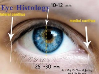
  • 1. Eye Histo lo gy Assoc. Prof. Dr. Karim Al-Jashamy
  • 2. -The superior and inferior tarsal plates are dense fibrous plates that give support and form to the eyelids. Tarsal glands produce oily secretions to prevent tears from leaking out, while the eyelids stay dry. - The obicularis oculi muscle contains both palpebral and orbital parts and acts to close the eyelids. - The levator palpebrae superioris muscle elevates the upper eyelid. It is a skeletal muscle under voluntary control.
  • 3. The eye • A complex and highly developed photosensitive organ that permits an accurate analysis of the form, light intensity, and color reflected from objects. • Each eye is composed of three concentric layers: an external layer that consists of the sclera and the cornea; a middle layer also called the vascular layer consisting of the choroid, ciliary body, and iris; and an inner layer of nerve tissue, the retina, which consists of an outer pigment epithelium and an inner retina proper. • The photosensitive retina proper is part of the central nervous system and communicates with the cerebrum through the optic nerve and extends forward to the ora serrata.
  • 4. Diagram, showing the structure of the eye, retina, fovea, and ciliary body. An enlarged diagram of the fovea is shown at the lower right: (1) axons of ganglion cells; (2) bipolar cells; (3) rods; (4) cones. Enlarged diagrams of the ciliary body (upper right) and retina (lower left)
  • 5. •The sclera consists of tough, dense connective tissue made up mainly of flat collagen bundles intersecting in various directions while remaining parallel to the surface of the organ, a moderate amount of ground substance, and a few fibroblasts. •The external surface of the sclera the episclera is connected by a loose system of thin collagen fibers to a dense layer of connective tissue called Tenon's capsule. Tenon's capsule comes into contact with the loose conjunctival stroma at the junction of the cornea with the sclera. • Between the sclera and the choroid is Section of choroid and sclera. The choroid is a highly vascular layer (arrowheads) of the suprachoroidal lamina, a thin layer connective tissue containing melanocytes that of loose connective tissue rich in prevent the reflection of incident light. melanocytes, fibroblasts, and elastic The sclera is a dense layer of connective fibers. tissue rich in fibers of collagen type I, arranged in parallel bundles.
  • 6. • The outer layer, or sclera, consists of dense fibrous connective tissue. – The sclera is "white" of the eye. – The sclera is continuous with the transparent substantia propria of the cornea. – The exposed front surface of the eye, including the cornea, is also covered by a thin, non-keratinized stratified squamous epithelium. 1 - cornea 2 - iris 3 - posterior chamber of the eye 4 - lens 5 - vitreous body 6 - ciliary body 7 - retina 8 - choroid 9 - sclera 10 - canal of Sclemm 11 - growth area of the lens 12 - anterior epithelium of the lens
  • 7. RETINA, CHOROID • 1 - 8 - retina 1 - optic nerve fibers 2 - ganglion cell layer 3 - inner plexiform layer 4 - inner nuclear layer 5 - outer plexiform layer 6 - outer nuclear layer 7 - outer processes of rods and cones 8 - pigmented epithelium 9 - choroid 10 - sclera
  • 8.
  • 9. • Cornea • colorless and transparent. • A transverse section of the cornea shows that it consists of five layers: epithelium, Bowman's membrane, stroma, Descemet's membrane, and endothelium. • The corneal epithelium is stratified, squamous, and nonkeratinized and consists of five or six layers of cells. • In the basal part of the epithelium are numerous mitotic figures that are responsible for the cornea's remarkable regenerative capacity • The surface corneal cells show microvilli protruding into the space filled by the precorneal tear film. • This epithelial tissue is covered by a protective layer of lipid and glycoprotein, about 7 m thick. The cornea has one of the richest sensory nerve supplies of any eye tissue.
  • 10. CORNEA •1 - anterior epithelium (stratified squamous epithelium) 2 - anterior basement (Bowman's) membrane 3 - substantia propria 4 - posterior basement (Descemet's) membrane 5 - posterior epithelium (simple squamous or endothelium)
  • 11. This layer, Bowman's membrane, consists of collagen fibers crossing at random, a condensation of the intercellular substance, and no cells . Bowman's membrane contributes greatly to the stability and strength of the cornea. • The stroma is formed of many layers of parallel collagen bundles that cross at approximately right angles to each other. The collagen fibrils within each lamella are parallel to each other and run the full width of the cornea. Between the several layers, the cytoplasmic extensions of fibroblasts are flattened like the wings of a butterfly. • Descemet's membrane is a thick homogeneous structure composed of fine collagenous filaments organized in a three-dimensional network. • The endothelium of the cornea is a simple squamous epithelium. These cells possess organelles for secretion that are characteristic of cells engaged in active transport and protein synthesis and that may be related to the synthesis and maintenance of Descemet's membrane. • The corneal endothelium and epithelium are responsible for maintaining the transparency of the cornea. Both layers are capable of transporting sodium ions toward their apical surfaces. Chloride ions and water follow passively, maintaining the corneal stroma in a relatively dehydrated state. This state, along with the regular orientation of the very thin collagen fibrils of the stroma, accounts for the transparency of the cornea.
  • 12. • The corneoscleral junction, or limbus, is an area of transition from the transparent collagen bundles of the cornea to the white opaque fibers of the sclera. It is highly vascularized, and its blood vessels assume an important role in corneal inflammatory processes. • The cornea, an avascular structure, receives its metabolites by diffusion from adjacent vessels and from the fluid of the anterior chamber of the eye. • In the region of the limbus in the stromal layer, irregular endothelium- lined channels, the trabecular meshwork, merge to form Schlemm's canal, which drains fluid from the anterior chamber of the eye. Schlemm's canal communicates externally with the venous system. • Middle, or Vascular, Layer • The middle (vascular) layer of the eye consists of three parts: choroid, ciliary body, and iris, known collectively as the uveal tract.
  • 13. - The ciliary body contains ciliary muscle that is composed of smooth muscle. Contraction and relaxation of the ciliary muscles change the tension of the zonular fibers, or suspensory ligaments, of the lens. This allows the lens to change shape, a process known as accommodation. - The ciliary processes are folds of connective tissue that are covered by two layers of epithelium. There is also a complex vasculature that cannot be seen easily. Fluid from these vessels is processed and transported by the epithelial cells to the posterior chamber as aqueous humor. The epithelial cells constitute the blood-aqueous barrier. -
  • 14. - The aqueous humor enters the anterior chamber through the pupil as it flows between the lens and the iris. - Aqueous humor leaves the anterior chamber through the trabecular meshwork and into the canal of Schlemm. - This is an endothelial lines, circumferentially arranged vessel that communicates with veins in the sclera and returns the aqueous humor back to the general circulation. - Obstruction of the trabecular meshwork and canals of Schlemm are thought to be the major cause of elevated intraocular pressure, which could then lead to glaucoma.
  • 15. - The iris is detailed here in higher magnification. Note the anterior and posterior chambers to help orient yourself. - The anterior surface of the iris contains loose, variably pigmented stroma. It is open to the circulating aqueous humor within the anterior chamber. - Two layers of heavily pigmented epithelium cover the posterior surface of the iris. - Note that the sphincter pupillae muscle can be easily seen near the pupil margin. It is smooth muscle controlled by parasympathetics. The dilator pupillae muscle is more difficult to identify, but it dilates the pupil upon sympathetic innervation.
  • 16. Lens • The lens consists of a lens capsule, the subcapsular epithelium and lens fibres. It does not contain blood vessels or nerves.