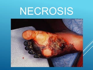
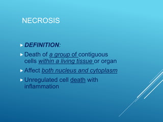
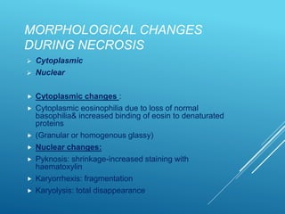
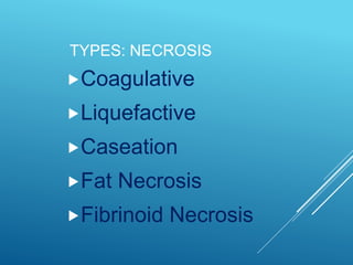
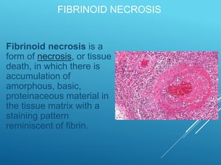
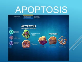
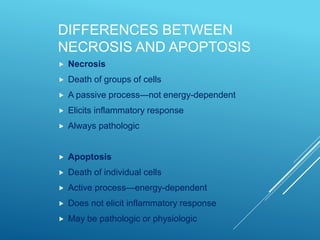
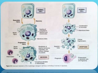
2. INTRODUCTION
“All organ injuries start with
structural or molecular
alterations in cells” concept
began by Virchow in 1800's
•modern study of disease
attempts to understand how cells
react to injury, often at the
subcellular or molecular level,
and how this is manifested in the
whole animal.
3. CLASSES OF CELL INJURY
Irreversible --> cell
death. Types:
Necrosis: pathological
(dissolution of dead
cells by its own
digestive enzymes)
Apoptosis:
PROGRAMMED cell
death
Reversible
If stressor removed, and
If cell damage mild: cells
survive
4. PROBABLE CAUSES
The first is the inability to reverse mitochondrial
dysfunction (lack of oxidative phosphorylation and ATP
generation) even after resolution of the original injury.
The second is the development of profound disturbances
in membrane function.
6. NECROSIS
DEFINITION:
Death of a group of contiguous
cells within a living tissue or organ
Affect both nucleus and cytoplasm
Unregulated cell death with
inflammation
7. MORPHOLOGICAL CHANGES
DURING NECROSIS
Cytoplasmic
Nuclear
Cytoplasmic changes :
Cytoplasmic eosinophilia due to loss of normal
basophilia& increased binding of eosin to denaturated
proteins
(Granular or homogenous glassy)
Nuclear changes:
Pyknosis: shrinkage-increased staining with
haematoxylin
Karyorrhexis: fragmentation
Karyolysis: total disappearance
9. COAGULATIVE NECROSIS
In: infarcts of kidney, heart, spleen
Gross: pale , yellow, opaque, firm
Mic.: All cellular details are lost but general
architecture of the tissue is preserved
Surrounding tissue----acute inflammation
Ex: Infarction - heart
Infarction - kidney
11. CASEATION NECROSIS
In: Tuberculosis
Necrotic tissue is partially liquefied---cheesy material (caseation)
Mic: Both cellular details & general architecture of dead tissue are
lost---structureless eosinophilic material
12. FAT NECROSIS
Traumatic: in female breast
Enzymatic: in acute hemorrhagic
pancreatitis
Gross: opaque & white
fat cells appear cloudy, surrounded by
chronic inflammatory cells, histiocytes,
foreign body giant cells
13. FIBRINOID NECROSIS
Fibrinoid necrosis is a
form of necrosis, or tissue
death, in which there is
accumulation of
amorphous, basic,
proteinaceous material in
the tissue matrix with a
staining pattern
reminiscent of fibrin.
15. APOPTOSIS
(programmed cell death)
Definition: death of individual cells
surrounded by viable cells
when a cell dies through activation of an
internally suicide program
It is an active process—energy
dependent
Does not elicit inflammatory response
May be physiologic or pathologic
16. APOPTOSIS CAUSES
PHYSIOLOGIC
During embryogenesis e.g. removal of
interdigital webs during embryonic
development of toes and fingers
Hormone-dependent e.g.
endometrial cellloss in menstruation
PATHOLOGIC
Irradiated tissues
Cell death induced by cytotoxic T-
lymphocytes
Viral infections e.g. viral hepatitis
Cell death in tumours
17. DIFFERENCES BETWEEN
NECROSIS AND APOPTOSIS
Necrosis
Death of groups of cells
A passive process—not energy-dependent
Elicits inflammatory response
Always pathologic
Apoptosis
Death of individual cells
Active process—energy-dependent
Does not elicit inflammatory response
May be pathologic or physiologic