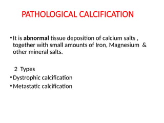
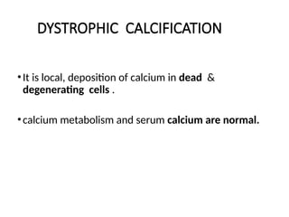
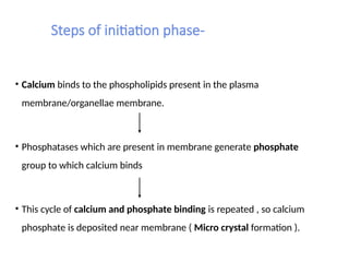
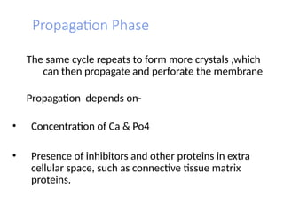
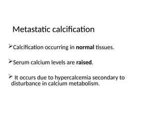
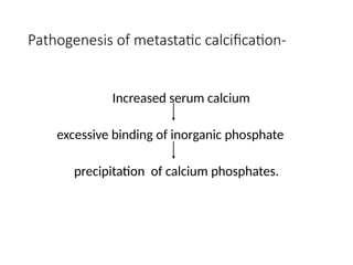
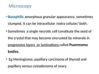
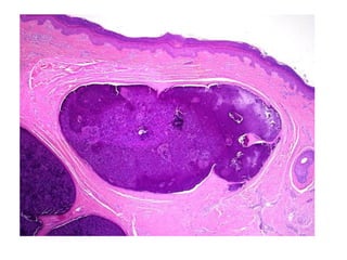
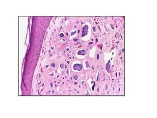
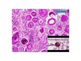
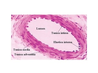
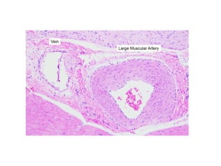
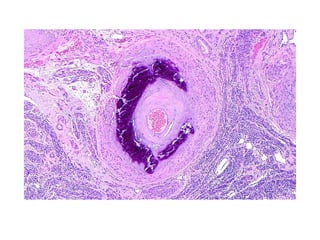
PATHOLOGICAL CALCIFICATION
Dr Payal Desai
PATHOLOGICAL CALCIFICATION
•It is abnormal tissue deposition of calcium salts ,
together with small amounts of Iron, Magnesium &
other mineral salts.
2 Types
•Dystrophic calcification
•Metastatic calcification
•It is local, deposition of calcium in dead &
degenerating cells .
•calcium metabolism and serum calcium are normal.
DYSTROPHIC CALCIFICATION
Calcification in Dead cells-
1. Necrosis-caseous , liquefactive and fat necrosis
2. Infarcts
3. Thrombi
4. Hematomas
5 dead parasites
Calcification in degenerated tissue-
1. Dense old scars
2. Atheromas
3. Stroma of tumors -Fibroid , Ca. breast , thyroid tumors
4. Monckenberg’s Sclerosis
5. Cyst walls
6. Old age- cartilage
Pathogenesis
a)Intiation phase
b)Propagation phase
Initiation phase (Enucleation phase)
Intracellular
• Deposition first occurs in mitochondria of dying cells.
Extracellular
• Deposition occurs in phospholipids of membrane bound
vesicles.
• Calcium is concentrated in these vesicles by process of
membrane facilitated calcification.
Steps of initiation phase-
• Calcium binds to the phospholipids present in the plasma
membrane/organellae membrane.
• Phosphatases which are present in membrane generate phosphate
group to which calcium binds
• This cycle of calcium and phosphate binding is repeated , so calcium
phosphate is deposited near membrane ( Micro crystal formation ).
Propagation Phase
The same cycle repeats to form more crystals ,which
can then propagate and perforate the membrane
Propagation depends on-
• Concentration of Ca & Po4
• Presence of inhibitors and other proteins in extra
cellular space, such as connective tissue matrix
proteins.
Calcification occurring in normal tissues.
Serum calcium levels are raised.
 It occurs due to hypercalcemia secondary to
disturbance in calcium metabolism.
Metastatic calcification
Pathogenesis of metastatic calcification-
Increased serum calcium
excessive binding of inorganic phosphate
precipitation of calcium phosphates.
1. Increased secretion of Parathyroid hormone  increased bone
resorption.
Eg..Parathyroid tumors and ectopic PTH secretion.
2. Destruction of bone tissue  multiple myeloma, diffuse skeletal
metastasis, prolonged immobilization.
3. Vitamin D related disorders  Vitamin D intoxication, William
syndrome.
4. Chronic renal failure.
5. Other  Aluminum intoxication, Milk alkali syndrome, Excessive intake
of antacids.
Causes of Metastatic calcification
Sites of Metastatic calcification
• Gastric mucosa – Acid secreting fundal glands
• Kidney ( Nephrocalcinosis ) – Basement membrane of tubular
epithelium
• Lungs – Alveolar walls
• Systemic arteries – Internal elastic lamina
• Pulmonary veins
• Cornea
MORPHOLOGY OF CALCIFICATION
(same for dystrophic and metastatic)
Morphology- same for dystrophic and metastatic
• Gross – Fine white granules or clumps, and often felt as
gritty deposits.
Microscopy
• Basophilic amorphous granular appearance, sometimes
clumped. It can be intracellular /extra cellular/ both.
• Sometimes a single necrotic cell constitute the seed of
the crystal that may become encrusted by minerals in
progressive layers or laminations called Psammoma
bodies.
• Eg Meningioma, papillary carcinoma of thyroid and
papillary serous cystadenoma of ovary.
Monckeberg’s Sclerosis
• Dystrophic calcification in the tunica media of large and medium
sized muscular arteries
• Site: Extremeties & genital tract
• Commonly seen in elderly (degenerative process)
• Little or no significance
Morphology
• Gross: thickened vessel, palpated clinically, lumen patent
• Microscopy:
- Deposition of basophilic calcium salts in form of granules
- Smooth muscle replaced by acellular hyalinized fibrous tissue
- Lumen normal, filled with red blood cells
- Tunica intima and adventitia spared
Path calcification for mbbs 2nd year pathology

Path calcification for mbbs 2nd year pathology

  • 1.
  • 2.
    PATHOLOGICAL CALCIFICATION •It isabnormal tissue deposition of calcium salts , together with small amounts of Iron, Magnesium & other mineral salts. 2 Types •Dystrophic calcification •Metastatic calcification
  • 3.
    •It is local,deposition of calcium in dead & degenerating cells . •calcium metabolism and serum calcium are normal. DYSTROPHIC CALCIFICATION
  • 4.
    Calcification in Deadcells- 1. Necrosis-caseous , liquefactive and fat necrosis 2. Infarcts 3. Thrombi 4. Hematomas 5 dead parasites Calcification in degenerated tissue- 1. Dense old scars 2. Atheromas 3. Stroma of tumors -Fibroid , Ca. breast , thyroid tumors 4. Monckenberg’s Sclerosis 5. Cyst walls 6. Old age- cartilage
  • 5.
  • 6.
    Initiation phase (Enucleationphase) Intracellular • Deposition first occurs in mitochondria of dying cells. Extracellular • Deposition occurs in phospholipids of membrane bound vesicles. • Calcium is concentrated in these vesicles by process of membrane facilitated calcification.
  • 7.
    Steps of initiationphase- • Calcium binds to the phospholipids present in the plasma membrane/organellae membrane. • Phosphatases which are present in membrane generate phosphate group to which calcium binds • This cycle of calcium and phosphate binding is repeated , so calcium phosphate is deposited near membrane ( Micro crystal formation ).
  • 8.
    Propagation Phase The samecycle repeats to form more crystals ,which can then propagate and perforate the membrane Propagation depends on- • Concentration of Ca & Po4 • Presence of inhibitors and other proteins in extra cellular space, such as connective tissue matrix proteins.
  • 9.
    Calcification occurring innormal tissues. Serum calcium levels are raised.  It occurs due to hypercalcemia secondary to disturbance in calcium metabolism. Metastatic calcification
  • 10.
    Pathogenesis of metastaticcalcification- Increased serum calcium excessive binding of inorganic phosphate precipitation of calcium phosphates.
  • 11.
    1. Increased secretionof Parathyroid hormone  increased bone resorption. Eg..Parathyroid tumors and ectopic PTH secretion. 2. Destruction of bone tissue  multiple myeloma, diffuse skeletal metastasis, prolonged immobilization. 3. Vitamin D related disorders  Vitamin D intoxication, William syndrome. 4. Chronic renal failure. 5. Other  Aluminum intoxication, Milk alkali syndrome, Excessive intake of antacids. Causes of Metastatic calcification
  • 12.
    Sites of Metastaticcalcification • Gastric mucosa – Acid secreting fundal glands • Kidney ( Nephrocalcinosis ) – Basement membrane of tubular epithelium • Lungs – Alveolar walls • Systemic arteries – Internal elastic lamina • Pulmonary veins • Cornea
  • 13.
    MORPHOLOGY OF CALCIFICATION (samefor dystrophic and metastatic)
  • 14.
    Morphology- same fordystrophic and metastatic • Gross – Fine white granules or clumps, and often felt as gritty deposits.
  • 15.
    Microscopy • Basophilic amorphousgranular appearance, sometimes clumped. It can be intracellular /extra cellular/ both. • Sometimes a single necrotic cell constitute the seed of the crystal that may become encrusted by minerals in progressive layers or laminations called Psammoma bodies. • Eg Meningioma, papillary carcinoma of thyroid and papillary serous cystadenoma of ovary.
  • 19.
    Monckeberg’s Sclerosis • Dystrophiccalcification in the tunica media of large and medium sized muscular arteries • Site: Extremeties & genital tract • Commonly seen in elderly (degenerative process) • Little or no significance
  • 20.
    Morphology • Gross: thickenedvessel, palpated clinically, lumen patent • Microscopy: - Deposition of basophilic calcium salts in form of granules - Smooth muscle replaced by acellular hyalinized fibrous tissue - Lumen normal, filled with red blood cells - Tunica intima and adventitia spared