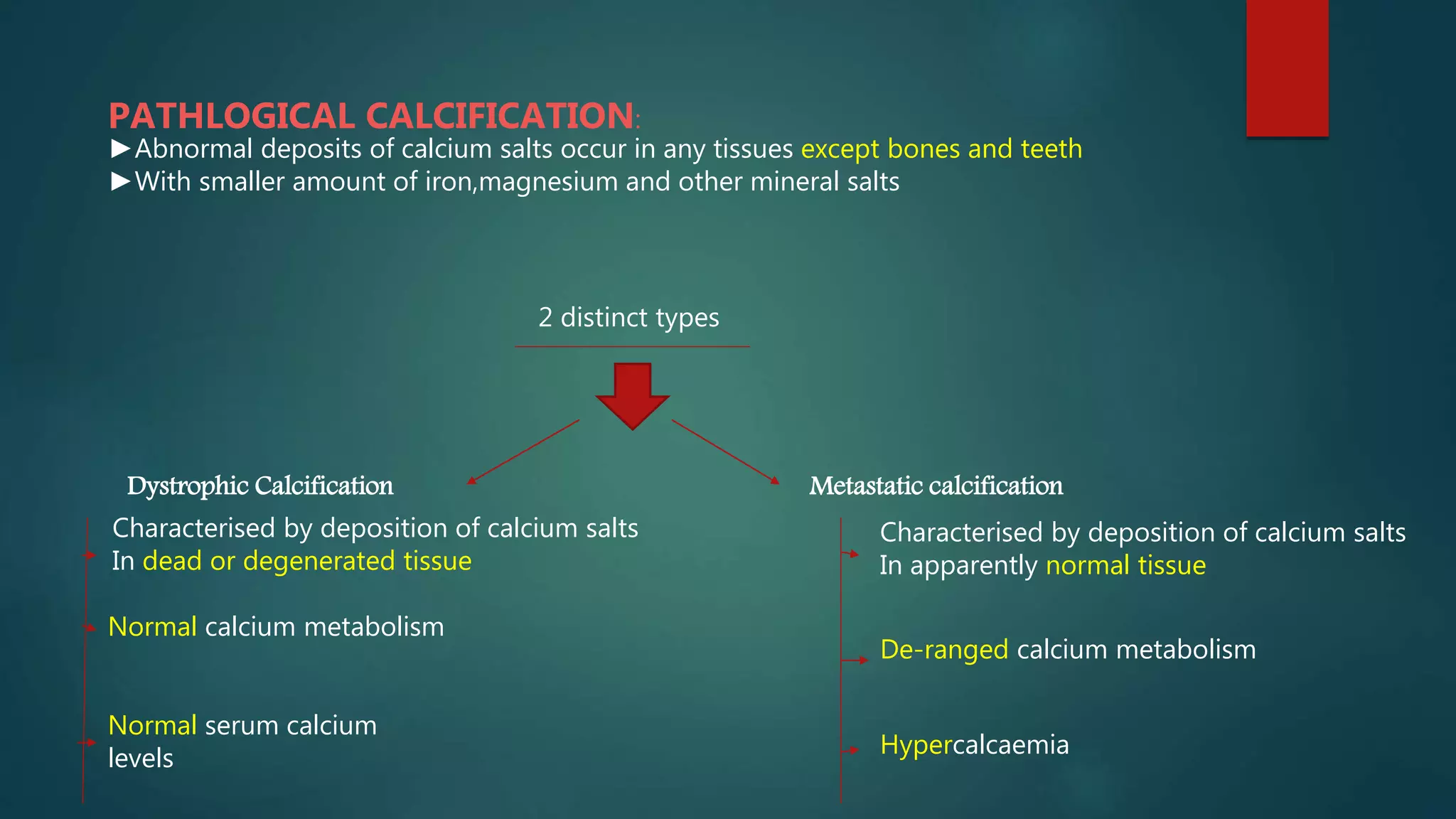
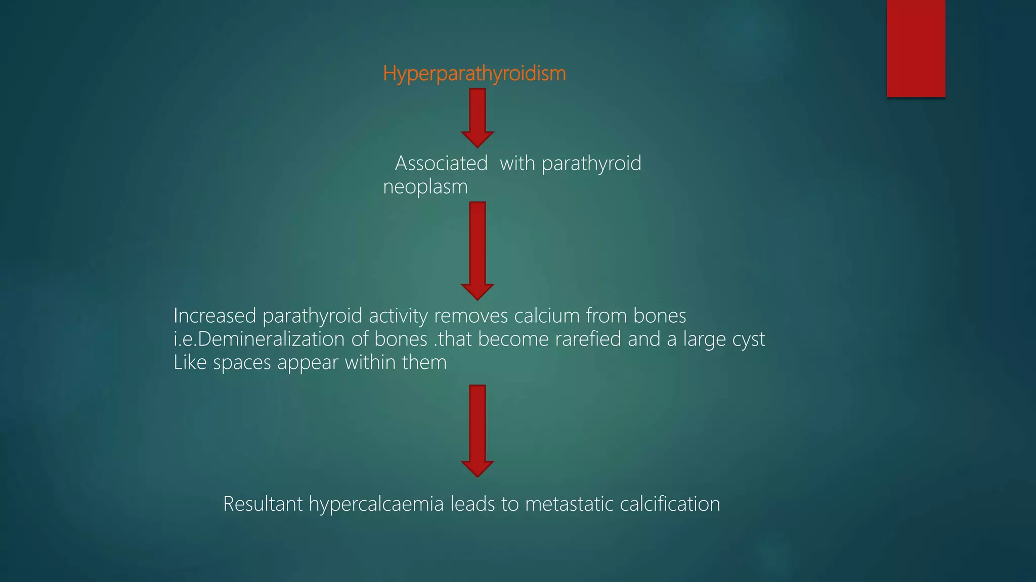
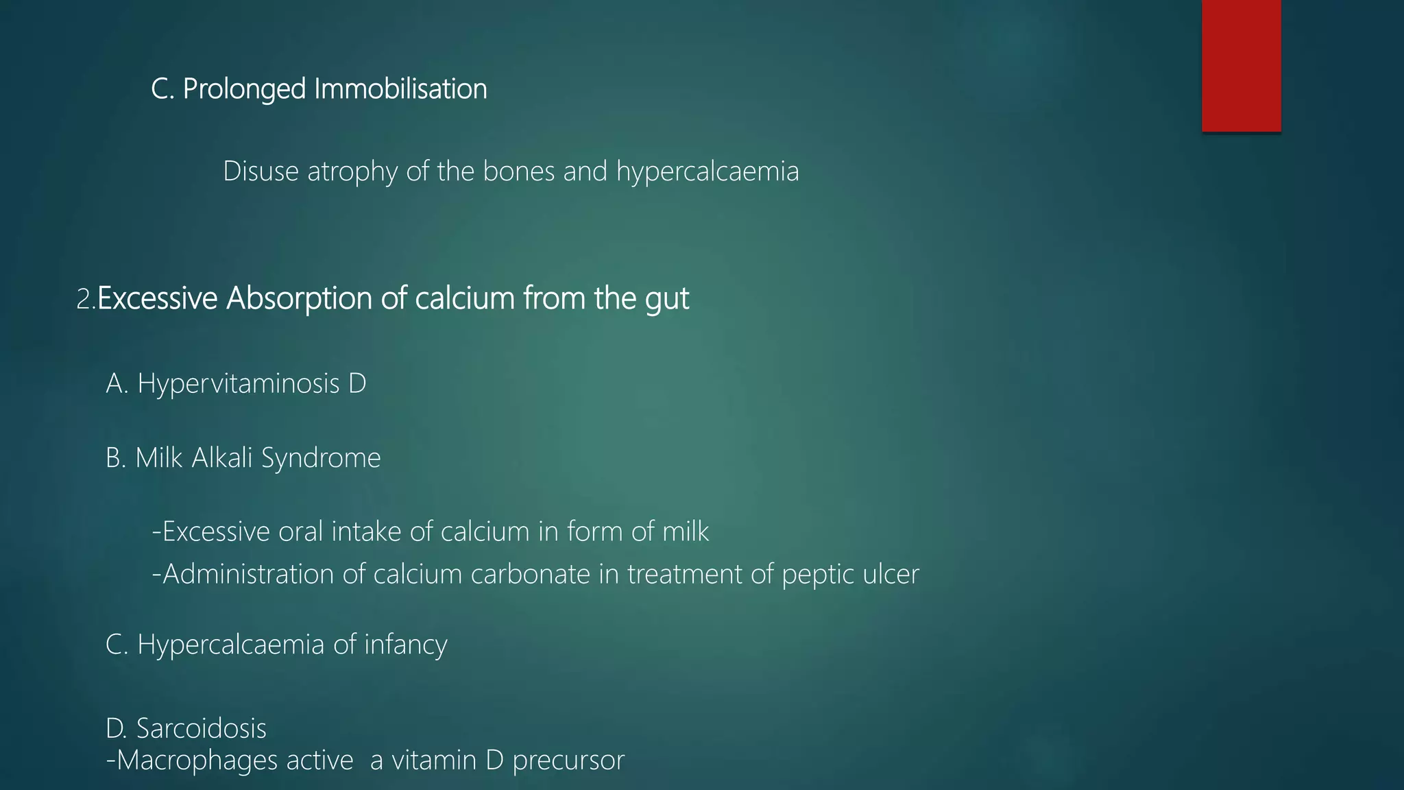
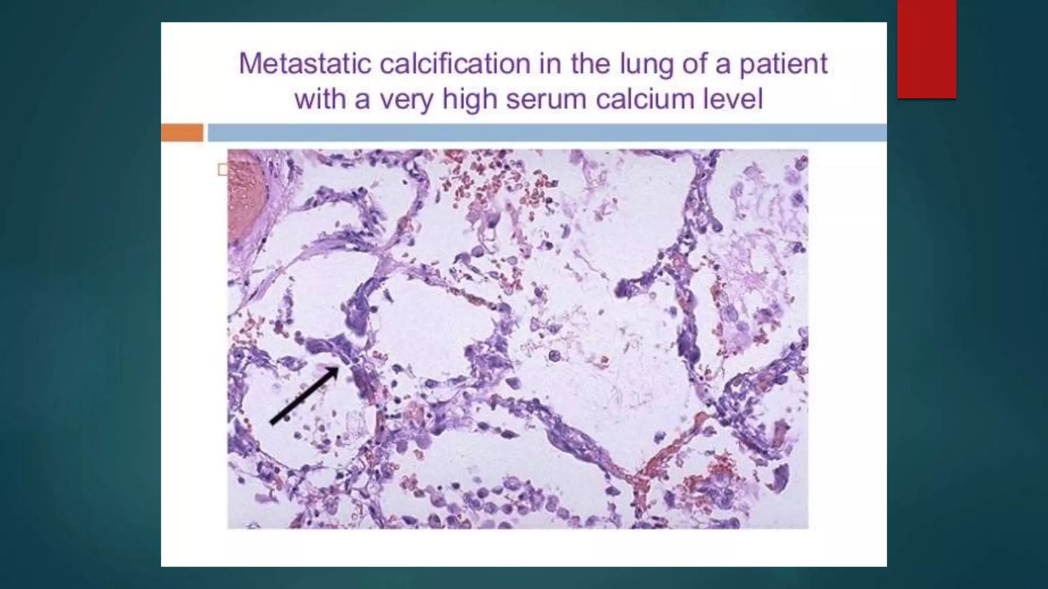
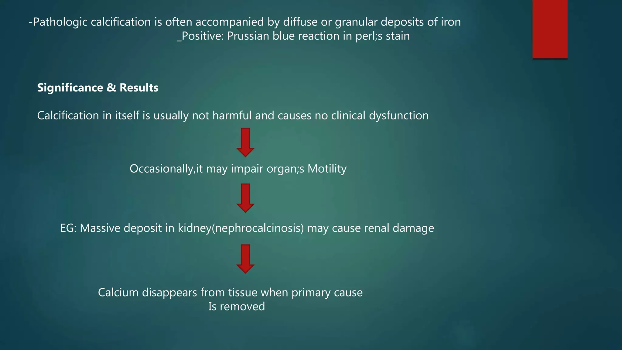
PATHOLOGICAL
CALCIFICATION
SUBMITTED TO:
Rajesh Sir
Pathology
Veterinary(B.V.Sc.&A.H)
AFU,Rampur
SUBMITTED BY:
Suman Bhattarai
Roll NO : 50(Fiftty)
Rampur,Chitwan,Nepal
PATHLOGICAL CALCIFICATION:
►Abnormal deposits of calcium salts occur in any tissues except bones and teeth
►With smaller amount of iron,magnesium and other mineral salts
2 distinct types
Dystrophic Calcification Metastatic calcification
Characterised by deposition of calcium salts
In dead or degenerated tissue
Normal calcium metabolism
Normal serum calcium
levels
Characterised by deposition of calcium salts
In apparently normal tissue
De-ranged calcium metabolism
Hypercalcaemia
Metastatic calcification
Reversible upon correction of metabolic disorder
Hypercalcaemia ( more than 12mg % )
May be due to the
Excessive mobilization of calcium from the bone
Excessice absorption of calcium from the gut
1)Excessive mobilization of calcium from bone:
A) Hyperparathyroidism
1.Primary
-Parathyroid adenoma
2.Secondary
-Parathyroid hyperplasia
-Chronic renal failure
Hyperparathyroidism
Associated with parathyroid
neoplasm
Increased parathyroid activity removes calcium from bones
i.e.Demineralization of bones .that become rarefied and a large cyst
Like spaces appear within them
Resultant hypercalcaemia leads to metastatic calcification
Renal Failure
Leads to phosphate retention
Causes increased activity of parathyroid gland
Called as Secondary hyperparathyroidism
Dimineralization of bones
Metastatic Calcification
B.Bony Destructive Lesions
-Multiple Myeloma
-Metastatic Carcinoma
-Leukemia
MYELOID TUMORS IN BONE MARROW
+
METASTATIC CANCER
causes
Demineralization of Skeleton
Blood stream is flooded with calcium
C. Prolonged Immobilisation
Disuse atrophy of the bones and hypercalcaemia
2.Excessive Absorption of calcium from the gut
A. Hypervitaminosis D
B. Milk Alkali Syndrome
-Excessive oral intake of calcium in form of milk
-Administration of calcium carbonate in treatment of peptic ulcer
C. Hypercalcaemia of infancy
D. Sarcoidosis
-Macrophages active a vitamin D precursor
Sites of Metastatic calcification
May occur in any normal tissue of body especially in those tihat
secrete or excrete acid substances.
Affected organs more commonly are:
1. Kidneys:(Secretes Hippuric acid)
Especially at basement membrane of tubular epithelium and
in tubular lumina causes nephrocalcinosis.
2.Lungs(C02)
especially in alveolar walls
3.Stomach(HCL)
On acid secreting fundal glands(gastric mucosa)
4.Blood Vessels
Especially on internal elastic lamina
5.Cornea
6.Synovium of joint causing pain and dysfunction
Pathogenesis:
Excessive binding of inorganic phosphate ions with the calcium ions i.e increased
Precipitates of Ca-phosphate due to hypercalcaemia at certain sites.
Eg.in lungs,stomach ,blood vessels and cornea.
Morphologically
-Ca-salts in all these sites, resembles those described in dystrophic calcification
-Etiology and pathogenesis of two are different
-Morphologically the deposits in both resemble normal minerals of bone.
H & E Stained Sections (Haematoxylin and eosin )
Macroscopy-
Microscopy-
Can vary in size,white granules which may be gritty.
-Ca-salts appear as deeply basophilic,irregular and granular clumps
-Depositss may be intracellular,extracellular or both.
-Occassionally ,heterotopic bone formation(ossification) may occur.
-Calcium deposits can be confirmed by special stains
_Silver Impregnation method of von-kossa producing black colour
_Alizarin red S that produces red staining
-Pathologic calcification is often accompanied by diffuse or granular deposits of iron
_Positive: Prussian blue reaction in perl;s stain
Significance & Results
Calcification in itself is usually not harmful and causes no clinical dysfunction
Occasionally,it may impair organ;s Motility
EG: Massive deposit in kidney(nephrocalcinosis) may cause renal damage
Calcium disappears from tissue when primary cause
Is removed

Pathological Calcification

  • 1.
  • 2.
    PATHLOGICAL CALCIFICATION: ►Abnormal depositsof calcium salts occur in any tissues except bones and teeth ►With smaller amount of iron,magnesium and other mineral salts 2 distinct types Dystrophic Calcification Metastatic calcification Characterised by deposition of calcium salts In dead or degenerated tissue Normal calcium metabolism Normal serum calcium levels Characterised by deposition of calcium salts In apparently normal tissue De-ranged calcium metabolism Hypercalcaemia
  • 3.
    Metastatic calcification Reversible uponcorrection of metabolic disorder Hypercalcaemia ( more than 12mg % ) May be due to the Excessive mobilization of calcium from the bone Excessice absorption of calcium from the gut 1)Excessive mobilization of calcium from bone: A) Hyperparathyroidism 1.Primary -Parathyroid adenoma 2.Secondary -Parathyroid hyperplasia -Chronic renal failure
  • 4.
    Hyperparathyroidism Associated with parathyroid neoplasm Increasedparathyroid activity removes calcium from bones i.e.Demineralization of bones .that become rarefied and a large cyst Like spaces appear within them Resultant hypercalcaemia leads to metastatic calcification
  • 5.
    Renal Failure Leads tophosphate retention Causes increased activity of parathyroid gland Called as Secondary hyperparathyroidism Dimineralization of bones Metastatic Calcification
  • 6.
    B.Bony Destructive Lesions -MultipleMyeloma -Metastatic Carcinoma -Leukemia MYELOID TUMORS IN BONE MARROW + METASTATIC CANCER causes Demineralization of Skeleton Blood stream is flooded with calcium
  • 7.
    C. Prolonged Immobilisation Disuseatrophy of the bones and hypercalcaemia 2.Excessive Absorption of calcium from the gut A. Hypervitaminosis D B. Milk Alkali Syndrome -Excessive oral intake of calcium in form of milk -Administration of calcium carbonate in treatment of peptic ulcer C. Hypercalcaemia of infancy D. Sarcoidosis -Macrophages active a vitamin D precursor
  • 8.
    Sites of Metastaticcalcification May occur in any normal tissue of body especially in those tihat secrete or excrete acid substances. Affected organs more commonly are: 1. Kidneys:(Secretes Hippuric acid) Especially at basement membrane of tubular epithelium and in tubular lumina causes nephrocalcinosis. 2.Lungs(C02) especially in alveolar walls 3.Stomach(HCL) On acid secreting fundal glands(gastric mucosa) 4.Blood Vessels Especially on internal elastic lamina 5.Cornea 6.Synovium of joint causing pain and dysfunction
  • 11.
    Pathogenesis: Excessive binding ofinorganic phosphate ions with the calcium ions i.e increased Precipitates of Ca-phosphate due to hypercalcaemia at certain sites. Eg.in lungs,stomach ,blood vessels and cornea. Morphologically -Ca-salts in all these sites, resembles those described in dystrophic calcification -Etiology and pathogenesis of two are different -Morphologically the deposits in both resemble normal minerals of bone. H & E Stained Sections (Haematoxylin and eosin ) Macroscopy- Microscopy- Can vary in size,white granules which may be gritty. -Ca-salts appear as deeply basophilic,irregular and granular clumps -Depositss may be intracellular,extracellular or both. -Occassionally ,heterotopic bone formation(ossification) may occur. -Calcium deposits can be confirmed by special stains _Silver Impregnation method of von-kossa producing black colour _Alizarin red S that produces red staining
  • 12.
    -Pathologic calcification isoften accompanied by diffuse or granular deposits of iron _Positive: Prussian blue reaction in perl;s stain Significance & Results Calcification in itself is usually not harmful and causes no clinical dysfunction Occasionally,it may impair organ;s Motility EG: Massive deposit in kidney(nephrocalcinosis) may cause renal damage Calcium disappears from tissue when primary cause Is removed