GASTROINTESTINAL PHYSIOLOGY (The Guyton and Hall Physiology)Maryam Fida
ANATOMY OF GASTROINTESTINAL TRACT
Consists of
Gastrointestinal (GI) tract
Accessory glandular organs
Anatomy and functions of the GI tract
GI tract
◦ mouth, pharynx, esophagus,
◦ stomach, small intestine,
◦ large intestine, anus
◦ Accessory Glandular Organs
salivary glands, liver, gallbladder, pancreas
Histology/organization of the Gut Wall
From esophagus to anus, GI tract has the same basic arrangement of tissues.
There are following layers from outer surface to inward.
Serosa
Longitudinal smooth muscle
Circular smooth muscle layer
Submucosa
Mucosa
Layers of Alimentary Canal
Gastrointestinal Smooth Muscle Functions as a syncytium
The individual smooth muscle fibers in the gastrointestinal tract are 200 to 500 micrometers in length and 2 to 10 micrometers in diameter, and they are arranged in bundles of as many as 100 parallel fibers.
In the LONGITUDINAL MUSCLE LAYER, the bundles extend longitudinally down the intestinal tract.
In the CIRCULAR MUSCLE Layer, they extend around the gut.
Within each bundle, the muscle fibers are electrically connected with one another through large numbers of GAP JUNCTION .
Because of these gap junction electrical signals that initiate muscle contractions can travel readily from one fiber to the next within each bundle but more rapidly along the length of the bundle than sideways.
Each muscle layer functions as a SYNCYTIUM . That is , when an action potential is elicited anywhere within the muscle mass, it generally travels in all directions in the muscle.
MAIN FUNCTIONS
1. Ingestion or consumption of food substances.
2. Breaking them in to small particles.
3. Transport of small particles to different areas of the digestive tract.
4. Secretion of necessary enzymes and other substances for digestion.
01.07.09(b): Tubular GI Tract - StomachOpen.Michigan
Slideshow is from the University of Michigan Medical School's M1 Gastrointestinal / Liver sequence
View additional course materials on Open.Michigan:
http://openmi.ch/med-m1gastro
Histology of Gall bladder and its formation which consist of mainly 3 layers which they are:
- Mucosa
- Muscularis / Fibromuscular layer
- Serosa / Adventitia
And you must note that there is no Muscularis mucosa
& Submucosa inside Gall bladder...
Prepared by Nahry Omer Muhammad, University of Sulaimany/Collage of Medicine
small intestine. parts . jujenum, ilieum, Malt, difference between jejunum and ilieum, mesentry, mesocolon, blood supply of small intetsine, arterial arcades, vesa recta, superior mesenteric vessles, meckels diverticulum,
The GI tract is a series of hollow organs joined in a long, twisting tube from the mouth to the anus. The hollow organs that make up the GI tract are the mouth, esophagus, stomach, small intestine, large intestine, and anus. The liver, pancreas, and gallbladder are the solid organs of the digestive system
Irritable bowel syndrome (IBS) is a group of symptoms, including pain discomfort in your abdomen combined with changes in your bowel movement patterns.
For More detail visit this link:
http://goo.gl/RaZhvc
Dr Vivek Baliga - The Basics Of Medical StatisticsDr Vivek Baliga
Medical statistics can be daunting. Understanding them is essential to understand any research paper. Here are some basic in medical statistics by Dr Vivek Baliga, Consultant Internal Medicine, Bangalore. Read more by Dr Vivek Baliga at http://drvivekbaliga.net
ECG In Ischemic Heart Disease - Dr Vivek Baliga ReviewDr Vivek Baliga
Dr Vivek Baliga Presentation on the role of ECG in the diagnosis of ischemic heart disease. Here, he covers the very basics in ECG diagnosis of heart disease. Suitable for medical students and physicians alike. For more health articles for patients, visit http://baligadiagnostics.com/category/dr-vivek-baliga/
study of structures and functions of the gastrointestinal tract
- histology of the oral cavity
- histology of the pharynx
- histology of the esophagus and stomach
- histology of intestine
- histology of the liver and pancreas
Gastrointestinal Tract (GIT)//DIGESTIVE SYSTEM Wasim Ak
The digestive tract or gastrointestinal tract ( GIT) is composed of mouth , pharynx, oesophagus, stomach , small intestine and large intestine .
This GIT will helps in digestion of food and absorption of needed nutrients into our body .
The mucose membrane lining of gastrointestinal tract is stratified squamous epithelium at the esophagus which slowly convert into simple columnar epithelium at the stomach until the anus it converts back into the stratified squamous epithelium at the lower half of the anal canal. The stratified epithelium is a wear and tear epithelium.
As it passes down from the small to large intestine, goblet cells increase because as it passes down water was absorb, goblet cells function to produce mucous.
This is just a rough idea, for better slides with more reference please PM the author at davidgqf@gmail.com.
1. F. Shammas / 04
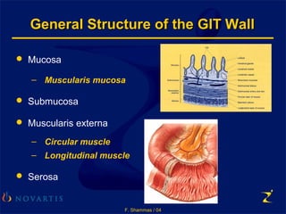
General Structure of the GIT WallGeneral Structure of the GIT Wall
Mucosa
– Muscularis mucosa
Submucosa
Muscularis externa
– Circular muscle
– Longitudinal muscle
Serosa
2. F. Shammas / 04
The Wall of the GI Tract
Layer Description Tissue type
Mucosa Moist lining
membrane
Esophagus: stratified
squamous epithelium
Elsewhere: simple epithelium,
some connective tissue,
smooth muscle and secretory
cells
Submucosa Soft connective
tissue layer
Blood vessels, nerves,
lymphatics
Muscularis externa
i. Muscularis mucosa Function unknown Thin layer of smooth muscle
ii. Circular muscle Muscle contraction causes
intestinal constriction
Smooth muscle
iii. Longitudinal muscle Muscle contraction causes
intestinal shortening
Smooth muscle
Serosa Production of serosal fluid Single layer of secretory cells
3. F. Shammas / 04
The Large IntestineThe Large Intestine
Sigmoid Colon, Rectum and AnusSigmoid Colon, Rectum and Anus
4. F. Shammas / 04
The Large IntestineThe Large Intestine
Anatomical DivisionsAnatomical Divisions
1. Caecum
2. Colon
• Ascending
• Transverse
• Descending
• Sigmoid
3. Rectum and anus
5. F. Shammas / 04
The Wall of the Colon
Submucosa
Circular
layer
Longitudinal
layer
Muscularis
externa
Intestinal
gland
Columnar
epithelium
6. F. Shammas / 04
The Accessory Organs of DigestionThe Accessory Organs of Digestion
The Salivary Glands
The Liver
The Gallbladder
The Pancreas
7. F. Shammas / 04
The Salivary GlandsThe Salivary Glands
The parotid gland
The Sublingual gland
The submandibular gland
The saliva contains Mucus, Ptyalin, Lysozymes
8. F. Shammas / 04
The Liver and GallbladderThe Liver and Gallbladder
9. F. Shammas / 04
The Biliary TractThe Biliary Tract
Common hepatic
duct
Cystic duct
Common bile
duct