Paranasal sinuses
•
26 likes•6,224 views

The Indian Dental Academy is the Leader in continuing dental education , training dentists in all aspects of dentistry and offering a wide range of dental certified courses in different formats.for more details please visit www.indiandentalacademy.com

Recommended
More Related Content
What's hot
What's hot (20)
Viewers also liked
Viewers also liked (20)
Similar to Paranasal sinuses
Similar to Paranasal sinuses (20)
More from Indian dental academy
More from Indian dental academy (20)
Recently uploaded
Recently uploaded (20)
Paranasal sinuses
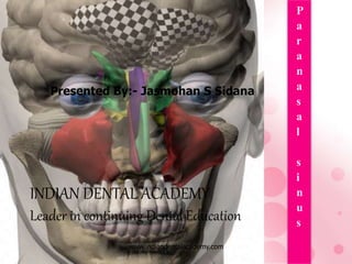
- 1. P a r a n a s a l s i n u s Presented By:- Jasmohan S Sidana INDIAN DENTAL ACADEMY Leader in continuing Dental Education www.indiandentalacademy.com
- 2. Learning Objectives • By the end of the presentation a learner should be able to :- 1. Enlist the Paranasal Sinuses. 2. Describe the Development of PNS. 3. Enumerate the Functions of Paranasal Sinuses. 4. Illustrate the Blood supply, Nerve Supply & Lymphatic drainage. 5. Enumerate the Diagnostic Methods in the Diseases of the Sinuses. 6. Describe the Developmental Anomalies, infections, Cysts & Tumors associated with Paranasal Sinuses along with the treatment. www.indiandentalacademy.com
- 4. Introduction: Sinus - cavity or a channel such as a cavity within a bone- a dilated channel for venous blood or one permitting the escape of purulent material. Paranasal sinus - air filled extension of the respiratory part of the nasal cavity into the frontal, ethmoidal, sphenoidal & maxillary cranial bone. www.indiandentalacademy.com
- 5. History: Galen (130-301 AD) made the first known description about the adult maxillary sinus Nathaniel Highmore in 1651 was the first to describe in detail the morphology of the maxillary sinus and advance the idea of pneumatization of the sinuses. Therefore maxillary sinus is also called as antrum of Highmore. www.indiandentalacademy.com
- 6. Development Sinuses begin their development at the third month of IUL as outpouchings of the mucous membrane of the nasal meatuses and the sphenoethmoidal recesses. The early paranasal sinuses expand into the walls and roof of the nasal fossae by growth of mucous membrane sacs into the maxillary, sphenoid, frontal and ethmoid bones. The sinuses enlarge variably and greatly from their initial small outpocketings but always retain their original communication with the nasal fossa through ostia. www.indiandentalacademy.com
- 7. Functions: Resonance of voice Lightening of the skull weight Production of bactericidal lysosome to the nasal cavity. Humidification and warming of inspired air and contribution to olfaction – controversial .It is possible that if air is arrested in the sinus for a certain time it quickly reaches body temperature and thus protects the internal structures particularly the brain against exposure to cold air. www.indiandentalacademy.com
- 8. There are four sets of paranasal sinuses namely; • Frontal sinus • Sphenoidal sinus • Ethmoidal sinus • Maxillary sinus Which are present in the respective bones. www.indiandentalacademy.com
- 9. Frontal Sinus : • These are two irregular cavities situated deep to glabella and superciliary arches between outer and inner tables of the bone. • Seen as furrows in frontal recess of middle meatus of nasal fossa at 3-4 months IU Height- 3.15cms Breadth- 2.5cms Depth- 1.8cms www.indiandentalacademy.com
- 10. Invades the bone at 2nd year of life. More developed in males. Radiographically visible at 6 years of age. They grow upward at an extremely variable rate until puberty. Even after puberty all the sinuses appear to increase slowly in size into old age. Two sinuses are separated from each other by a thin bony septum which is often deflected to one or the other side. www.indiandentalacademy.com
- 11. Each sinus communicates with the middle meatus of the nose by a passage called the frontonasal canal. Subsequent enlargement is the result of atrophic changes in the bone www.indiandentalacademy.com
- 12. Blood supply: Supra orbital artery and Anterior ethmoidal arteries Venous Drainage: Into the anastomotic vein between supraorbital and superior ophthalmic veins Lymphatics: To the submandibular nodes www.indiandentalacademy.com
- 13. Sphenoidal Sinus : Height- 2cms Breadth- 1.8cms Depth- 2cms • Two large irregular cavities enclosed in the body of sphenoid bone. • Right and left sinuses are separated from each other by a deflected bony septum. www.indiandentalacademy.com
- 14. • The two sinuses therefore are usually asymmetrical and often partially subdivided by additional bony septa. • Commence at 4th month of IUL by invading posterior part of nasal capsule into the body of the sphenoidal bone. • It continues growing into early adulthood and may invade the wings and rarely the pterygoid plates of the sphenoid bone • Sinus may extend into the lingual and basilar part of occipital bone. www.indiandentalacademy.com
- 15. • Sinus opens into the sphenoethmoidal recess of the lateral wall of the nose. • Radiographically visible at four years of age only. • By 8th year it extends to the hypophyseal fossa. Relations: • Above - optic chiasma and hypophysis cerebri. • Each side – Internal Carotid Artery and Cavernous sinus.www.indiandentalacademy.com
- 16. • Blood supply: Posterior ethmoidal arteries • Lymphatics: To the retropharyngeal nodes. • Nerve supply: Posterior ethmoidal nerve and orbital branches of the pterygoid ganglion. www.indiandentalacademy.com
- 17. Ethmoidal sinus: • Occupy the labrynth of ethmoidal bone. • Ethmoidal labyrinths are two very light cubical masses which enclose a large number of air cells arranged in three groups, Anterior, middle and posterior ethmoidal sinuses. www.indiandentalacademy.com
- 18. • Many of these cells are incomplete and are closed by the related bones (frontal, maxillary, lacrimal, sphenoid and palatal). • Invade the ethmoid bone from the 5th month of IUL and may also be of a clinically significant size at birth. • Grow variably into irregular contour until puberty. • The most anterior of the ethmoidal cells grow upward into the frontal bone and may form the frontal sinuses retaining their origin from the middle meatus of the nose as the fronto-nasal duct.www.indiandentalacademy.com
- 19. • Anterior sinus: Consists of around 11-12 air cells. Opens into the middle meatus at the anterior part of hiatus semilunaris. • Middle sinus: Consists of around 1-7 air cells. Opens to middle meatus by 1 or more opening above ethmoidal bulla. • Posterior sinus: Consists of around 1-7 air cells. Opens to superior meatus of nasal cavity.www.indiandentalacademy.com
- 20. Lymphatic drainage and blood and nerve supply • Anterior : Anterior ethmoidal nerve and vessels. Submandibular nodes. • Middle : Posterior ethmoidal nerve and vessels and the orbital branches of the pterygopalatine ganglion. Submandibular nodes. • Posterior : Posterior ethmoidal nerve and vessels and the orbital branches of the pterygopalatine ganglion. Retropharyngeal nodes.www.indiandentalacademy.com
- 21. Maxillary Sinus: Height - 3.5cms Breadth – 2.5cms Depth – 3.2cms • First sinus to develop. • The maxillary sinus is the pneumatic space that is lodged inside the body of maxilla and that communicates with the environment by way of middle nasal meatus. www.indiandentalacademy.com
- 22. • Four sides related to surface of maxilla in the following manner Anterior: Facial surface of body Posterior: Infratemporal surface Superior: Orbital surface Inferior: Alveolar and zygomatic processes • Pyramidal in shape with the base directed medially towards the lateral wall of nose and the apex directed laterally into the zygomatic process of maxilla. www.indiandentalacademy.com
- 23. Maxillary Sinus contd: • The base of the sinus, which is thinnest of all walls presents a perforation, the ostium, at the level of middle nasal meatus. • In majority of the cases the ostium presents at the posterior one third of hiatus semilunaris • 23% accessory ostia seen in the middle meatus. www.indiandentalacademy.com
- 24. • Pneumatisation of the maxillary sinus is the earliest to start, at 3 months of IUL. • The rapid and continuous growth of the sinus after birth brings its walls in close proximity to the roots of maxillary molars and its floor below its osteal opening. • It expands and modifies until the eruption of all permanent teeth. www.indiandentalacademy.com
- 25. Radiographically • Identified in most newborns (caldwell’s view). • At the latest it is seen at 5 months (water’s view). www.indiandentalacademy.com
- 26. • Blood supply: Facial, maxillary, infraorbital and greater palatine arteries • Venous Drainage: Into the facial vein and the pterygoid plexus of veins • Lymphatics: To the submandibular nodes • Nerve supply: Infraorbital, anterior, middle and posterior superior alveolar nerves www.indiandentalacademy.com
- 27. Histology: • Microscopically in a sinus 3 layers can be seen The epithelial layer Basal lamina Subepithelial layer including periosteum www.indiandentalacademy.com
- 28. • Epithelium is pseudostratified columnar ciliated which is derived from the olfactory epithelium of the middle nasal meatus and therefore undergoes the same pattern of differentiation as does the respiratory segment of the nasal epithelium. • Most cells are columnar ciliated cells. • In addition there are columnar non-ciliated cells, mucous producing, secretory goblet cells. www.indiandentalacademy.com
- 30. DIAGNOSTIC METHODS IN DISEASES OF THE SINUSES. www.indiandentalacademy.com
- 31. 1.ELICITING SINUS TENDERNESS 2. TRANSILLUMINATION: In maxillary and frontal sinusitis. 3. RADIOLOGICAL EXAMINATION. 4. DIAGNOSTIC PROOF PUNCTURE. 5. SINOSCOPY 6. ECHOSINOGRAPHY: detecting sinus pathology by ultra sound 7. CT SCAN www.indiandentalacademy.com
- 32. Eliciting Sinus Tenderness: • Maxillary sinus is palpated in the small area of the anterior wall of the maxillary sinus just lateral to the ala naris. • Frontal sinuses are palpated by pressing the finger superiorly at the medial end of the superior orbital margin. • Ethmoidal sinuses are palpated with the thumb in the inner canthus of one eye and the index finger in the other and pushing posteriorly, posterior to the lacrimal bone and squeezing. www.indiandentalacademy.com
- 33. Transillumination Purpose To detect obstruction of the sinuses or openings to the sinuses How it works Since light is able to pass through the delicate skin covering the hollow sinus cavities, a light source held against the upper cheek will produce a red dot on the palate if the sinuses are normal (filled with air rather than obstructed). www.indiandentalacademy.com
- 34. • Test procedure The examiner presses the light source against the patient’s upper cheek, close to the nose, asks him to open his mouth widely, and looks at palate to see if the light passes through. www.indiandentalacademy.com
- 35. Advantages It's simple, quick, and noninvasive. It's inexpensive. • Disadvantages It detects obstruction of the sinuses but not its cause. Factors affecting results The presence of fluids, pus, or other debris in the sinus cavities. Interpretation If the sinus cavities are obstructed (by a tumor, infection, or inflammation due to an allergic reaction), no red dot will be seen. X-rays or other tests must be performed to establish the cause. www.indiandentalacademy.com
- 36. Radiological examination Caldwell Water’s view lateral submento vertex OPG www.indiandentalacademy.com
- 37. Computed Tomography • Great details of sinus structure • Demonstraton of solid masses like osteoma Antroliths malignancy www.indiandentalacademy.com
- 38. Sinoscopy • endoscopic examination of the maxillary sinus • Using a fibre optic sinoscope. Detects early pathology, particularly malignancy. Done through intranasal antrostomy www.indiandentalacademy.com
- 39. Applied anatomy Developmental anomalies Sinusitis Referred pain Infections of dental origin Cysts and tumors Trauma (Fractures) Accidental opening Oro-antral fistula www.indiandentalacademy.com
- 40. Developmental anomalies Agenesis: • Complete absence or aplasia or hypoplasia - Altered development or under development of the maxillary sinus occurs either alone or in association with other anomalies. • Eg.Cleft palate, high palate, septal deformity, absence of concha, mandibulofacial dysostosis, malformation of external nose, and pathologic conditions of the nasal cavity as a whole. . www.indiandentalacademy.com
- 41. Absence of frontal and spenoidal sinus • Seen in Down's syndrome(trisomy 21). • Diminution or absence of sinuses seen in Apert's syndrome. www.indiandentalacademy.com
- 42. Supernumerary maxillary sinus: • Occurrence of two completely separated sinuses on the same side. • Most likely initiated by outpocketing of the nasal mucosa into the primordium of the maxillary body from two points either in the middle nasal meatus or in the middle and superior, or middle and inferior nasal meatuses respectively. • Consequently the result is two permanently separated ostia of the sinus www.indiandentalacademy.com
- 43. Pituitary gigantism: All sinuses assume a much larger volume than in healthy individuals. Pituitary dwarfism: The sinus size is much smaller. Congenital syphilis: Pneumatic process is greatly suppressed resulting in small sinus. www.indiandentalacademy.com
- 44. Sinusitis • Inflammation of the mucosal lining of the sinus. • Inflammation of most or all of paranasal sinuses simultaneously is called Pansinusitis. • Classified as: Acute Subacute Chronic www.indiandentalacademy.com
- 45. Acute • Causative organism : Streptococcus pneumoniae and hemophilus influenza. Main signs • Tenderness over the cheek . • Teeth may become sore and painful. TOP positive. • Anaesthesia of of cheek www.indiandentalacademy.com
- 46. • Patient gives history of cold 3-4 days prior to the attack • Nasal discharge may be initially thin, watery and serous but soon it becomes mucopurulent in form, dripping into the nasopharynx and causing a constant irritation. • sinusitis that develops from infected teeth the secretion has foul odour. • General toxemia develops with the disease, producing chills, sweats, elevation of temperature, dizziness and nausea. • Difficulty in breathing. www.indiandentalacademy.com
- 47. Diagnosis: • mainly by the signs and symptoms. • Radiographs • Transillumination Treatment : • Bed rest ,fluids and maintainance of oral hygiene. • Antibiotics & Analgesics • Nasal decongestants • Mucolytic agents www.indiandentalacademy.com
- 48. Chronic sinusitis • Causative organism - Anaerobic bacteria - Branhamella catarrhalis and -lactum producing strains of H.influenzae. Causes : • Repeated attack of acute sinusitis • Single attack of long duration • Persistent dental focus • Chronic rhinitis www.indiandentalacademy.com
- 49. • Chronic infection in frontal or ethmoidal sinuses • Fatigue • Overindulgence, worries, dietary deficiency, lack of sleep • Allergies • Endocrine imbalance and debilitating diseases Symptoms : Pain and tendernesss Unilateral foul discharge through posterior nares cacosmia www.indiandentalacademy.com
- 50. Diagnosis : • History • Symptoms • X-ray - waters view –hyperplasia and multiple polyps Treatment • Removal of cause • Antral regime • Establish adequate drainage - by intranasal antrostomy. • Endoscopic nasal surgery/caldwell luc. Complications of Maxillary Sinusitis : • Acute cellulitis • Osteitis • Rarely infection may spread to the orbit. www.indiandentalacademy.com
- 51. Fronto Ethmoidal Sinusitis • Symptoms : Same as maxillary sinusitis. Diagnosis : • Nasal endoscopy • CT scan • X-ray - Water's view Treatment : • Mainly antibiotics and nasal decongestants • Small incision made below the medial end of the eyebrow Sialastic tube is left in it for drainage. • In chronic sinusitis - Osteoplastic flap procedure www.indiandentalacademy.com
- 52. Complications : • Rare, but if occurs are serious • May spread to other sinus • Orbital cellulitis - may extend to form extraperiosteal abscess & may lead to blindness • Intracranial spread of infection-CST,meningitis, subdural empyema, brain abscess Treatment : • I.V. broad spectrum antibiotics and decompression by external approach. www.indiandentalacademy.com
- 53. Referred Pain: • Tooth ache may be a symptom of sinusitis • Superior alveolar nerve runs for a considerable distance in the walls of the antrum. • Progressive expansion of the sinus in older persons cause resorption of bone and thus the connective tissue covering the structures of the canal are brought in direct contact with the muco-periosteum of the sinus. This will cause involvement of dental nerves if inflammation of sinus occurs. • Examination of teeth by cold stimulation reveals that not one tooth but an entire group of teeth are hypersensitive.www.indiandentalacademy.com
- 54. Infections of Dental Origin: • 10 - 15% of all the pathological conditions involving maxillary sinus are of dental origin. It includes : • Accidental opening during extraction • Displacement of roots or whole teeth during extraction. • Infections introduced through the abscessed tooth through the antral floor www.indiandentalacademy.com
- 55. • Granuloma, cyst or a tumor may invade the sinus • Empyema of the sinus may also occur as a result of too active curettage of the root sockets after extraction. www.indiandentalacademy.com
- 56. Cysts and Tumors • Dentigerous cyst • Cyst of mucosa of sinus lining • Benign and malignant neoplasms • Antral rhinoliths • Polyps • Angiomas, myomas, fibromas and central giant cell granulomas seldom invade the sinus. • Cystic odontomas • Osteoma. www.indiandentalacademy.com
- 57. Cysts and Tumors • Ameloblastoma • Epidermoid carcinoma Symptoms • Initially asymptomatic • Teeth may become loose and pain if extraction is done which fails to heal • Metastasis to vital organs may cause death • Often swelling of the face Treatment - surgical www.indiandentalacademy.com
- 58. Trauma • Fracture of maxilla with associated crushing of sinus region may occur. • Zygoma may be forced into the sinus • An acute infection may follow because of retention of an accumulation of blood in the sinus Treatment • Most of the time treatment is symptomatic. www.indiandentalacademy.com
- 59. Accidental Opening • Mostly occurs during extraction of maxillary second premolar and molars. Symptoms: 5 E’s • Escape of fluids • Epistaxis (unilateral) • Escape of air • Enhanced column of air • Excruciating pain www.indiandentalacademy.com
- 60. Diagnosis : • Ask the patient to compress the nostrils and blow the nose gently. • If an opening has occurred through the membrane lining the sinus, the blood in the tooth socket will bubble. Treatment : • If the opening is small , avoid irrigation, vigorous mouth washing, frequent blowing of the nose, majority of cases a good clot will form and organize and normal healing will occur. • Ideal is primary closure + Antibiotic prophylaxis. www.indiandentalacademy.com
- 61. • Probing of the socket must be avoided to prevent infection. • If floor of the antrum is completely disrupted then immediately closure is done This prevents infection at the sinus. • If the tip of the root is pushed into the anturm try to remove it through the socket. If unsuccessful then stop the procedure, encourage healing. • Take a radiograph • Remove it through Cald Well Luc procedure www.indiandentalacademy.com
- 62. Oroantral Fistula • Fistula : Communication between two organs / structures which is lined by epithelium. Both the ends are opened. Oro-Antral Fistula : • Communication between the oral cavity and antrum. • To call it as a fistula it has to be chronic. www.indiandentalacademy.com
- 63. Causes: Untreated accidental opening during extraction Perforation due to any tumour, cyst Injudicious use of instruments Extensive trauma to face Surgery Osteomylitis of sinus Gumma involving palate Infected implants www.indiandentalacademy.com
- 64. Symptoms 5 p’s • Pain – but minimal • Persistent, purulent or mucopurulent unilateral nasal discharge • Post nasal drip • Possible sequelae of general systemic toxemic condition • Popping out of an antral polyp www.indiandentalacademy.com
- 65. Treatment • There will be chronic sinusitis, it should be treated first • An acrylic plate should be given • Irrigate the sinus • Antral regime • Once the sinusitis subsides then only treat the fistula • Surgical closure of fistula www.indiandentalacademy.com
- 66. Surgical closure Types of Flaps : • Palatal flap • Buccal flap • Combinaion of both Causes for Failure of Closure : • Poor flap technique • Asepsis not maintained • Removal of sutures • Sinusitis not treated www.indiandentalacademy.com
- 67. Cald Well Luc Operation • By George Walter Caldwell, USA(1983) & Henri Luc, France(1889) • It is a technique to gain access to the maxillary sinus through the canine fossa region. Indications : • Removal of teeth, root fragments, cysts, neoplasms, chronic maxillary sinusitis. • Management of hematomas of the antrum with active bleeding through the nose. • Trauma to the maxilla or when the floor of the orbit has dropped. www.indiandentalacademy.com
- 68. Procedure • Done under LA with sedation or GA • Semilunar incision in buccal vestibule from canine to 2nd molar just above gingival attachment • Mucoperiosteal flap elevated till infraorbital ridge.window in anterior wall of maxillary sinus • Opening enlarged with Rongeur forceps www.indiandentalacademy.com
- 69. • Pus sucked away, irrigation, inspection, removal • Iodoform ribbon gauze pack & Suture • Antibiotics, analgesics, nasal drops • Pack removal on the 5th day www.indiandentalacademy.com
- 70. Antrostomy : • It is a technique of making an artificial ostium to the maxillary sinus in the inferior meatus of the nose. • uses a combination of 300&700 rigid endoscopes. www.indiandentalacademy.com
- 71. • The principle behind it is that the ostium of the sinus is at as higher level than the floor of the sinus. Therefore making an opening at a lower level improves the drainage. • But the recent concept is even though the opening is made at a lower level the cilia of the cells tend to beat the secretions towards the direction of original ostium. Therefore now a days antrostomy is rarely done www.indiandentalacademy.com
- 72. CONCLUSION : • There is a great importance of paranasal sinuses especially maxillary sinus in dentistry. • Extensive vascular and neural connections between maxillary sinus and oral structures. • Close proximity of premolar and molar roots to the sinus. www.indiandentalacademy.com
- 73. • To differentiate whether the disease is of dental origin or from the sinus and treatment of the diseases of the sinus if it is of dental origin. • Referral of the patients with diseases of sinus origin to ENT. www.indiandentalacademy.com
- 74. REFERENCES : • Human Anatomy - By B.D. Chaurasia, 3rd Volume, 4th edition, Pg.168-173. • Oral Histology and Embryology - By Orban, 12th edition, Pg.313-321. • Short Practices of Surgery - By Bailey and Love, 24th edition, Pg.674-686. • Text book of Oral and Maxillofacial Surgery - By Neelima Anil Malik, 1st edition, Pg.525-545. • Text book of Oral and Maxillofacial Surgery – By Gustav o Kruger, 6th edition, Pg. 281-295 • David A Gowan.The Maxillary sinus and its dental implications,1 st edition • Peterson ,Ellis,Hupp,Tucker.Oral and maxillofacial surgery,4th edition • Brand,Issel Hard.Anatomy of orofacial structures,3rd edition. www.indiandentalacademy.com
- 75. Do not get upset by people or situations. They are powerless without your reaction. THANK YOU www.indiandentalacademy.com