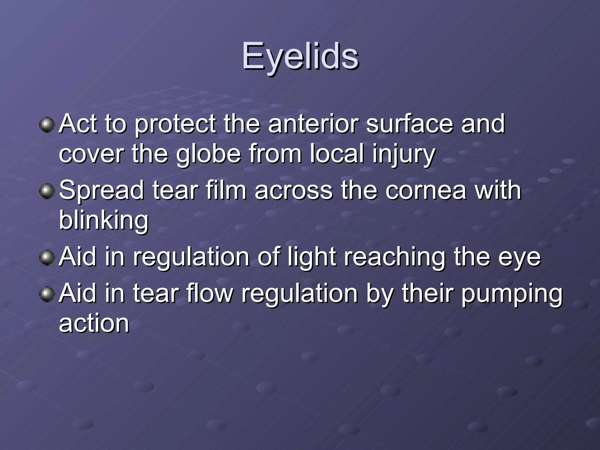
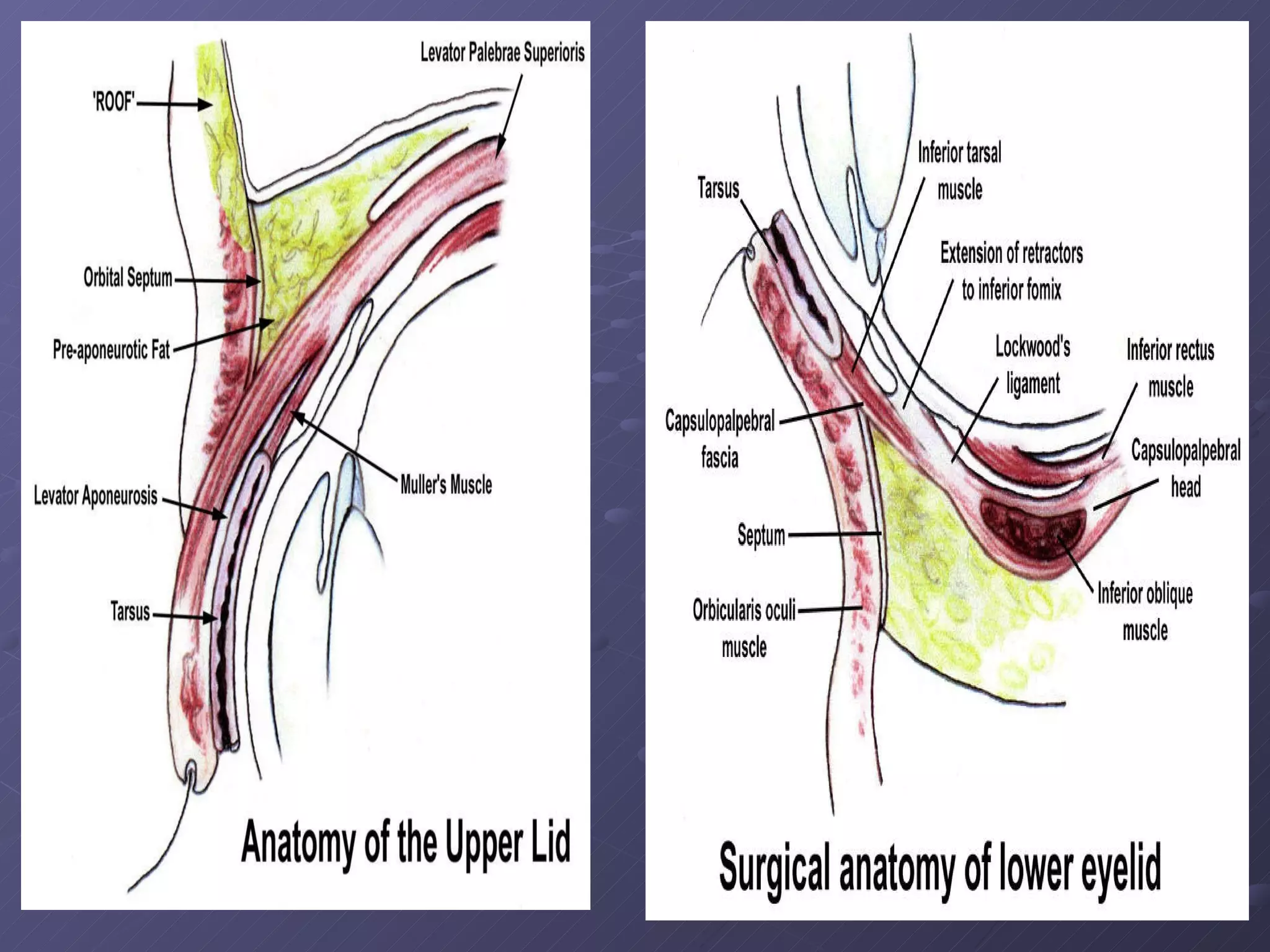
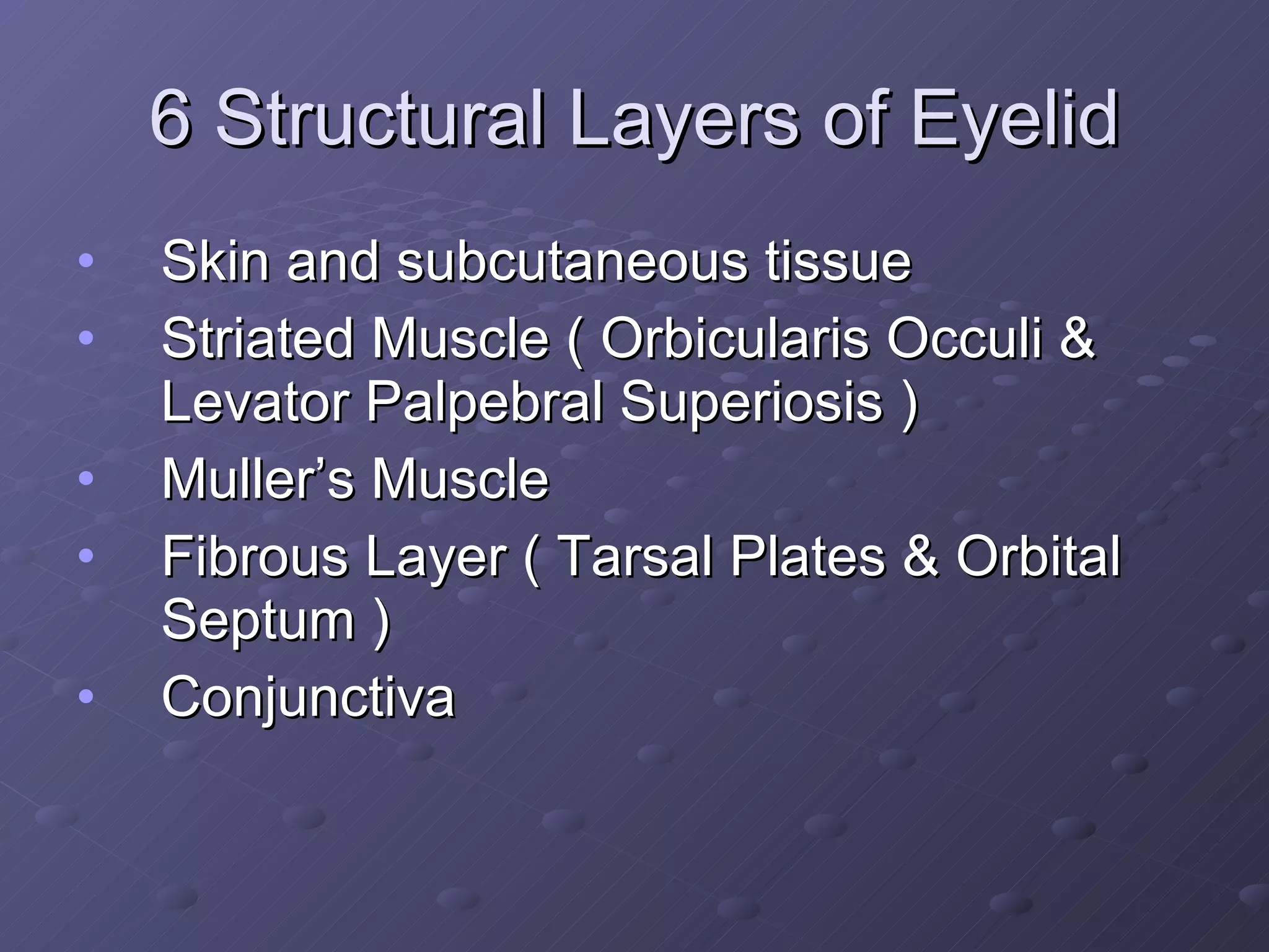
ANATOMY OF E Y E L I D S FEIRUZ HAMID  UMAR BAHASYWEN  (A126670)
Eyelids Act to protect the anterior surface and cover the globe from local injury Spread tear film across the cornea with blinking Aid in regulation of light reaching the eye  Aid in tear flow regulation by their pumping action
 
6 Structural Layers of Eyelid Skin and subcutaneous tissue Striated Muscle ( Orbicularis Occuli & Levator Palpebral Superiosis )  Muller’s Muscle Fibrous Layer ( Tarsal Plates & Orbital Septum ) Conjunctiva
1. Skin and Subcutaneous Tissue  Skin : thinnest of the body 2 layers : Epidermis  - superficial keratinising stratified squamous epithelium - keratonocyte developed from basal cell layer Dermis - located below epidermis - vascular connective tissue - hair follicles, sebum gland and sweat gland Upper eyelid crease : caused by attachment of levator aponeurosis to pretarsal orbicularis bundle and skin
2. Striated Muscle a. Orbiccularis Occuli Superficial muscles of facial expression  Close the eyelids by moving down the upper lid to meet the lower lid and palpebral fissure tighten up from the outer to the inner canthus 300 msec to complete ; 1 blink / 3 sec Forms the thickess of the eyelid The muscle divided into the  orbital  and  palpebral  parts, with the latter being divided further into the  preseptal  and  pretarsal  portions.  Palpebral : blinking, winking ( spontaneous ) Pretarsal : forced closure Both of the palpebral and orbital portion is innervated by facial nerve
cont.. b. Levator Palpebral Superiosis Originates at the inferior aspect of lesser wing of sphenoid bone, just above the optic foramen It is a skeletal muscle that aided by tarsal muscle Maintain the position of the upper eyelid Innervated by oculomotor nerve
the anatomy of of orbicularis occuli muscle A : Frontalis B : Corrugator superciliaris C : Procerus D : Orbital orbicularis E : Preseptal orbicularis F : Pretarsal orbicularis.
3. Muller’s Muscle Originate from the undersurface of the levator in the region of the aponeurotic-muscle junction, and insert into the superior margin of the tarsus.  To assist levator palpebral superiosis and raise the upper lid Innervated by sympathetic nervous system Fatty infiltration may occur, giving the muscle a yellowish color, due to aging
4. Fibrous Layer a. Orbital Septum Fibrous tissue, arises from periosteum of the orbital margin to the edge of tarsal plates Laterally attached to the orbital margin Hollowed by blood vassels, nerves, and aponeurosis of the levator Fuse with : - levator aponeurosis ( to cover  the tarsal plate ) - capsulopalpebral orbital  septum complex Weak and thin with age
cont.. b. Tarsal Plates Dense connective tissue that responsible for the integrity of the lids Skeleton of eyelid Each tarsus approximately 25 - 29 mm long and 1 mm thick Upper plate ~ 10 – 12 mm high Lower eyelid ~ 4 mm high
5. Conjunctiva A clear mucous membrane Non-keratinizing squamous epithelium and goblet cell Posterior layer of the eyelids Goblet cell – producing mucous component of tear film
Other Anatomical Consideration Lid Margin Mucocutaneous junction (grey line) 2mm wide ; 30 mm long Eyelashes One of the hairs that grow at the edge of the eyelid Protect the eye Upper lid : 100 – 150 eyelashes Lower lid : 50 – 75 eyelashes Eyelashes take about 7 – 8 weeks to grow back if pulled out Meibomian Glands Also known as tarsal gland. Originated in tarsus Located at the rim of the eyelids inside the tarsal plate Responsible for the supply of meibum, and oily substance that prevents evaporation of the eye’s tear film. Upper : 25 glands Lower : 20 glands
… Thank You …

Anatomy of Eyelids

  • 1.
    ANATOMY OF EY E L I D S FEIRUZ HAMID UMAR BAHASYWEN (A126670)
  • 2.
    Eyelids Act toprotect the anterior surface and cover the globe from local injury Spread tear film across the cornea with blinking Aid in regulation of light reaching the eye Aid in tear flow regulation by their pumping action
  • 3.
  • 4.
    6 Structural Layersof Eyelid Skin and subcutaneous tissue Striated Muscle ( Orbicularis Occuli & Levator Palpebral Superiosis ) Muller’s Muscle Fibrous Layer ( Tarsal Plates & Orbital Septum ) Conjunctiva
  • 5.
    1. Skin andSubcutaneous Tissue Skin : thinnest of the body 2 layers : Epidermis - superficial keratinising stratified squamous epithelium - keratonocyte developed from basal cell layer Dermis - located below epidermis - vascular connective tissue - hair follicles, sebum gland and sweat gland Upper eyelid crease : caused by attachment of levator aponeurosis to pretarsal orbicularis bundle and skin
  • 6.
    2. Striated Musclea. Orbiccularis Occuli Superficial muscles of facial expression Close the eyelids by moving down the upper lid to meet the lower lid and palpebral fissure tighten up from the outer to the inner canthus 300 msec to complete ; 1 blink / 3 sec Forms the thickess of the eyelid The muscle divided into the orbital and palpebral parts, with the latter being divided further into the preseptal and pretarsal portions. Palpebral : blinking, winking ( spontaneous ) Pretarsal : forced closure Both of the palpebral and orbital portion is innervated by facial nerve
  • 7.
    cont.. b. LevatorPalpebral Superiosis Originates at the inferior aspect of lesser wing of sphenoid bone, just above the optic foramen It is a skeletal muscle that aided by tarsal muscle Maintain the position of the upper eyelid Innervated by oculomotor nerve
  • 8.
    the anatomy ofof orbicularis occuli muscle A : Frontalis B : Corrugator superciliaris C : Procerus D : Orbital orbicularis E : Preseptal orbicularis F : Pretarsal orbicularis.
  • 9.
    3. Muller’s MuscleOriginate from the undersurface of the levator in the region of the aponeurotic-muscle junction, and insert into the superior margin of the tarsus. To assist levator palpebral superiosis and raise the upper lid Innervated by sympathetic nervous system Fatty infiltration may occur, giving the muscle a yellowish color, due to aging
  • 10.
    4. Fibrous Layera. Orbital Septum Fibrous tissue, arises from periosteum of the orbital margin to the edge of tarsal plates Laterally attached to the orbital margin Hollowed by blood vassels, nerves, and aponeurosis of the levator Fuse with : - levator aponeurosis ( to cover the tarsal plate ) - capsulopalpebral orbital septum complex Weak and thin with age
  • 11.
    cont.. b. TarsalPlates Dense connective tissue that responsible for the integrity of the lids Skeleton of eyelid Each tarsus approximately 25 - 29 mm long and 1 mm thick Upper plate ~ 10 – 12 mm high Lower eyelid ~ 4 mm high
  • 12.
    5. Conjunctiva Aclear mucous membrane Non-keratinizing squamous epithelium and goblet cell Posterior layer of the eyelids Goblet cell – producing mucous component of tear film
  • 13.
    Other Anatomical ConsiderationLid Margin Mucocutaneous junction (grey line) 2mm wide ; 30 mm long Eyelashes One of the hairs that grow at the edge of the eyelid Protect the eye Upper lid : 100 – 150 eyelashes Lower lid : 50 – 75 eyelashes Eyelashes take about 7 – 8 weeks to grow back if pulled out Meibomian Glands Also known as tarsal gland. Originated in tarsus Located at the rim of the eyelids inside the tarsal plate Responsible for the supply of meibum, and oily substance that prevents evaporation of the eye’s tear film. Upper : 25 glands Lower : 20 glands
  • 14.