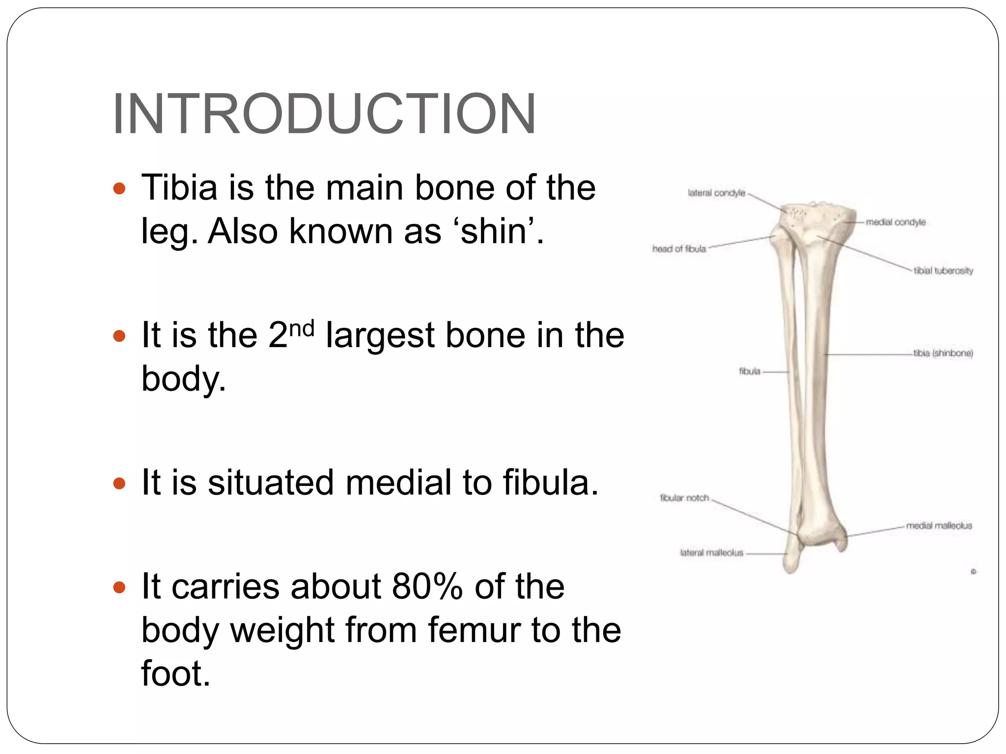
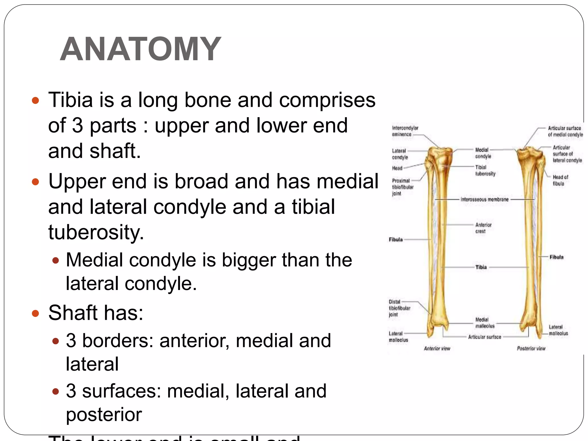
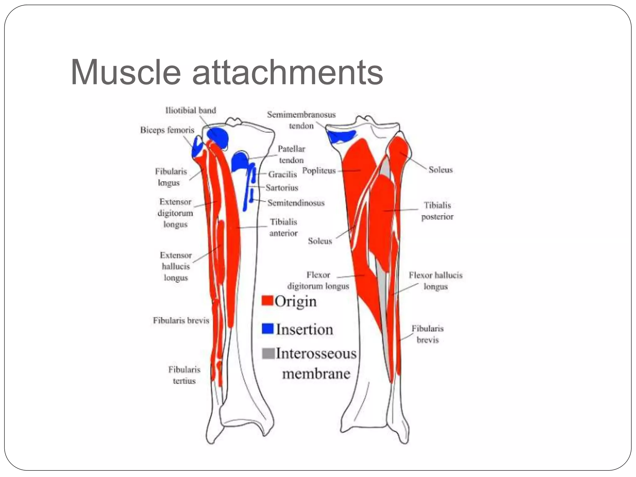
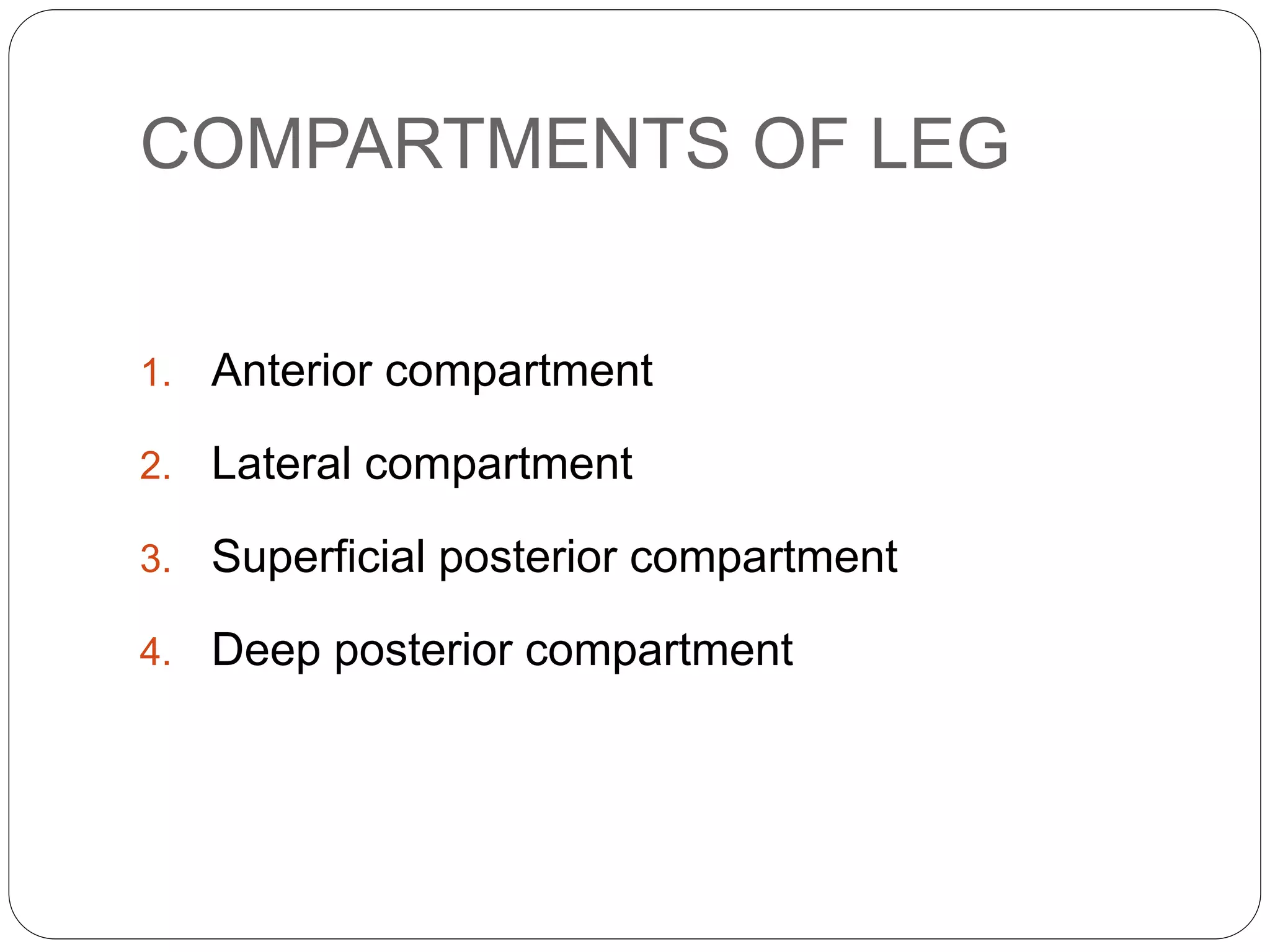
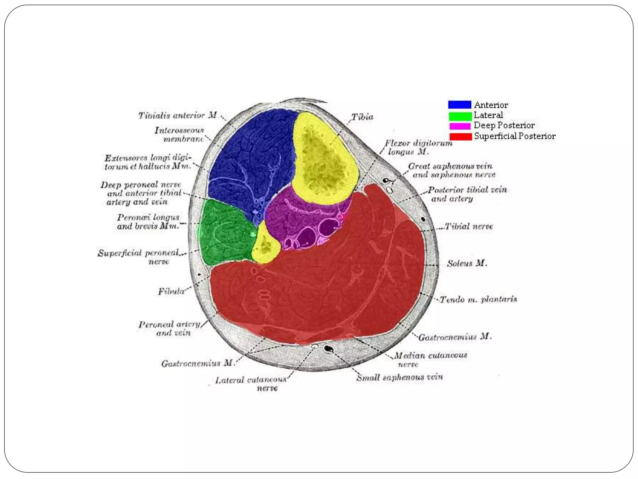
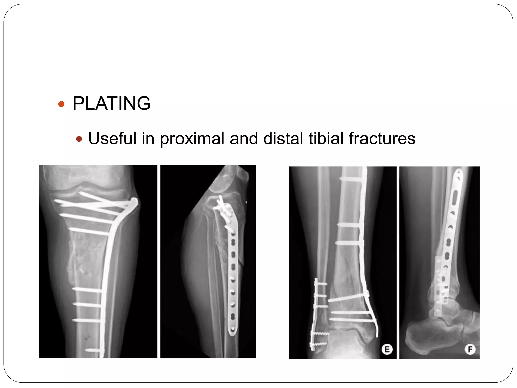
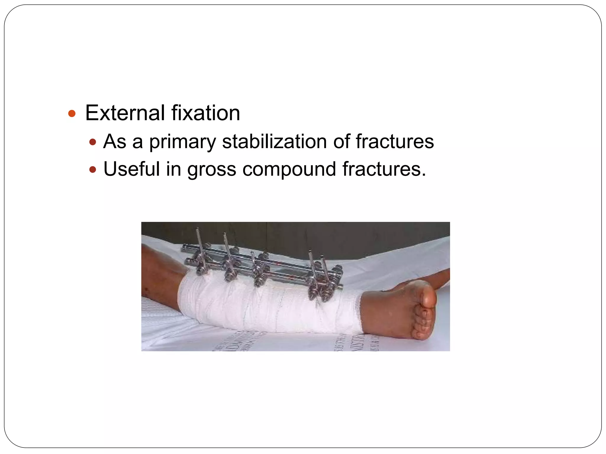
Call Girls In Andheri East Call 9920874524 Book Hot And Sexy Girls
Fracture shaft of tibia
1. BY: DR. BIPUL BORTHAKUR,
PROFESSOR,
DEPT. OF ORTHOPAEDICS, SMCH
FRACTURE SHAFT OF TIBIA
2. INTRODUCTION
Tibia is the main bone of the
leg. Also known as ‘shin’.
It is the 2nd largest bone in the
body.
It is situated medial to fibula.
It carries about 80% of the
body weight from femur to the
foot.
3. ANATOMY
Tibia is a long bone and comprises
of 3 parts : upper and lower end
and shaft.
Upper end is broad and has medial
and lateral condyle and a tibial
tuberosity.
Medial condyle is bigger than the
lateral condyle.
Shaft has:
3 borders: anterior, medial and
lateral
3 surfaces: medial, lateral and
posterior
4. ANATOMICAL POSITION AND
SIDE DETERMINATION
It is hold in a manner that:
Its expanded end with condyles faces
upward.
Its tibial tuberosity and sharp anterior
border faces anteriorly.
Medial malleolus is on the medial side.
The superior surface (tibial plateau) is
placed horizontal.
9. FRACTURE SHAFT OF TIBIA
Most common long bone fracture.
Accounts for about 4% of all fractures.
It is usually associated with fibula fracture.
10. MECHANISM OF FRACTURE
1. Low energy pattern
Result of torsion injury
Indirect or stress fracture
2. High energy pattern
Motor vehicle crash
Gunshot injury
Associated with fibula fracture
11. Associated conditions
Soft tissue injury (in open wounds)
Compartment syndrome
Bone loss
Ipsilateral fibula fracture
Ligamentous injury
Ipsilateral femur fracture resulting in “floating
knee”
13. PRESENTATION
Pain and swelling
Inability to walk
Deformity
Contusion/ wound
Abnormal mobility
Bony crepitus
14. INVESTIGATION
Radiographs
Full length AP and Lateral views of the affected leg
AP and Lateral views of the ipsilateral knee and
ankle
CT scan
To rule out any intraarticular fracture extension
15. MANAGEMENT
Resuscitation of the patient as per ATLS
guidelines
Airway
Breathing
Circulation
Disability
Exposure
Volume stabilization with IV fluids or blood
transfusion
Proper immobilization of the limb with Above-
knee splint.
16. TREATMENT
NON-OPERATIVE
Closed reduction/cast
immobilization
Indications
Closed low energy fractures and
undisplaced or acceptable
alignment
<5 degree valgus-varus
angulation
<10 degree antero-posterior
angulation
17. Technique
Long leg cast and convert to functional patellar
tendon bearing cast after 4 weeks
Complication:
Malunion
Non union
Risk of compartment syndrome
21. External fixation
As a primary stabilization of fractures
Useful in gross compound fractures.
22. COMPARTMENT SYNDROME
Compartment syndrome occurs when excessive
pressure builds up inside an enclosed muscle space
in the body.
Compartment syndrome usually results from bleeding
or swelling after an injury.
The dangerously high pressure in compartment
syndrome impedes the flow of blood to and from the
affected tissues.
It can be an emergency, requiring surgery to prevent
permanent injury.