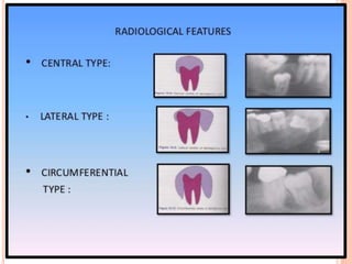
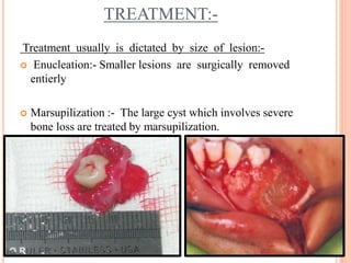
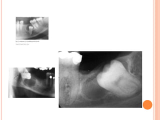
PRESENTED BY :-
HIMANI THAWALE
(INTERN)
DENTIGEROUS
CYST
DEFINATION:-
 Dentigerous cyst can be defined as an odantogenic
cyst that surrounds the crown of an impacted
tooth; caused by fluid accumulation between the
reduced enamel epithelium and the enamel surface
resulting in the cyst in which the crown is located
within the lumen.
PREVALANCE:-
 This is one of the most common type of
developmental odontogenic cyst - 20% of all jaw cyst.
 10% of impacted teeth have formed a dentigerous
cyst.
 Frequency in the general population has been
estimated at 1.44 cyst for every 100 unerupted teeth.
CLINICAL FEATURES:
 Age:- second and third decade of life.
 Sex:- male prediliction
 Most common site:- 1) mandibular third molar
2) maxillary third molar
3) maxillary cuspid areas
(since these are most commonly impacted teeth)
 Most dentigerous cyst are solitary.
 Bilateral and multiple cyst are usually found in
association with number of syndromes including:-
- Cleidocranial dysplasia
- Maroteaux- lamy syndrome.
 Dentigerous cyst is capable of becoming an aggressive
lesion.
 Cystic involvement of an unerupted mandibular third
molar may result in “hollowing out” of entire ramus
 In case cyst is associated with a maxillary cuspid ,
expansion of anterior maxilla often occurs & may
superficially resemble an acute sinusitis or cellulitis.
 No pain or discomfort is associated with the cyst unless
it become secondarily infected.
RADIOGRAPHIC FEATURES:-
 Three radiological variations of dentigerous cyst are seen
radiographically:-
 Central type:-
In this type the crown is enveloped symmetrically.
 Lateral type:-
This type of radiographic appearance results from
dilation of follicles on one aspect of crown.
 Circumferential type:-
This type results when the follicle expands in a manner in
which the entire tooth appears to be involved by the cyst
HISTOLOGIC FEATURES:-
 It is usually composed of thin connective tissue wall with a
thin layer of stratified squamous epithelium lining the lumen.
 Rete peg formation generally absent unless it is secondarily
infected.
 Inflammatory cells and infiltration of connective tissue is
common.
 Presence of rushton bodies within the lining epitehlium is
usually seen with the cyst exhibiting inflammation.
 The content of cystic lumen is thin, watery yellow fluid
,ocasionaaly blood tinged
TREATMENT:-
Treatment usually is dictated by size of lesion:-
 Enucleation:- Smaller lesions are surgically removed
entierly
 Marsupilization :- The large cyst which involves severe
bone loss are treated by marsupilization.
COMPLICATIONS:-
 Ameloblastoma
 Squamous cell carcinoma
 Mucoepidermoid carcinoma
Dentigerous cyst
Dentigerous cyst

Dentigerous cyst

  • 1.
    PRESENTED BY :- HIMANITHAWALE (INTERN) DENTIGEROUS CYST
  • 2.
    DEFINATION:-  Dentigerous cystcan be defined as an odantogenic cyst that surrounds the crown of an impacted tooth; caused by fluid accumulation between the reduced enamel epithelium and the enamel surface resulting in the cyst in which the crown is located within the lumen.
  • 3.
    PREVALANCE:-  This isone of the most common type of developmental odontogenic cyst - 20% of all jaw cyst.  10% of impacted teeth have formed a dentigerous cyst.  Frequency in the general population has been estimated at 1.44 cyst for every 100 unerupted teeth.
  • 4.
    CLINICAL FEATURES:  Age:-second and third decade of life.  Sex:- male prediliction  Most common site:- 1) mandibular third molar 2) maxillary third molar 3) maxillary cuspid areas (since these are most commonly impacted teeth)  Most dentigerous cyst are solitary.
  • 5.
     Bilateral andmultiple cyst are usually found in association with number of syndromes including:- - Cleidocranial dysplasia - Maroteaux- lamy syndrome.  Dentigerous cyst is capable of becoming an aggressive lesion.  Cystic involvement of an unerupted mandibular third molar may result in “hollowing out” of entire ramus  In case cyst is associated with a maxillary cuspid , expansion of anterior maxilla often occurs & may superficially resemble an acute sinusitis or cellulitis.  No pain or discomfort is associated with the cyst unless it become secondarily infected.
  • 6.
    RADIOGRAPHIC FEATURES:-  Threeradiological variations of dentigerous cyst are seen radiographically:-  Central type:- In this type the crown is enveloped symmetrically.  Lateral type:- This type of radiographic appearance results from dilation of follicles on one aspect of crown.  Circumferential type:- This type results when the follicle expands in a manner in which the entire tooth appears to be involved by the cyst
  • 8.
    HISTOLOGIC FEATURES:-  Itis usually composed of thin connective tissue wall with a thin layer of stratified squamous epithelium lining the lumen.  Rete peg formation generally absent unless it is secondarily infected.  Inflammatory cells and infiltration of connective tissue is common.  Presence of rushton bodies within the lining epitehlium is usually seen with the cyst exhibiting inflammation.  The content of cystic lumen is thin, watery yellow fluid ,ocasionaaly blood tinged
  • 9.
    TREATMENT:- Treatment usually isdictated by size of lesion:-  Enucleation:- Smaller lesions are surgically removed entierly  Marsupilization :- The large cyst which involves severe bone loss are treated by marsupilization.
  • 10.
    COMPLICATIONS:-  Ameloblastoma  Squamouscell carcinoma  Mucoepidermoid carcinoma