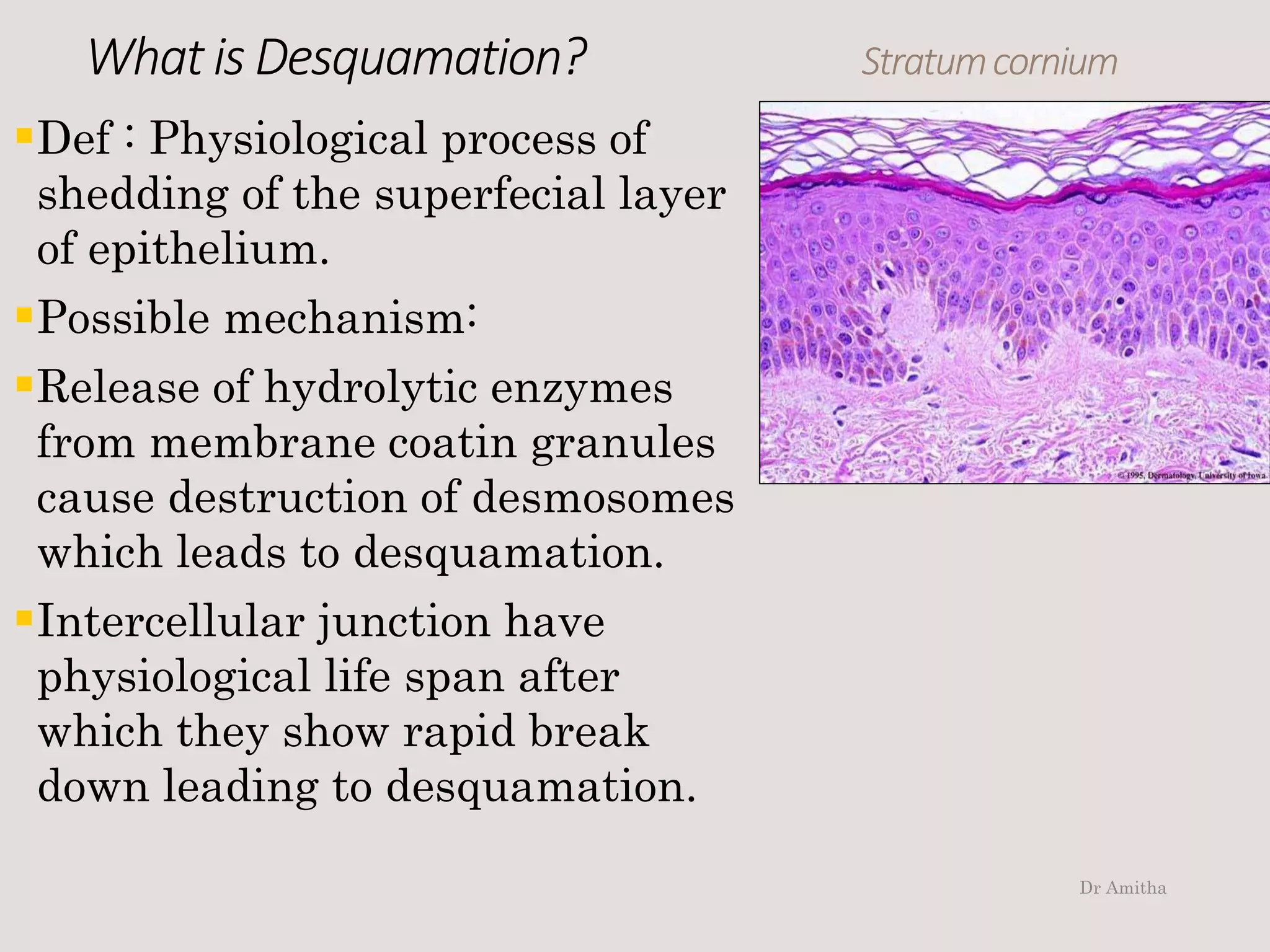
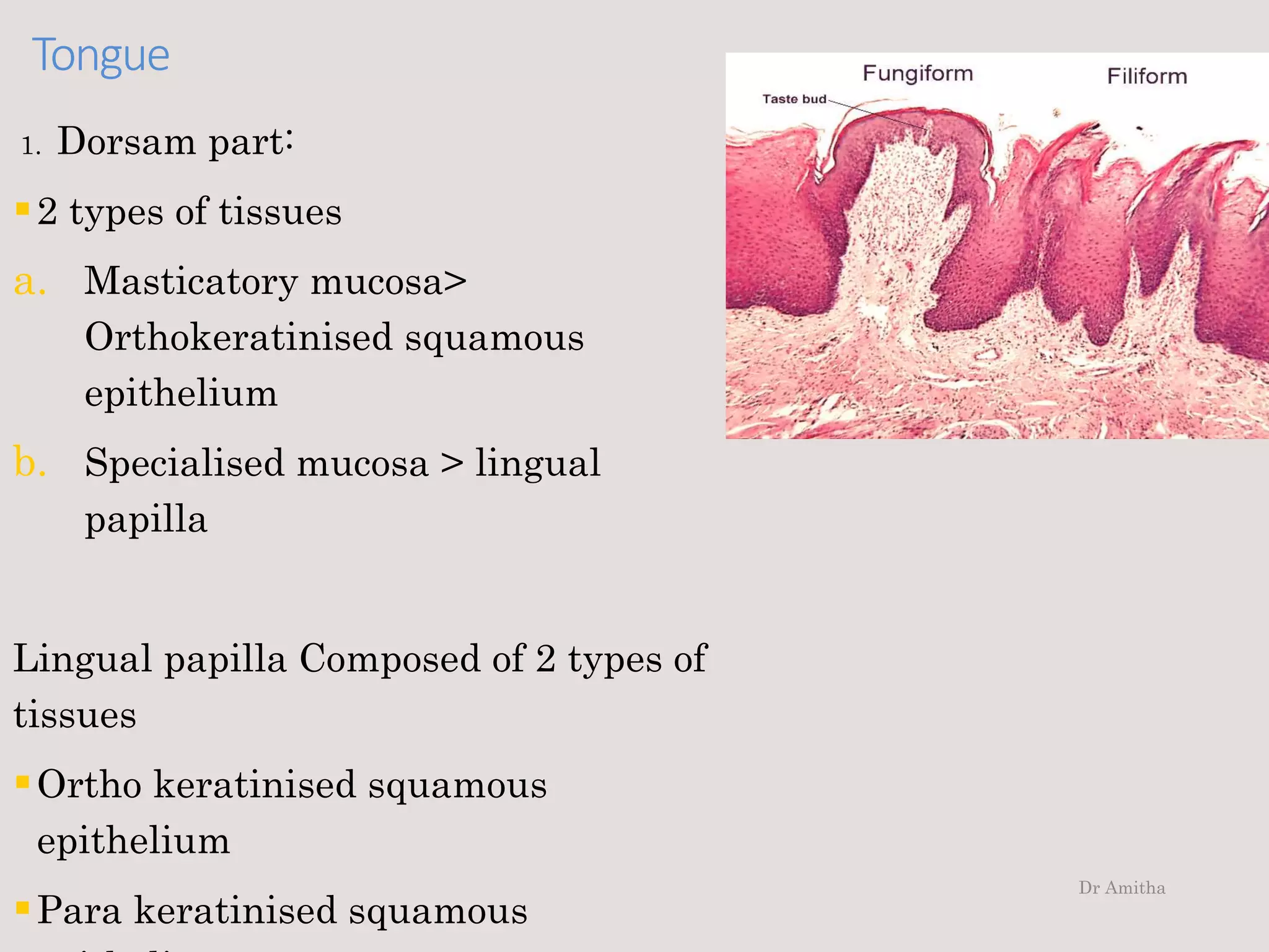
Oral Mucous Membrane
Dr Amitha G, BDS, MDS
Dept of oral and maxillofacial pathology
Dr Amitha
Types Of Oral Mucosa :
Dr Amitha
Oralmucosa
A.
Lining
Mucosa
Buccal, labial,
alveolar
mucosa.
Ventral part
of the tongue
Floor of the
mouth
Soft palate
Non
keratinised
squamous
Epithelium
B.
Masticatory
Mucosa
Attached
gingiva
Hard palate
Dorsam of
the tongue
Ortho
Keratinis
ed
squamous
epthelium
Para
keratinise
d
epitheliu
m
C.
Specialised
Mucosa
Dorsam of the
tongue
Lateral
border of the
tongue
Keratin
ised
mucosa
Dr Amitha
In oral mucosa
2 main patterns: keratinisation and non keratinisation.
Kertinisation: mucosal surface result from formation of a
surface layer of keratin and process of maturation is
called kertinisation
Its tough, resistant to abrasion and tightly bound to
lamina propria.
Shows 4 layers:
1. Stratum basele
2. Stratum spinosam
3. Stratum granulosam
4. Stratum corneum
Dr Amitha
Cells in detail:
1. Basallayer:
Its called proliferative/
germinative layer.
Single layered of cuboidal/
coloumnar cells
Rest on basement
membrane
 this cells have capability
to to undergo mitotic
division
Dr Amitha
Basallayer
Cells have: basophilic cytoplasam
Centrally placed large hyper
chromatic nucleus
Nucleus occupies 1/3rd of the cells
with evenly distributed chromatin
and 2-3 nucleoli.
Nucleus is arranged perpendicular
to basement membrane
It’s a least differenteated cells of
epithelium.
Cells are rich in RER, mitocondria,
golgi complex, lysosomes - involved
in protein syntesis. Dr Amitha
It has 2 groupof cells Basallayer
1st group cells are serrated with
protoplasmic processes at basal
region
Heavily packed with
tonofilaments
2nd group cells are stem cells
which undergo division and
provide cells for maturing
compartment.
Cells are attached to each other
by desmosomes, Basement by
hemidesmosomes.
It contain few tonofilaments
Dr Amitha
2. StratumSpinosum:
Seen above the basal layer
Several rows of polyhedral
cells
Cells pass from basal to
prickle cell layer it will
descrease in basophelia
which makes border distinct.
Cells larger than basal cells
Centrally placed nucleus
Nuclear cytoplasmic ration is
1:6
Dr Amitha
StratumSpinosum:
Why its called Pickle cells?
Pickle cells due to
histological section
Cells have spiny/ prickly
apperance.
During staining cells shrink
remaining contact only in
intercellular atatchment.
Showing spiny/ prickly
apperance.
Dr Amitha
StratumSpinosum:Nucleus has evenly distributed
chromatin with 2-3 nucleoli
Cytoplasm is rich in
oraganelles for protein
synthesis
Concentration of
tonafilaments increases to
form bundles
Cells attach to each other by
desmosomes
Number of desmosomes and
intra cellular space is more Dr Amitha
StratumSpinosum:
Size of desmosomes is wider in prickle cell layer
than in basal cell layer as it moves to upper
layer desmosomes becomes smaller.
As cells mature and move superfecially they
increase in size and becomes more flattened
with flattened nucleus.
Cells of upper part of prickle cell layer show
new cytoplasmic organelles called odland
bodies.
Dr Amitha
What is Odland bodies ?
They are also known as: Membrane coating granules
Cytoplasmic lamellated bodies
Keratinosomes
Microgranules
Cementosomes
In keratinised epethelium odland bodies apper a ovoid membrane
bound oraganelle
Length: 0.25 microns
It has series of internal lamellae with alternating electron lucent
and dense bands.
Derived from golgibodies
Size donot increase but density increases as it moves to
superficial layer.
Stratum
Spinosum
Dr Amitha
3. Stratumgranulosam
It composed of few layers of
flattened cells
Seen above the stratum
spinosam
Cytoplasam filled with
basophilic granules called
keratohyline granules
hence the name.
Nucleus – flattened with
long axis parallel to outer
surface of epithelium.
Dr Amitha
StratumgranulosamGranular layer cells :
Size of the cells increases
Cells are flatter with long axis parallel to
epithelial surface
Nucleus is flattened shows pyknotic changes.
Cells retain capasity of protein syntesis and
decrease in cytoplasm oragnelle
More of tonofilaments
Odland bodies fuse to superfecial cell
membrane and discharge content into
intercellular space
It has lipid rich permeability barrier that
limits movements of substance through
intercellular space.
Dr Amitha
Stratumgranulosam
Desmosomes maintain there structure in layer but
intercellular contact layer of desmosomes becomes more
condensed
Cytoplasm shows keratohyaline granules
As they moves through granular layer granules are
angular/ irregular
Synthesized by ribosomes
Contain sulphur rich protein fillagrin and loricrin which
provide embedding matrix for tonafilaments
It contain protein involucrin provide consituents cell
membrane thickening and makes it resistent to
chemical solvents.
Dr Amitha
4. Stratumcorneum:
Superficial layer found in the
keratinized epithelium
Composed of keratin squames
which is larger and flatter
than cells of Stratum
granulosum
In histologic section:
layer appear eosinophillic
amorphous
As cells reach cornified layer
nucleus undergoes Dr Amitha
Stratumcornium
If nucleus is absent in
superficial layer, This pattern
maturation is called
orthokertinization
If pyknotic nucleus is retained in
all or some some squamous its
called parakeratization. Eg :
Gingiva
It’s a karatohyaline granules in
stratum granulosum is less
prominent
Dr Amitha
Stratumcornium
This layer composed of cells resembling hexogonal discs
called squames
It contain keratin tonofilaments, embadded in matrix
as an envelop
Keratin fills interior of shrunken cells
Cellular oragnelles are lost
Nucleus may remain picknotic or completely lost
Cell membrane thickened
Desmosomes can be recognised but it becomes less
distinct
Cells moves to superfecial layer desmosomes tend to
degenarate resulting in desquamation of cells
Dr Amitha
What is Desquamation? Stratumcornium
Def : Physiological process of
shedding of the superfecial layer
of epithelium.
Possible mechanism:
Release of hydrolytic enzymes
from membrane coatin granules
cause destruction of desmosomes
which leads to desquamation.
Intercellular junction have
physiological life span after
which they show rapid break
down leading to desquamation.
Dr Amitha
Non keratinizedepithelium
There are 3 layers
1. Stratum basale
2. Stratum intermedium
3. Stratum superficiale
* Stratum cornum (Absent )
•Straum basele and straum intermedium is same as
keratinised epithelium.
•Difference is only seen in stratum superficiale.
Dr Amitha
Stratumsuperficiale:
Most superfecial layer
Increase in size of cells
Composed of few layer of flattened cells
Nucleus is flattened with long axis parallel to outer
surface of epithelium.
Has pyknotic nucleus
Dr Amitha
Stratumsuperficiale
Cytoplasmic organelles decrease in number
Lesser capacity to produce protein
Increase In tonofilaments in unbundled form.
Cells surface become regular and closely
approximated with each other
Desmosomes decrease in size and number
Intercellular space become wider and irregular
Intercellular contact layer of desmosomes
becomes more condensed.
Dr Amitha
Stratumsuperficiale
Superficial layer shows nucleus and cytoplasmic
organellae
This cells ultimately undergoes desquamation.
Dr Amitha
Non Kertinised vs Kertinised
Dr Amitha
Liningmucosa
It contains non keratinised epithelium
Includes:
Lining mucosa of cheek
Alveolar mucosa
Vestibular mucosa
Vestibule
Floor of the mouth
Ventral aspect of tongue
Soft palate
Dr Amitha
Liningmucosa–NonKeratinised
Labial/Buccal mucosa > Thick non keratinised
Alveolar mucosa > Thin non keratinised
Ventral tongue > Thin non kertinised
Soft palate > Thin non keratinised
Masicatory mucosa- ( ortho/ para keratinised)
Hard palate >Thick ortho keratinised
Attached mucosa > Thick keratinised
Ortho keratinised (or)
Para keratinised
Dr Amitha
Tongue
It composed of 2 region:
Base
Body
Dr Amitha
Tongue
1. Dorsam part:
2 types of tissues
a. Masticatory mucosa>
Orthokeratinised squamous
epithelium
b. Specialised mucosa > lingual
papilla
Lingual papilla Composed of 2 types of
tissues
Ortho keratinised squamous
epithelium
Para keratinised squamous
Dr Amitha
Lingual papilla
Dr Amitha
tongue
2. Ventral part:
Non kertinised squamous epithelium
Dr Amitha
Thereare4typesoflingualpapilla Tongue
1. Filliform papilla>
Most comman
Dr Amitha
Tongue
2. Fungiform papilla> Mushroom
like ( red dots)
Seen on dorsum surface of
tongue
Dr Amitha
Tongue
3. Folliate papilla >
Vertical Ridges
Seen on lateral tongue
Dr Amitha
tongue
4. Circumvellate papilla >
Located on sulcus terminalis
 Divides anterior and
posterior part of tongue
 Here we can see salivary
glands called Von ebners
salivary gland
 Produce serous saliva
Dr Amitha
TongueTongue have sensory function:
Sweet, Salty, Sour, Bitter
Taste buds are present in
1. funfiform
2. folliate
3. circumvellate
Dr Amitha
Tastebuds Tongue
Have Taste cells
Supporting cells
Taste pores
Sensory nerve > Cns
Dr Amitha
Epithelium of lip and cheek
Non Keratinised mucosa
Epithelial ridges are seen btw epithelium and connective
tissue
Lamina propria is thick
Less dense collagen fibers
It has distinct submucosa that contain mixed salivary
gland, fat cells etc
Mucosa is stratchable
Underlying musculature is well adapted for contraction
and relaxation.
Dr Amitha
Epithelium of lip and cheek
Ventral band of collagen fibers with elastic fibers
are found extending from lamina propria to fascia
covering the underlying muscle
This provides attachments btw mucosa and
muscle.
Thus folding of mucosa is prevented while
relaxation and avoid mucosa being caught
between teeth
They are tightly bound to underlyning structure.
Dr Amitha
Dr Amitha
Vestibular mucosa andAlveolar mucosa
Vestibular mucosa
lines vestibule its ‘v’ shaped
Separates alveolar mucosa from lip and cheek.
Vestibular mucosa continues with alveolar mucosa
which lines alveolar bone
Alveolar mucosa apper reddish and extend upto
mucogingival junction which seperates it from gingival
mucosa.
Dr Amitha
Vestibularmucosa
Loosely attached to underlyning structure.
They permit easy movement of lip and cheek.
Median and lateral labial frenum are seen as folds of
mucous membrane containing loose connective tissue.
Dr Amitha
Alveolar mucosa
Thin non keratinised epithlium
Epithelial connective tissue junction is flat with small
rete ridges or connective tissue may be absent some
times.
AM is loosely attached to underlying bone by loose
connective tissue
Contain minor salivary gland.
Dr Amitha
Floor of the mouth and ventralsurfaceof tongue
Floor of the mouth
Horseshoe shaped beneath tongue
Mucosa sharing floor of the mouth and ventral tongue
share many comman features
Mucosa is thin with non keratinised epithelium.
Epithelial rete ridges and connective tissues papillae
are short.
Connective tissue shows rich blood supply.
Submucosa contain adipose tissue and minor salivary
gland
Dr Amitha
Verticalaspectof tongue
Submucosa is very thin or even absent wher mucosa
will tightly bound to underlyining musculature.
This thin epithelium lining and rich blood supply
permit that rapid absorption of medicine administered
sublingually.
Dr Amitha
Vermilion borderof lip
Its transitional zone
Its btw skin covering external
surface of lip and labial mucosa
lining inner aspect.
Skin- Keratinised Stratified
Squamous Epithelium
with hair follicles, sweat
gland, sebaceous gland.
Dr Amitha
Labialmucosa
Labial mucosa- Non Keratinised Stratified Squamous
Epithelium
Connective tissue beneath labial mucosa shows minor
salivary glands.
Center most region of lip shows orbicularis oris muscle
Dr Amitha
Labialmucosa
Transitional zone has thin lining epithelium with mild
keratinisation on surface.
There are long connective tissue papillae reaching high
into epithelium carrying capillary loops.
Its more reddish compared to labial mucosa.
Underlying connective tissue is a characteristic devoid
which causes mucosa to dry up.
Dr Amitha
Gingiva :
Its parakeratinised stratified squamous epithelium
and connective tissue.
Covering epithelium showa charectaristic variation
in different region.
Its categorized as:
Epithelium covering
1. oral region of gingiva.
2. Sulcular epithelium
3. Junctional epitehlium
Dr Amitha
OralEpithelium
Epithelium lining oral region
Keratinised or Parakeratinised Stratified
Squamous Epithelium.
Gingival epithelium is parakeratinised in
75% population.
Stipplings are seen as heavy epithelial ridges
which reprasents free gingival grooves and
depression correspond to center of epithelial
ridges.
Dr Amitha
OralepitheliumEpithelial connective tissue
interface is irregular with
numerous long narrow ridges
interdigitating with long
connective tissue papillae.
Extensive digitation increases
with strength to withstand
masticatory stress.
Long branching rete ridges
helps in identification of
gingiva from other part of oral
mucosa.
Dr Amitha
SulcularEpithelium
It lines gingival sulcus
Extends from coronal limit of junctional
epithelium to crest of gingival margin.
Thin layer of Non Kertinised Epithelium.
Junction btw epithelium and connective
tissue is flat without rete ridges.
Lack of keratinisation is due to inflammation
of connective tissue.
Dr Amitha
JunctionalEpithelium
Part of gingival epithelium that attached to
cervical part of tooth forming junction btw
tooth and gingiva.
Stratified Squamous Epithelium
Apper as triangular strip with 15-30 layer
thickness at cervical portion
3-4 cell layer thickness at apical margin
It consist of flat cells which arranged parallel
to tooth surface.
Dr Amitha
JunctionalEpithelium
Cells have less desmosomal junctions and more
intercellualr spaces.
This helps in migration of of polymorpho nuclear
leucocytes(PNML’s) into epithelium and sulcus.
Epithelium and connective tissue interface is flat.
Most important feature is:
Presence of basal lamina on both sides
i.e at the junction of epithelium and connective
tissue
And surface adjasent to the tooth. Dr Amitha
JunctionalEpithelium
Basal lamina on the surface attached to tooth by
hemidesmosomes.
Juctional epithelium shows high turmover rate
Cells from basal layer migrates 2-3 layer of
junctional epithelium and join migratory root in
coroanl direction and finally exfoliate at gingival
sulcus.
Dr Amitha
Gingivalconnectivetissue
Connective tissue beneath gingival epithelium is
lamina propria with papillary and reticular layer.
Connective tissue consist of dense collagenous
tissue arranged in bundles of fibers which helps
in maintaining the integrity of supporting
apparatus of tooth.
Called gingival fibers of periodontal ligament.
It consist of collagen, oxytalan, elastic fibers.
Includes:
Dento-gingival fibers, dento-periosteal fibers,
alveologingival fibers, circular fibers. Dr Amitha
Palate
Forms roof of the oral
cavity
Divides into Immovable
hard palate anteriorly
Has hard bony support
Movable soft palate
posteriorly
Has only fibrous tissue
Dr Amitha
Hardpalate
Keratinised stratified squamous mucosa
Its has 4 different layers
Stratum basale, stratum spinosum, stratum
granulosam, stratum corneum.
It has functional adaptaion to bear masticatory stress.
Dr Amitha
Hardpalate
Cells show more tonofilaments
Increased number and length of desmosomes.
Epithelium and connective tissue interface is
irregular with long regular epithelial ridges with
interdigitating with connective tissue papillae.
Lamina propria is dense throughout hard palate
Thicker in anterior than in posterior region.
Rugae : connective tissue core is dense with
interwoven collagen fiber
Incisive or palatine papilla contain dense connective
tissue.
Dr Amitha
Hardpalate
Remnants of nasopaltine ducts which is lined by
pseudo stratified squamous epithelium.
Small hyline ducts are seen around ducts opening.
Dr Amitha
Hardpalate
Structure of sub mucosa varies in different
region of palate.
Sub mucosa is absent in peripheral zone of
palate adjusent to teeth.
i.e gingival zone and in mid palatine raphae.
Lamina propria is tightly bound to periosteum
of bone ( mucoperiosteal attachment)
Dr Amitha
Hardpalate
In between gingival and mid palatine raphae,
palate has distint submucosa.
Submucosa is Thicker in posterior
Thinner in anterior region.
Anterior region is filled with adipose tissue
Posterior region with mucous gland.
 Anteriolateral part is referred to as fatty zone
Posterolateral part as glandular zone.
Dr Amitha
Hardpalate
Mucosa is tightly fixed to underlying bone and its
immobile.
It has dense vertical band of connective tissue which
attaches mucosa firmly to periosteum of palatal bone.
Dense band of connective tissue are at right angle to
surface and divide submucosa into compartments.
Dr Amitha
Hardpalate
Wedge shaped area where alveolar process joins
horizontal plate of hard palate contain loose connective
tissue which has large vessels and nerves.
Thickness of loose connective tissue gradually increases
from anterior region of palate to posterior region.
Dr Amitha
Soft palate
Lined by Non Keratinised
stratified Epithelium.
It shows few taste buds
Lamina propria is highly
vascular (SP apper red)
Dr Amitha
Softpalate
Epithilium and connective
tissue interface with thick
and short rete ridges and
connective tissue papillae.
Elastic fibers are found btw
lamina propria and
submucosa.
Submucosa composed of loose
connective tissue which
contain minor salivary gland.
Dr Amitha
Thank you
Dr Amitha

Oral Mucous Membrane

  • 1.
    Oral Mucous Membrane DrAmitha G, BDS, MDS Dept of oral and maxillofacial pathology Dr Amitha
  • 2.
    Types Of OralMucosa : Dr Amitha
  • 3.
    Oralmucosa A. Lining Mucosa Buccal, labial, alveolar mucosa. Ventral part ofthe tongue Floor of the mouth Soft palate Non keratinised squamous Epithelium B. Masticatory Mucosa Attached gingiva Hard palate Dorsam of the tongue Ortho Keratinis ed squamous epthelium Para keratinise d epitheliu m C. Specialised Mucosa Dorsam of the tongue Lateral border of the tongue Keratin ised mucosa Dr Amitha
  • 4.
    In oral mucosa 2main patterns: keratinisation and non keratinisation. Kertinisation: mucosal surface result from formation of a surface layer of keratin and process of maturation is called kertinisation Its tough, resistant to abrasion and tightly bound to lamina propria. Shows 4 layers: 1. Stratum basele 2. Stratum spinosam 3. Stratum granulosam 4. Stratum corneum Dr Amitha
  • 5.
    Cells in detail: 1.Basallayer: Its called proliferative/ germinative layer. Single layered of cuboidal/ coloumnar cells Rest on basement membrane  this cells have capability to to undergo mitotic division Dr Amitha
  • 6.
    Basallayer Cells have: basophiliccytoplasam Centrally placed large hyper chromatic nucleus Nucleus occupies 1/3rd of the cells with evenly distributed chromatin and 2-3 nucleoli. Nucleus is arranged perpendicular to basement membrane It’s a least differenteated cells of epithelium. Cells are rich in RER, mitocondria, golgi complex, lysosomes - involved in protein syntesis. Dr Amitha
  • 7.
    It has 2groupof cells Basallayer 1st group cells are serrated with protoplasmic processes at basal region Heavily packed with tonofilaments 2nd group cells are stem cells which undergo division and provide cells for maturing compartment. Cells are attached to each other by desmosomes, Basement by hemidesmosomes. It contain few tonofilaments Dr Amitha
  • 8.
    2. StratumSpinosum: Seen abovethe basal layer Several rows of polyhedral cells Cells pass from basal to prickle cell layer it will descrease in basophelia which makes border distinct. Cells larger than basal cells Centrally placed nucleus Nuclear cytoplasmic ration is 1:6 Dr Amitha
  • 9.
    StratumSpinosum: Why its calledPickle cells? Pickle cells due to histological section Cells have spiny/ prickly apperance. During staining cells shrink remaining contact only in intercellular atatchment. Showing spiny/ prickly apperance. Dr Amitha
  • 10.
    StratumSpinosum:Nucleus has evenlydistributed chromatin with 2-3 nucleoli Cytoplasm is rich in oraganelles for protein synthesis Concentration of tonafilaments increases to form bundles Cells attach to each other by desmosomes Number of desmosomes and intra cellular space is more Dr Amitha
  • 11.
    StratumSpinosum: Size of desmosomesis wider in prickle cell layer than in basal cell layer as it moves to upper layer desmosomes becomes smaller. As cells mature and move superfecially they increase in size and becomes more flattened with flattened nucleus. Cells of upper part of prickle cell layer show new cytoplasmic organelles called odland bodies. Dr Amitha
  • 12.
    What is Odlandbodies ? They are also known as: Membrane coating granules Cytoplasmic lamellated bodies Keratinosomes Microgranules Cementosomes In keratinised epethelium odland bodies apper a ovoid membrane bound oraganelle Length: 0.25 microns It has series of internal lamellae with alternating electron lucent and dense bands. Derived from golgibodies Size donot increase but density increases as it moves to superficial layer. Stratum Spinosum Dr Amitha
  • 13.
    3. Stratumgranulosam It composedof few layers of flattened cells Seen above the stratum spinosam Cytoplasam filled with basophilic granules called keratohyline granules hence the name. Nucleus – flattened with long axis parallel to outer surface of epithelium. Dr Amitha
  • 14.
    StratumgranulosamGranular layer cells: Size of the cells increases Cells are flatter with long axis parallel to epithelial surface Nucleus is flattened shows pyknotic changes. Cells retain capasity of protein syntesis and decrease in cytoplasm oragnelle More of tonofilaments Odland bodies fuse to superfecial cell membrane and discharge content into intercellular space It has lipid rich permeability barrier that limits movements of substance through intercellular space. Dr Amitha
  • 15.
    Stratumgranulosam Desmosomes maintain therestructure in layer but intercellular contact layer of desmosomes becomes more condensed Cytoplasm shows keratohyaline granules As they moves through granular layer granules are angular/ irregular Synthesized by ribosomes Contain sulphur rich protein fillagrin and loricrin which provide embedding matrix for tonafilaments It contain protein involucrin provide consituents cell membrane thickening and makes it resistent to chemical solvents. Dr Amitha
  • 16.
    4. Stratumcorneum: Superficial layerfound in the keratinized epithelium Composed of keratin squames which is larger and flatter than cells of Stratum granulosum In histologic section: layer appear eosinophillic amorphous As cells reach cornified layer nucleus undergoes Dr Amitha
  • 17.
    Stratumcornium If nucleus isabsent in superficial layer, This pattern maturation is called orthokertinization If pyknotic nucleus is retained in all or some some squamous its called parakeratization. Eg : Gingiva It’s a karatohyaline granules in stratum granulosum is less prominent Dr Amitha
  • 18.
    Stratumcornium This layer composedof cells resembling hexogonal discs called squames It contain keratin tonofilaments, embadded in matrix as an envelop Keratin fills interior of shrunken cells Cellular oragnelles are lost Nucleus may remain picknotic or completely lost Cell membrane thickened Desmosomes can be recognised but it becomes less distinct Cells moves to superfecial layer desmosomes tend to degenarate resulting in desquamation of cells Dr Amitha
  • 19.
    What is Desquamation?Stratumcornium Def : Physiological process of shedding of the superfecial layer of epithelium. Possible mechanism: Release of hydrolytic enzymes from membrane coatin granules cause destruction of desmosomes which leads to desquamation. Intercellular junction have physiological life span after which they show rapid break down leading to desquamation. Dr Amitha
  • 20.
    Non keratinizedepithelium There are3 layers 1. Stratum basale 2. Stratum intermedium 3. Stratum superficiale * Stratum cornum (Absent ) •Straum basele and straum intermedium is same as keratinised epithelium. •Difference is only seen in stratum superficiale. Dr Amitha
  • 21.
    Stratumsuperficiale: Most superfecial layer Increasein size of cells Composed of few layer of flattened cells Nucleus is flattened with long axis parallel to outer surface of epithelium. Has pyknotic nucleus Dr Amitha
  • 22.
    Stratumsuperficiale Cytoplasmic organelles decreasein number Lesser capacity to produce protein Increase In tonofilaments in unbundled form. Cells surface become regular and closely approximated with each other Desmosomes decrease in size and number Intercellular space become wider and irregular Intercellular contact layer of desmosomes becomes more condensed. Dr Amitha
  • 23.
    Stratumsuperficiale Superficial layer showsnucleus and cytoplasmic organellae This cells ultimately undergoes desquamation. Dr Amitha
  • 24.
    Non Kertinised vsKertinised Dr Amitha
  • 25.
    Liningmucosa It contains nonkeratinised epithelium Includes: Lining mucosa of cheek Alveolar mucosa Vestibular mucosa Vestibule Floor of the mouth Ventral aspect of tongue Soft palate Dr Amitha
  • 26.
    Liningmucosa–NonKeratinised Labial/Buccal mucosa >Thick non keratinised Alveolar mucosa > Thin non keratinised Ventral tongue > Thin non kertinised Soft palate > Thin non keratinised Masicatory mucosa- ( ortho/ para keratinised) Hard palate >Thick ortho keratinised Attached mucosa > Thick keratinised Ortho keratinised (or) Para keratinised Dr Amitha
  • 27.
    Tongue It composed of2 region: Base Body Dr Amitha
  • 28.
    Tongue 1. Dorsam part: 2types of tissues a. Masticatory mucosa> Orthokeratinised squamous epithelium b. Specialised mucosa > lingual papilla Lingual papilla Composed of 2 types of tissues Ortho keratinised squamous epithelium Para keratinised squamous Dr Amitha
  • 29.
  • 30.
    tongue 2. Ventral part: Nonkertinised squamous epithelium Dr Amitha
  • 31.
  • 32.
    Tongue 2. Fungiform papilla>Mushroom like ( red dots) Seen on dorsum surface of tongue Dr Amitha
  • 33.
    Tongue 3. Folliate papilla> Vertical Ridges Seen on lateral tongue Dr Amitha
  • 34.
    tongue 4. Circumvellate papilla> Located on sulcus terminalis  Divides anterior and posterior part of tongue  Here we can see salivary glands called Von ebners salivary gland  Produce serous saliva Dr Amitha
  • 35.
    TongueTongue have sensoryfunction: Sweet, Salty, Sour, Bitter Taste buds are present in 1. funfiform 2. folliate 3. circumvellate Dr Amitha
  • 36.
    Tastebuds Tongue Have Tastecells Supporting cells Taste pores Sensory nerve > Cns Dr Amitha
  • 37.
    Epithelium of lipand cheek Non Keratinised mucosa Epithelial ridges are seen btw epithelium and connective tissue Lamina propria is thick Less dense collagen fibers It has distinct submucosa that contain mixed salivary gland, fat cells etc Mucosa is stratchable Underlying musculature is well adapted for contraction and relaxation. Dr Amitha
  • 38.
    Epithelium of lipand cheek Ventral band of collagen fibers with elastic fibers are found extending from lamina propria to fascia covering the underlying muscle This provides attachments btw mucosa and muscle. Thus folding of mucosa is prevented while relaxation and avoid mucosa being caught between teeth They are tightly bound to underlyning structure. Dr Amitha
  • 39.
  • 40.
    Vestibular mucosa andAlveolarmucosa Vestibular mucosa lines vestibule its ‘v’ shaped Separates alveolar mucosa from lip and cheek. Vestibular mucosa continues with alveolar mucosa which lines alveolar bone Alveolar mucosa apper reddish and extend upto mucogingival junction which seperates it from gingival mucosa. Dr Amitha
  • 41.
    Vestibularmucosa Loosely attached tounderlyning structure. They permit easy movement of lip and cheek. Median and lateral labial frenum are seen as folds of mucous membrane containing loose connective tissue. Dr Amitha
  • 42.
    Alveolar mucosa Thin nonkeratinised epithlium Epithelial connective tissue junction is flat with small rete ridges or connective tissue may be absent some times. AM is loosely attached to underlying bone by loose connective tissue Contain minor salivary gland. Dr Amitha
  • 43.
    Floor of themouth and ventralsurfaceof tongue Floor of the mouth Horseshoe shaped beneath tongue Mucosa sharing floor of the mouth and ventral tongue share many comman features Mucosa is thin with non keratinised epithelium. Epithelial rete ridges and connective tissues papillae are short. Connective tissue shows rich blood supply. Submucosa contain adipose tissue and minor salivary gland Dr Amitha
  • 44.
    Verticalaspectof tongue Submucosa isvery thin or even absent wher mucosa will tightly bound to underlyining musculature. This thin epithelium lining and rich blood supply permit that rapid absorption of medicine administered sublingually. Dr Amitha
  • 45.
    Vermilion borderof lip Itstransitional zone Its btw skin covering external surface of lip and labial mucosa lining inner aspect. Skin- Keratinised Stratified Squamous Epithelium with hair follicles, sweat gland, sebaceous gland. Dr Amitha
  • 46.
    Labialmucosa Labial mucosa- NonKeratinised Stratified Squamous Epithelium Connective tissue beneath labial mucosa shows minor salivary glands. Center most region of lip shows orbicularis oris muscle Dr Amitha
  • 47.
    Labialmucosa Transitional zone hasthin lining epithelium with mild keratinisation on surface. There are long connective tissue papillae reaching high into epithelium carrying capillary loops. Its more reddish compared to labial mucosa. Underlying connective tissue is a characteristic devoid which causes mucosa to dry up. Dr Amitha
  • 48.
    Gingiva : Its parakeratinisedstratified squamous epithelium and connective tissue. Covering epithelium showa charectaristic variation in different region. Its categorized as: Epithelium covering 1. oral region of gingiva. 2. Sulcular epithelium 3. Junctional epitehlium Dr Amitha
  • 49.
    OralEpithelium Epithelium lining oralregion Keratinised or Parakeratinised Stratified Squamous Epithelium. Gingival epithelium is parakeratinised in 75% population. Stipplings are seen as heavy epithelial ridges which reprasents free gingival grooves and depression correspond to center of epithelial ridges. Dr Amitha
  • 50.
    OralepitheliumEpithelial connective tissue interfaceis irregular with numerous long narrow ridges interdigitating with long connective tissue papillae. Extensive digitation increases with strength to withstand masticatory stress. Long branching rete ridges helps in identification of gingiva from other part of oral mucosa. Dr Amitha
  • 51.
    SulcularEpithelium It lines gingivalsulcus Extends from coronal limit of junctional epithelium to crest of gingival margin. Thin layer of Non Kertinised Epithelium. Junction btw epithelium and connective tissue is flat without rete ridges. Lack of keratinisation is due to inflammation of connective tissue. Dr Amitha
  • 52.
    JunctionalEpithelium Part of gingivalepithelium that attached to cervical part of tooth forming junction btw tooth and gingiva. Stratified Squamous Epithelium Apper as triangular strip with 15-30 layer thickness at cervical portion 3-4 cell layer thickness at apical margin It consist of flat cells which arranged parallel to tooth surface. Dr Amitha
  • 53.
    JunctionalEpithelium Cells have lessdesmosomal junctions and more intercellualr spaces. This helps in migration of of polymorpho nuclear leucocytes(PNML’s) into epithelium and sulcus. Epithelium and connective tissue interface is flat. Most important feature is: Presence of basal lamina on both sides i.e at the junction of epithelium and connective tissue And surface adjasent to the tooth. Dr Amitha
  • 54.
    JunctionalEpithelium Basal lamina onthe surface attached to tooth by hemidesmosomes. Juctional epithelium shows high turmover rate Cells from basal layer migrates 2-3 layer of junctional epithelium and join migratory root in coroanl direction and finally exfoliate at gingival sulcus. Dr Amitha
  • 55.
    Gingivalconnectivetissue Connective tissue beneathgingival epithelium is lamina propria with papillary and reticular layer. Connective tissue consist of dense collagenous tissue arranged in bundles of fibers which helps in maintaining the integrity of supporting apparatus of tooth. Called gingival fibers of periodontal ligament. It consist of collagen, oxytalan, elastic fibers. Includes: Dento-gingival fibers, dento-periosteal fibers, alveologingival fibers, circular fibers. Dr Amitha
  • 56.
    Palate Forms roof ofthe oral cavity Divides into Immovable hard palate anteriorly Has hard bony support Movable soft palate posteriorly Has only fibrous tissue Dr Amitha
  • 57.
    Hardpalate Keratinised stratified squamousmucosa Its has 4 different layers Stratum basale, stratum spinosum, stratum granulosam, stratum corneum. It has functional adaptaion to bear masticatory stress. Dr Amitha
  • 58.
    Hardpalate Cells show moretonofilaments Increased number and length of desmosomes. Epithelium and connective tissue interface is irregular with long regular epithelial ridges with interdigitating with connective tissue papillae. Lamina propria is dense throughout hard palate Thicker in anterior than in posterior region. Rugae : connective tissue core is dense with interwoven collagen fiber Incisive or palatine papilla contain dense connective tissue. Dr Amitha
  • 59.
    Hardpalate Remnants of nasopaltineducts which is lined by pseudo stratified squamous epithelium. Small hyline ducts are seen around ducts opening. Dr Amitha
  • 60.
    Hardpalate Structure of submucosa varies in different region of palate. Sub mucosa is absent in peripheral zone of palate adjusent to teeth. i.e gingival zone and in mid palatine raphae. Lamina propria is tightly bound to periosteum of bone ( mucoperiosteal attachment) Dr Amitha
  • 61.
    Hardpalate In between gingivaland mid palatine raphae, palate has distint submucosa. Submucosa is Thicker in posterior Thinner in anterior region. Anterior region is filled with adipose tissue Posterior region with mucous gland.  Anteriolateral part is referred to as fatty zone Posterolateral part as glandular zone. Dr Amitha
  • 62.
    Hardpalate Mucosa is tightlyfixed to underlying bone and its immobile. It has dense vertical band of connective tissue which attaches mucosa firmly to periosteum of palatal bone. Dense band of connective tissue are at right angle to surface and divide submucosa into compartments. Dr Amitha
  • 63.
    Hardpalate Wedge shaped areawhere alveolar process joins horizontal plate of hard palate contain loose connective tissue which has large vessels and nerves. Thickness of loose connective tissue gradually increases from anterior region of palate to posterior region. Dr Amitha
  • 64.
    Soft palate Lined byNon Keratinised stratified Epithelium. It shows few taste buds Lamina propria is highly vascular (SP apper red) Dr Amitha
  • 65.
    Softpalate Epithilium and connective tissueinterface with thick and short rete ridges and connective tissue papillae. Elastic fibers are found btw lamina propria and submucosa. Submucosa composed of loose connective tissue which contain minor salivary gland. Dr Amitha
  • 66.