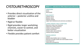
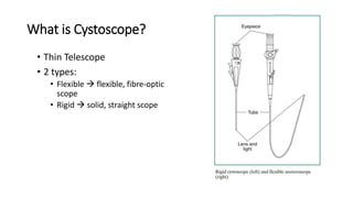
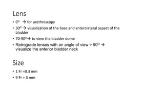
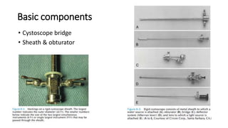
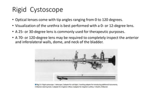
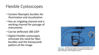
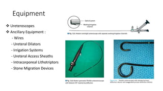
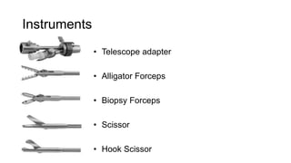
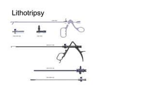
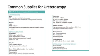
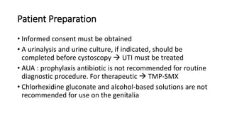
Cystoscopy and ureteroscopy are endoscopic procedures used to visualize the bladder and upper urinary tract. Cystoscopy can be performed rigidly or flexibly, with rigid cystoscopes providing better optics and larger working channels while flexible cystoscopes are more comfortable for patients. Important components of cystoscopes include the telescope lens, bridge, and sheath. Rigid and flexible ureteroscopes are used to inspect the ureters and kidneys. Proper patient preparation, use of irrigation, and careful technique help ensure safe and effective visualization and potential treatment of conditions like stones.
CYSTOURETHROSCOPY
• Provides directvisualization of the
anterior – posterior urethra and
bladder
• Rigid or flexible
• Rigid provides larger workshing
channels, easier to control, and
better visualization
• Flexible provides patient comfort
3.
What is Cystoscope?
•Thin Telescope
• 2 types:
• Flexible flexible, fibre-optic
scope
• Rigid solid, straight scope
4.
Lens
• 0O for urethroscopy
• 30O visualization of the base and anterolateral aspect of the
bladder
• 70-90O to view the bladder dome
• Retrograde lenses with an angle of view > 90O
visualize the anterior bladder neck
Size
• 1 Fr =0.3 mm
• 9 Fr = 3 mm
Rigid Cystoscope
• Opticallenses come with tip angles ranging from 0 to 120 degrees.
• Visualization of the urethra is best performed with a 0- or 12-degree lens.
• A 25- or 30-degree lens is commonly used for therapeutic purposes.
• A 70- or 120-degree lens may be required to completely inspect the anterior
and inferolateral walls, dome, and neck of the bladder.
7.
Flexible Cystoscopes
• Containfiberoptic bundles for
illumination and visualization
• Has an irrigating channel and a
working channel for passage of
instruments
• Can be deflected 180-220o
• Digital Flexible cystoscopes
eliminate the need for fiber
bundles and the honeycomb
pattern of the image
8.
What to choose?
Advantagesof rigid endoscope:
• Better optics
• Larger working channel allows greater
versatility in passage of instruments
• Larger lumen for water flow, thus improving
visualization
• Ease of manipulation and maintaining
orientation during inspection within the
bladder
• Better for evacuating clots
Advantages of flexible endoscope:
• Greater comfort for the patient
• Ability to perform the procedure with the
patient in the supine position
• Ease of passing the instrument over an
elevated bladder neck
• Ability to inspect at any angle with
deflection of the tip of the instrumen
9.
UPPER URINARY TRACTENDOSCOPY
Direct visualization of the upper urinary tract, facilitating both
diagnostic and therapeutic interventions
Indication :
• Treatment of nephrolithiasis
• Evaluation of lateralizing hematuria, abnormal urinary cytology,
and abnormal upper tract imaging studies
Patient Preparation
• Informedconsent must be obtained
• A urinalysis and urine culture, if indicated, should be
completed before cystoscopy UTI must be treated
• AUA : prophylaxis antibiotic is not recommended for routine
diagnostic procedure. For therapeutic TMP-SMX
• Chlorhexidine gluconate and alcohol-based solutions are not
recommended for use on the genitalia
• In women,rigid cystourethroscope insertion is safest using the
sheath obturator
• In men, the penis should be angled 45 - 90° relative to the
abdominal wall while the scope is passed through the anterior
urethra.
• Visualization of the lateral walls is accomplished by rotating the
cystoscope while keeping the camera orientation fixed.
• If a Foley catheter is to be placed after the procedure, it is best to
leave the bladder at least partially full before removing the
cystoscope.
Ureteroscopy Technique
19.
Ureteroscopy Technique
• Uppertract imaging IVP or helical CT scan
• UTI are treated preoperatively
• Routine preoperative antibiotic is give to all patients
fluoroquinolones or based on antibiotic sensitivity test
• Patient in cystolithotomy position
21.
Semirigid Ureteroscopy Technique
•Maneuvering the tip of the ureteroscope next to the guidewire posterolaterally
and elevate the wire propping the ureteral orifice open to allow scope
passage.
• If the intramural ureter is too tight to allow safe passage of the ureteroscope, a
dilating balloon catheter can be used.
22.
If the ureteroscopedoes not pass easily
1) rotate the beak of the ureteroscope while engaging the ureteral orifice
and providing adequate irrigation;
2) pass a super-stiff wire into the ureter through the working channel,
rotate the ureteroscope so that the tip lies between the two wires, and
guide the ureteroscope into the ureter between the two “railroading”
wires (Fig. 8–10);
3) Dilate the ureteral orifice with a 6/10-Fr coaxial dilator over a
guidewire;
4) dilate the ureteral orifice with a 4-mm radially expanding balloon
dilator; or
5) if the preceding measures fail, place an indwelling ureteral stent and
allow for passive dilation for 2 weeks.
24.
• Normal salineshould be used for irrigation
to prevent accumulation and absorption of
hypotonic solution and resultant TUR
syndrome.
• Do not activate the laser unless the tip of the
fiber is seen to be contact with the stone
Flexible Ureteroscopy Technique
25.
References
1. Duty BD,Conlin MJ. Principles of Urologic Endoscopy. In: Campbell-Walsh’s Urology.
Netherland: Elsevier. 2015
2. Hofenfellner R, Stolzenburg JU. Manual Endourology. Leipzig: Springer. 2005
3. Smith A, Badlani G, Preminger GM, Kavoussi LR (editors). Smith’s Textbook of
Endourology. 3rd ed. Oxford: Wiley – Blackwell. 2007
4. University college hospital at Westmoreland street. Cystoscopy: urology
directorate.University colleg London Hospital. NHS foundation trust.
5. Chancellor MB, Steers WD. Cystoscopy and ureteroscopy. NIH publication. 2014
6. Narance D. Urology Overview: part II. Cystoscope processing and handling
procedures. CIS self study lesson plan. Med Central Health System.
26.
Care and Sterilization
•Scraped inner working channel deflection of flexible
ureteroscope when the fiber is introduced
• Fired fiber within the working channel to prevent the tip
must be seen in the central portion of the field of view
• Should be cleased with warm water and a non abrasive
detergent after each use