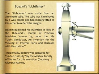
The document describes the history and development of endoscopy from its origins with Bozzini's "Lichtleiter" in 1806 to modern innovations. Key developments include:
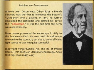
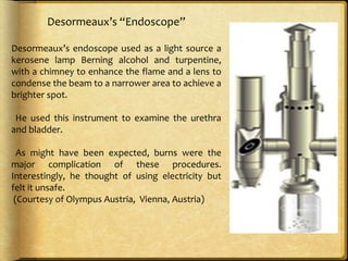
- Desormeaux introducing Bozzini's device into patients and coining the term "endoscope" in 1853.
- Czerny constructing the first rigid endoscope in 1880 and using Edison's light bulb, allowing angling and air insufflation.
- Kelling pioneering laparoscopy in 1901 after being introduced to endoscopy by Mikulicz-Radecki.
- Hopkins inventing the rigid rod-lens system for scopes in 1959, allowing their widespread adoption after partnering with
Minimally invasive surgery uses small incisions and miniaturized imaging systems to perform major operations with less trauma than traditional open surgery. The techniques were developed starting in the early 1900s and improved with advances like rod lens endoscopes, flexible instruments, and fluoroscopic imaging. Laparoscopic surgery involves inflating the abdominal cavity with gas to provide space to see and operate. Thoracoscopy may require deflating one lung. Other minimally invasive techniques provide access through subcutaneous tissues or body cavities without requiring incisions into organs. Endoluminal and intraluminal procedures operate from within lumens like blood vessels or the digestive tract.
laparoscopy is recent advancing area in the field of general surgery. the identification and underlying mechanism of action of each laparoscopic instrument is necessary for their handling ans use.
1. The document discusses the basics of laparoscopy including the laparoscopic tower setup, ergonomics, port placement, entry techniques, energy devices, and complications.
2. Key aspects of ergonomics include following the straight line principle with the tower, monitor, and instruments, proper table height, and triangulating or sectoring port placement depending on whether the surgeon stands ipsilateral or contralateral.
3. Safe entry techniques include closed insertion with a Veress needle or open insertion with direct trocar placement, with Palmer's point being an alternative to umbilical entry.
Laparoscopy involves using small incisions and a camera to visualize the inside of the abdomen. It has several advantages over open surgery such as less pain, shorter hospital stays, and quicker recovery times. Some of the key equipment used in laparoscopy include rod lens systems and fiber optic cables for optics, trocars for abdominal access, and insufflators to inflate the abdomen with gas. Potential risks include injuries from trocars or pneumoperitoneum as well as effects of the pneumoperitoneum on respiratory and renal systems. Common procedures now performed laparoscopically include cholecystectomy, appendisectomy, hernia repair, and some cancer staging.
This document provides an overview of basics of laparoscopy in gynecology. It describes the key components and steps of laparoscopy including pneumoperitoneum creation using Veress needle or open technique, trocar placement using safe entry techniques, and use of laparoscopic instruments. It discusses the imaging system including light source, camera, monitor and recording systems. Advantages of laparoscopy over open surgery and indications are highlighted. Potential complications are also reviewed.
This document provides an overview of laparoscopic instrumentation used in minimally invasive surgery. It discusses the key components needed, including optical devices like telescopes and cameras, equipment for insufflation, trocars and other instruments for accessing the surgical site. A variety of operative instruments are described, such as graspers, dissectors, scissors, and bowel/lung clamps. Energy sources like electrosurgery, ultrasonic devices, and staplers are also covered. The document concludes with a discussion of instruments for tissue approximation and hemostasis, including clip appliers and mechanical staplers, as well as some miscellaneous tools.
This document discusses the instrumentation used in laparoscopic surgery. It describes the key components of the optical chain including the endoscope, light cable, light source, camera system, and monitor. It also discusses the gas insufflation apparatus, including the insufflator and carbon dioxide cylinder. Specific instruments are explained in more detail such as fiber optic cables and the different types of light sources used including halogen, halogen halide, and xenon lamps. Risks and limitations of laparoscopic surgery are also mentioned.
Laparoscopy in gynaecology presented by drs igbodike emeka philip and dr rotimiigbodikeobgyn
This slide will be helpful if the presentation revolves around laparoscopy in gynaecological practice. Kindly like , clip and share the slide. it is free!
This document discusses the evolution of laparoscopic surgery and laparoscopic instruments from ancient times to modern times. It describes some of the early pioneers who developed early endoscopic instruments in the 18th-19th centuries like Philip Bozzini and Maximilian Nitze. It then discusses the development of modern laparoscopy in the early 20th century with pioneers like Jacobaeus coining the term "laparoscopy" and Veress developing pneumoperitoneum. The document highlights the crucial inventions of the rod lens system by Harold Hopkins in the 1950s and videolaparoscopy by Camran Nezhat in the 1980s that enabled complex laparoscopic surgery. It also discusses some of the challenges
This presentation will help u know with the history,present and coming up trends in laparoscopy .Also it is an acquaintance presentation regarding laparoscopy.
This document provides an overview of minimal invasive surgery. It defines minimal invasive surgery as aiming to accomplish surgical goals with minimal somatic and psychological trauma using reduced wound access. The core principles of laparoscopic surgery are summarized as insufflate, visualize, identify, triangulate, retract, operate, and seal. The main types of minimal invasive surgery are then described. The advantages are outlined as reduced wound size, pain, and recovery time while the disadvantages include loss of tactile feedback and reliance on new techniques. Preoperative evaluation, contraindications, operative problems, postoperative care, discharge criteria and basic surgical principles are also summarized. Robotic surgery is introduced as computer-enhanced surgical devices that provide steadier images and fewer incisions
This document discusses various laparoscopy equipment used in minimally invasive surgeries. It describes key components like laparoscopes, trocars, insufflators, and various surgical instruments. A laparoscopic surgeon needs to be technically proficient in operating the equipment and understanding the principles of the instruments being used, as the procedures are technologically dependent and any emergency requires quick problem-solving skills without overreliance on technical support.
Laparoscopy involves using small incisions and a camera to perform abdominal surgeries. It was pioneered in the early 1900s and has since been used for procedures like cholecystectomy and appendectomy. Advantages include less pain, scarring and faster recovery compared to open surgeries. Proper patient positioning, insufflation, trocars and energy devices are required. Complications can include injuries from access and cautery. Recent advances include natural orifice translumenal endoscopic surgery and single-incision laparoscopic surgery.
This document discusses the principles and techniques of laparoscopic surgery. It begins with an introduction to minimal access surgery and its aims of reducing somatic and psychological trauma while allowing for shorter hospital stays and faster recovery. The document then covers the categories of minimally invasive procedures and diagnostic and therapeutic applications of laparoscopic surgery. It provides details on preoperative evaluation and preparation, creating pneumoperitoneum, intraoperative equipment and techniques, postoperative care, and examples of common laparoscopic procedures like cholecystectomy, hernia repair, and fundoplication. Throughout it includes diagrams to illustrate surgical anatomy and procedure steps.
Robotic surgery uses robotic systems to assist surgeons with complex procedures. Some key points:
- Early systems included ROBODOC in 1985 for hip replacements and AESOP in 1994 for positioning the endoscope. The Da Vinci system, introduced in 2000, is now the most widely used system.
- Systems can be tele-surgical like Da Vinci where the surgeon controls the robot remotely, shared-control where the robot provides feedback, or supervisory where the robot executes pre-planned motions autonomously under surgeon oversight.
- The Da Vinci system allows the surgeon to sit at a console several feet from the patient with magnified 3D HD vision and wristed instruments that mimic hand movements with
What is MIS?
A minimally invasive medical procedure is defined as one that is carried out by entering the body through the skin or through a body cavity or anatomical opening, but with the smallest damage possible to these struct uresIncludes laparoscopic, endoscopic, and other approaches.
Why MIS?
Decreased patient pain
Decreased patient recovery period
Possible decrease in inflammatory response in the patient which may prove to have a better outcome in oncologic operations.
Distant future
In the distant future, there will be a para- digm shift with the development of non-inva- sive surgical techniques in combination with nanotechnologies and a new era in the devel- opment of surgery, and subsequently in surgi- cal techniques, will be opened.
Nanotechnology is an umbrella term for materials and devices that operate at the nanoskill (1 billionth of a meter). In terms of scale, a nanometer is approximately one 1/8000 of a human hair or 10 times the diam- eter of a hydrogen atom. The size of the device can vary but starts from a ten thou- sand-logic element system that will occupy a cube of no more than one hundred nanome- ters. This is a volume slightly larger than 0.001 cubic microns. This would be sufficient to hold a small computer. For example, if red blood cells are approximately eight microns in diameter, the 100 nanomicroprocessor will be 80 times smaller than a red blood cell. Devices this size could easily fit into the circulatory system and could even conceivably enter indi- vidual cells.
This document provides an overview of minimal invasive surgery (MIS), also known as laparoscopic surgery. It discusses the history and development of laparoscopic techniques, advantages like less pain and faster recovery compared to open surgery. Prerequisites for MIS include patient fitness, instruments used like laparoscopes and trocars. Risks include injuries from trocars and potential complications from carbon dioxide insufflation like gas embolism. The document outlines general principles of MIS including visualization, triangulation and sealing blood vessels. Both basic procedures like laparoscopic cholecystectomy and more advanced surgeries are discussed.
This document discusses the basic principles of laparoscopy. It describes the key differences between laparoscopic and open surgery for both patients and surgeons. For patients, laparoscopic surgery results in less pain, faster recovery times, and quicker return to normal activities due to smaller incisions. For surgeons, laparoscopy provides a magnified view but with altered tactile response and two-dimensional images. The document outlines the typical laparoscopic setup including the endoscope, light source, camera, monitor, insufflator, trocars and various instruments. It also lists some common laparoscopic procedures that can be used for diagnostic and operative purposes.
Two years before surgery, the surgeon must prepare themselves through skills training, observing other surgeons, and ensuring the operating room (OR) and team are ready. One week before, the surgeon confirms the indication for surgery, reviews contraindications and counsels the patient. The day before, the patient receives instructions and the surgeon ensures the OR and team are prepared. On the day of surgery, the surgeon obtains consent, checks equipment and the anesthetized patient is brought to the OR. Special considerations are discussed for obese, pregnant and high risk patients.
It has not changed the nature of disease
The basic principles of good surgery still apply,including appropriate case selection, excellent exposure,adequate retraction and a high level technical expertise
If a procedure makes no sense with conventional access, it will make no sense with a minimal access approach
The cleaner and gentler the act of operation, the less the patient suffers, the smoother and quicker his convalescence,the more exquisite his healed wound.
We actually do not know what is there stored for us, but we believe that laparoscopy is trending towards advancement and nano and robotic technology is going to replace in future.
3D cameras have come into existence and various newer technologies are being invented.
This document discusses ergonomics principles for laparoscopic surgery. It notes that laparoscopic surgery provides less painful surgery for patients but is more demanding on surgeons. It covers topics such as instrument triangulation, patient positioning, table height, tactile limitations, port positioning, and ergonomic principles. The concept of ergonomics and problems at laparoscopy like visual axis vs motor axis are assessed. Management of ergonomic problems and who is more ergonomic are discussed.
Ergonomics is vital for efficient laparoscopic surgery. The key ergonomic principles for surgeons include:
1) Maintaining straight line visibility between the surgical site, instruments, and monitor using triangulation to allow coaxial alignment of the visual and motor axes.
2) Positioning instruments at angles of 30-60 degrees for manipulation and 60 degrees for elevation to reduce strain.
3) Adopting a relaxed stance with straight back, shoulders neutral, and elbows bent to minimize fatigue.
4) Considering equipment design with articulating instruments, adjustable tables and monitors to optimize ergonomics.
This presentation of introduction of laparoscopic surgery made by Dr. R.K. Mishra Director and chief surgeon World Laparoscopy Hospital. Dr. Mishra in this presentation has explained present pas and future of laparoscopic surgery. Laparoscopy is a surgical procedure which uses a special surgical instrument called a laparoscope to look inside the body, or to perform certain operations. World Laparoscopy Hospital is the center of excellence for laparoscopic and da vinci robotic surgery training and considered as one of the best institute in the world. For more detail about laparoscopic surgery please visit: http://www.laparoscopyhospital.com
Laparoscopy: Historic, Present and Emerging TrendsGeorge S. Ferzli
The document provides a historical overview of laparoscopy from its origins in ancient Greece and Rome to modern developments. Key events and innovators are discussed, including the first laparoscopic procedures in the early 20th century and developments of critical tools like trocars, insufflators, and improved optics. The document also outlines current standard laparoscopic procedures like cholecystectomy and discusses trends in bariatric surgery like the increasing popularity and safety of laparoscopic Roux-en-Y gastric bypass.
This document provides an overview of basic principles in gynecologic laparoscopy. It discusses the history and pioneers of laparoscopy. The basic prerequisites, anatomy, setup, instruments, patient positioning, trocar placement, indications, and procedures are described. Key steps for procedures like salpingectomy and treatment of endometriosis are outlined. Both advantages like quick recovery and potential complications are reviewed. The goal is to educate on fundamentals of performing laparoscopy safely and effectively.
The slit lamp or biomicroscope combines two components:
• a bright focal source of light shone through a slit of variable width or height
• a microscope, usually binocular
Although it began as an instrument more commonly used by ophthalmologists, its development was overseen by several optical physicists and it came to be a staple item of equipment of contact lens opticians.
For centuries, artists and anatomists have worked together to create detailed illustrations of human anatomy through paintings and models, improving understanding of the body's structure and function. Key figures who advanced anatomical knowledge through their work include Andreas Vesalius in the 16th century, Leonardo Da Vinci who studied human development, and Galen in the 2nd century whose drawings informed anatomical art for years. Photography later became an important tool in anatomy for teaching, learning, and research applications like medical documentation and histology.
For centuries, artists and anatomists have worked together to create detailed illustrations of human anatomy through paintings and models, improving understanding of the body's structure and function. Key figures who advanced anatomical knowledge through their work include Andreas Vesalius in the 16th century, Leonardo Da Vinci who studied fetal development, and Galen in the 2nd century whose drawings set conventions for anatomical artists. Photography, microscopy, ultrasound, CT scans, and other technologies also allow detailed examination and documentation of anatomy for teaching, research, and medical purposes.
Flexible and capsule endoscopes have evolved significantly since their invention in the early 1800s. Flexible endoscopes use fiber optic bundles or video cameras to examine internal organs and cavities. Capsule endoscopes are wireless devices that can examine the small intestine. Both have advantages like allowing minimally invasive diagnosis and treatment. Flexible endoscopes come in different types for specific organs like the esophagus, stomach, lungs and bladder. They are used to detect cancers, infections, and other medical issues. Capsule endoscopes are pill-sized cameras that can provide images of the small intestine over 8 hours to diagnose conditions like bleeding and inflammation. Endoscopy has advanced medicine by enabling improved visualization of the inside
FOL fibre optic laryngoscopy for ear nose and throat.pdfDilip Biswas
Flexible and capsule endoscopes have evolved significantly since their invention in the early 1800s. Flexible endoscopes use fiber optic bundles or video cameras to examine internal organs and cavities. Capsule endoscopes are wireless devices that can examine the small intestine. Both have advantages like allowing minimally invasive diagnosis and treatment. Flexible endoscopes come in different types for specific organs like the esophagus, stomach, lungs and bladder. They are used to detect cancers, infections, and other abnormalities. Capsule endoscopes are pill-sized cameras that can provide images of the small intestine over 8 hours to diagnose conditions like bleeding and inflammation. Endoscopy has advanced medicine by enabling improved non-surgical visualization
Bright field microscopy, Principle and applicationsKAUSHAL SAHU
This document discusses the basics of light microscopy. It begins with a brief history, noting that the first compound microscope was created in 1590, while Antonie van Leeuwenhoek discovered microorganisms and sperm cells using a simple microscope in the 1630s. The basic components of light microscopes are described, including the objective lenses, eyepieces, stage, and condenser. Brightfield microscopy is explained in more detail, noting that it uses transmitted white light and staining to increase contrast. Applications include viewing stained bacteria, tissue sections, and algae. While brightfield microscopy is simple to use, its disadvantages include low contrast and an inability to see transparent, unstained samples clearly.
The document discusses the history of the microscope from its origins in ancient Rome to modern developments. It notes that while the Romans were the first to observe magnification using glass, the microscope was more fully developed in the late 16th/early 17th centuries by Dutch lens grinders Jansen and Van Leeuwenhoek. Key developments included compound microscopes with multiple lenses, improved magnification from 50x to 270x, and biological discoveries using microscopes. The document outlines the timeline of microscope history and classifications of microscopes based on optics, structure, application, and more modern developments like electron microscopes.
The document discusses the history and evolution of endoscopic and laparoscopic surgery from the 1800s to present day. Key developments include the first use of endoscopes in the 1800s, advancements in instrumentation and video technology in the 1900s enabling more complex procedures, and the explosion of laparoscopic surgeries starting in the late 1980s with procedures like laparoscopic cholecystectomy. The future of laparoscopy may include improvements like 3D imaging to enhance the surgical experience.
Indian Dental Academy: will be one of the most relevant and exciting training center with best faculty and flexible training programs for dental professionals who wish to advance in their dental practice,Offers certified courses in Dental implants,Orthodontics,Endodontics,Cosmetic Dentistry, Prosthetic Dentistry, Periodontics and General Dentistry.
This document provides a history of laparoscopy and key developments in the field. It discusses early developments in the 1800s and 1900s using cystoscopes and insufflation. The first laparoscopy was performed in 1901 by George Kelling. Developments continued through the 1900s including the introduction of trocars and telescopes. Laparoscopic cholecystectomy was first performed in 1985 but was not widely adopted until the late 1980s. The document also discusses current standard laparoscopic procedures and emerging technologies like fluorescent cholangiography and natural orifice transluminal endoscopic surgery (NOTES).
This document provides an overview of light microscopy and the brightfield microscope. It begins with the history and basic components of microscopes. It then describes the key features of light microscopy and different types including brightfield, darkfield, phase contrast, and differential interference contrast microscopy. The document focuses on brightfield microscopy, explaining that it uses transmitted white light and lenses to produce magnified images of stained specimens against a bright background. It discusses the principles, advantages, disadvantages and applications of brightfield microscopy. In conclusion, the document emphasizes the crucial role of microscopy in fields like cell biology and medical sciences.
1) The document discusses the history and basic principles of microbiology. It describes early pioneers like Antonie van Leeuwenhoek who first observed microbes under a microscope.
2) Louis Pasteur and Robert Koch made major contributions, with Pasteur developing pasteurization and vaccines and Koch connecting specific microbes to diseases like anthrax, cholera, and tuberculosis.
3) The document also discusses the history of the theory of spontaneous generation and how scientists like Redi, Needham, and Spallanzani through experiments eventually disproved this theory through experiments showing microbes come from other microbes and not non-living matter.
The document provides a historical overview of computed tomography (CT) scanning. It describes how CT scanning was developed in the 1970s by Godfrey Hounsfield and Allan Cormack, who were later awarded the Nobel Prize. It outlines the key events in the development of CT technology, from the early prototype scanners to modern multi-detector CT machines. The document also discusses the impact of CT scanning in medical imaging and different scan types like spiral CT.
The document provides a historical overview of computed tomography (CT) scanning. It describes how CT scanning was developed in the 1970s by Godfrey Hounsfield and Allan Cormack, who were later awarded the Nobel Prize. It outlines the key events in the development of CT technology, including the creation of the first CT scanner by Hounsfield in 1971 and its introduction to medical use. It also summarizes the different generations of CT scanners and how they have improved imaging capabilities over time.
The document provides a historical overview of computed tomography (CT) scanning. It describes how CT scanning was developed in the 1970s by Godfrey Hounsfield and Allan Cormack, who were later awarded the Nobel Prize. It outlines the key events in the development of CT technology, including the creation of the first CT scanner by Hounsfield in 1971 and the introduction of multi-detector CT scanners. The document also discusses the impact of CT scanning in medical imaging and different types of CT scans.
X-ray imaging uses X-rays that are generated by an X-ray tube and pass through the body. Differences in absorption of the X-rays creates a shadow-like image that can be detected by either screens or digital detectors. There are several types of medical X-rays including analog X-rays, digital X-rays, mammograms, CT scans, and fluoroscopy. Image detection has transitioned from screen-film systems to digital detectors like photostimulable phosphor plates and flat panel detectors. Flat panel detectors directly convert X-rays to a digital image and come in two main types - direct detectors using materials like selenium or indirect detectors using a scintillator to first convert X-rays to light,
General x-ray machine and fluoroscopy
the presentation to medical workers
contain simple explanation about radiation protection in the radiology department
Endoscopy is a minimally invasive medical procedure that uses an endoscope to examine the interior of a body cavity or organ. An endoscope is a thin, flexible or rigid tube with a light source and lens that transmits images to a monitor. Modern endoscopes can be either fiber optic or video-based. A variety of tools can be passed through channels in the endoscope, including biopsy forceps, snares, and brushes. Endoscopy has evolved greatly since its origins in the early 19th century, and is now commonly used to diagnose and treat many gastrointestinal and respiratory conditions.
How to Fix the Import Error in the Odoo 17Celine George
An import error occurs when a program fails to import a module or library, disrupting its execution. In languages like Python, this issue arises when the specified module cannot be found or accessed, hindering the program's functionality. Resolving import errors is crucial for maintaining smooth software operation and uninterrupted development processes.
Executive Directors Chat Leveraging AI for Diversity, Equity, and InclusionTechSoup
Let’s explore the intersection of technology and equity in the final session of our DEI series. Discover how AI tools, like ChatGPT, can be used to support and enhance your nonprofit's DEI initiatives. Participants will gain insights into practical AI applications and get tips for leveraging technology to advance their DEI goals.
How to Manage Your Lost Opportunities in Odoo 17 CRMCeline George
Odoo 17 CRM allows us to track why we lose sales opportunities with "Lost Reasons." This helps analyze our sales process and identify areas for improvement. Here's how to configure lost reasons in Odoo 17 CRM
This presentation includes basic of PCOS their pathology and treatment and also Ayurveda correlation of PCOS and Ayurvedic line of treatment mentioned in classics.
বাংলাদেশের অর্থনৈতিক সমীক্ষা ২০২৪ [Bangladesh Economic Review 2024 Bangla.pdf] কম্পিউটার , ট্যাব ও স্মার্ট ফোন ভার্সন সহ সম্পূর্ণ বাংলা ই-বুক বা pdf বই " সুচিপত্র ...বুকমার্ক মেনু 🔖 ও হাইপার লিংক মেনু 📝👆 যুক্ত ..
আমাদের সবার জন্য খুব খুব গুরুত্বপূর্ণ একটি বই ..বিসিএস, ব্যাংক, ইউনিভার্সিটি ভর্তি ও যে কোন প্রতিযোগিতা মূলক পরীক্ষার জন্য এর খুব ইম্পরট্যান্ট একটি বিষয় ...তাছাড়া বাংলাদেশের সাম্প্রতিক যে কোন ডাটা বা তথ্য এই বইতে পাবেন ...
তাই একজন নাগরিক হিসাবে এই তথ্য গুলো আপনার জানা প্রয়োজন ...।
বিসিএস ও ব্যাংক এর লিখিত পরীক্ষা ...+এছাড়া মাধ্যমিক ও উচ্চমাধ্যমিকের স্টুডেন্টদের জন্য অনেক কাজে আসবে ...
This slide is special for master students (MIBS & MIFB) in UUM. Also useful for readers who are interested in the topic of contemporary Islamic banking.
Exploiting Artificial Intelligence for Empowering Researchers and Faculty, In...Dr. Vinod Kumar Kanvaria
Exploiting Artificial Intelligence for Empowering Researchers and Faculty,
International FDP on Fundamentals of Research in Social Sciences
at Integral University, Lucknow, 06.06.2024
By Dr. Vinod Kumar Kanvaria
A review of the growth of the Israel Genealogy Research Association Database Collection for the last 12 months. Our collection is now passed the 3 million mark and still growing. See which archives have contributed the most. See the different types of records we have, and which years have had records added. You can also see what we have for the future.
2. Bozzini’s “Lichtleiter”
The “Lichtleiter” was made from an
aluminum tube. The tube was illuminated
by a wax candle and had mirrors fitted to
it in order to reflect the images.
Bozzini published his invention in 1806 in
the Hufeland’s Journal of Practical
Medicine, Volume 24, under the title
“Light Conductor, An Invention for the
Viewing of Internal Parts and Diseases
with Illustration.”
Incidentally, Bozzini was censured for
“undue curiosity” by the Medical Faculty
of Vienna for this invention. (Courtesy of
Olympus Austria,
3. Antoine Jean Desormeaux
Antoine Jean Desormeaux (1815–1894), a French
Surgeon, was the first to introduce the Bozzini’s
“Lichtleiter” into a patient. In 1853, he further
developed the Lichtleiter and termed his device
the “Endoscope.” It was the first time this term
was used in history.
Desormeaux presented the endoscope in 1865 to
the Academy in Paris. He even used his endoscope
to examine the stomach; but due to an insufficient
light source he was not quite successful.
(Copyright Verger-Kuhnke AB. The life of Philipp
Bozzini (1773-1809), an idealist of endoscopy. Actas
Urol Esp. 2007;31:437-444)
4. Desormeaux’s “Endoscope”
Desormeaux’s endoscope used as a light source a
kerosene lamp Berning alcohol and turpentine,
with a chimney to enhance the flame and a lens to
condense the beam to a narrower area to achieve a
brighter spot.
He used this instrument to examine the urethra
and bladder.
As might have been expected, burns were the
major complication of these procedures.
Interestingly, he thought of using electricity but
felt it unsafe.
(Courtesy of Olympus Austria, Vienna, Austria)
5. Johannes Freiherr
surgeon of Polish-Lithuanian descent born in
Bukowina, Romania, constructed the first rigid
endoscope in 1880 and was the first to use
Edison’s light bulb for his gastroscope in
practice.
He modified the instrument so that it could be
angled by 30Åã near to its lower third to
achieve better visualization.
He added a separate channel for air
insufflation. In one of the first interventional
endoscopic procedures, he pushed a large
swallowed bone from the esophagus into the
stomach, thus avoiding surgery. (Copyright:
surgeon in the evolution of flexible endoscopy.
Surg
Endosc 2007: 21; 838-853 Springer Verlag)
6. Georg Kelling (1866–1945), a German
physician
from Dresden, was introduced to
endoscopy and gastrointestinal surgery
when he worked with Professor Mikulicz-
Radecki at the Royal Surgical Clinic in
Breslau, Germany. In with the help of
Nitze’s cystoscope, and coined this
laparoscopic examination “celioscopy.”
He used air filtered through sterile cotton
to create pneumoperitoneum in dogs. For
insufflation he used a trocar Developed by
Alfred Fiedler, an internist from Dresden.
(Copyright: Hatzinger M: Georg Kelling
(1866–1945) Der Erfinder der modernen
Laparoskopie. Urologe A 2006; 45 (7):868-71
Springer Verlag)
7. Harold Horace Hopkins
Hopkins (1918–1994) obtained a degree in physics and
mathematics at Leicester University in 1939. After the war,
in 1947, Hopkins became a research fellow at Imperial
College, London, UK.
Hopkins invented the rigid rod-lens system for scopes,
which allows double light transmission, requires short and
thin spacer tubes, and gives a larger and clearer aperture.
He filed a patent for the rod-lens system in 1959.
However, the English and American companies to whom he
offered the system displayed little interest.
The situation changed however in 1965 when Professor
George Berci, who recognized the potential of this
invention, introduced Hopkins to Karl Storz to manufacture
the scopes. (Courtesy of William P. Didusch Center for
Urologic History, American Urological Association, MD,
USA)
8. Kurt Karl Stephan Semm
Kurt Karl Stephan Semm (1927–2003) was born in Munich,
Germany, where he also studied medicine at the Ludwigs-
Maximillian University. In 1958, he wrote his medical
thesis under the guidance of Nobelmlaureate Adolf
Butenandt. Semm began his career in gynecology under
Professor Fikentscher in Munich. In 1970s, as the Head of
Gynecology in Kiel, introduced an;
1- Automatic insufflation device capable of monitoring
intra-abdominal pressures,
2- Endoscopic loop sutures,
3-Extra- and intracorporeal suturing techniques.
4-Created the pelvi- trainer. He performed the first
laparoscopic appendectomy
in 1982. (Courtesy of Monika Bals-Pratsch MD,
Zentrum für Gyn.kologie, Universit.t Regensburg,
Germany)
10. Central to the instrumentation is the scope. Its
backbone is the rod lens system designed by
Hopkins.
The shaft of scopes houses both light fibers
and viewing optics.
The viewing optic consist of three distinct
parts:
o The objective lens,
o Rod lenses,
o ocular lens.
11. Field of View
The field of view (also field of vision)
is the angular extent of the
observable area that is seen at any
given moment. The field of view in
scopes for endoscopic surgery can
vary from 600 to 820 depending up
on the type of instrument. Wider
angles of view provide a greater
depth of field in the image with
better utilization of illumination. A
smaller field of view allows the
scope to be farther from the tissue,
for the same to be observed.
12. Angle of View
The angle of view in scopes can vary with
respect to the central axis view are
designated as 00 and provide a straight
view of the structure in question. Scopes
are also available with a 50, 250, 300, 450,
and even 700 angle of view, allowing
utilization of the scopes much as a
periscope. The off-axis scopes enable one
to observe
down into the gutters and up the anterior
abdominal wall as well as sideways. Off-axis
scopes are difficult to work with; however,
they provide an excellent means of
obtaining close inspection of tissues
at difficult angles and positions.
13. Scope Size and Screen Image
The decrease in the size of scopes was an important
factor in the advancement of minimally invasive
surgery in the pediatric age group. Although scopes
are available in sizes from 1.9 mm to 12 mm in
diameter, the majority of the procedures are
performed using 5- or 10-mm scopes.
When compared to the reduced view obtained in the
previous generation of scopes (left), modern5-mm,
full-screen scopes provide a bright, distortion- free,
full-screen image (right). In addition, the image size
in modern 5-mm scope is equivalent to that obtained
by the previous-generation
10-mm scope. (Courtesy of Richard Wolf, Knittlingen,
Germany)
14. Charge Coupled Device (CCD)
Video Cameras Scope cameras are available in either
single-chip
three-chip versions (one chip offers 300,000 pixels/cm2).
In single-chip CCD cameras, all the three primary colors (red,
blue and green) are sensed by a single chip. In three-chip CCD
cameras, there are three chips for separate capture and
processing of the primary colors.
Single-chip CCD cameras produce images of 450 lines/inch
resolution and are ideal for outpatient surgery. On the other
hand, three-chip CCD cameras have high fidelity with
unprecedented color reproduction to produce images of 750
lines/ inch resolution that can be viewed optimally on flat-panel
screens and are best suited for endoscopic surgery.
(Courtesy of RichardWolf, Knittlingen , Germany)
Light source:
Light-Source Generators and Transmission Pathways There
are two commonly utilized light sources: halogen and xenon.
A schematic overview of light transmission is outlined12
15. The Concept of White Balancing
White balancing should be performed before
inserting the camera inside the abdominal cavity. This
is necessary before commencing surgery to diminish
the added impurities of color that may be introduced
due to a variety of reasons such as:
(1) voltage difference,
(2) staining of the tip by cleaners,
(3) scratches and wear of the eyepiece.
White balancing is achieved by keeping a white
object in front of the scope and activating the
appropriate button on the video system or camera.
The camera senses the white object as its reference
to adjust all of the primary colors (red, blueand
green). (Courtesy of Richard Wolf, Knittlingen,
Germany)
16.
17. A three-CCD camera is a camera whose
imaging system uses three separate charge-coupled
devices (CCDs),
each one taking a separate measurement of
the primary colors, red, green, or blue light.
Light coming into the lens is split by a
trichroic prism assembly, which directs the
appropriate wavelength ranges of light to
their respective CCDs.
The system is employed by still cameras,
telecine systems, professional video cameras
and some prosumer video cameras.
18. Rods and Cones
The retina contains two types of photoreceptors,
Rods
cones.
The rods are more numerous, some 120 million, and are more sensitive than the
cones
they are not sensitive to color. The 6 to 7 million cones provide the eye's color
sensitivity and they are much more concentrated in the central yellow spot known as
the macula. In the center of that region is the " fovea centralis ", a 0.3 mm diameter
rod-free area with very thin, densely packed cones.
The experimental evidence suggests that among the cones there are three different
types of color reception. Response curves for the three types of cones have been
determined. Since the perception of color depends on the firing of these three types
of nerve cells, it follows that visible color can be mapped in terms of three
numbers called tristimulus values.
Color perception has been successfully modeled in terms of tristimulus values and
mapped on the CIE chromaticity diagram.
19. Rods Do Not See Red!
The light response of the rods peaks sharply in the blue; they respond very little
to red light. This leads to some interesting phenomena:
Red rose at twilight: In bright light, the color-sensitive cones are predominant
and we see a brilliant red rose with somewhat more subdued green leaves. But
at twilight, the less-sensitive cones begin to shut down for the night, and most
of the vision comes from the rods. The rods pick up the green from the leaves
much more strongly than the red from the petals, so the green leaves become
brighter than the red petals!
The ship captain has red instrument lights. Since the rods do not respond to red,
the captain can gain full dark-adapted vision with the rods with which to watch
for icebergs and other obstacles outside. It would be undesirable to examine
anything with white light even for a moment, because the attainment of
optimum night-vision may take up to a half-hour. Red lights do not spoil it.
These phenomena arise from the nature of the rod-dominated dark-adapted
vision, called scotopic vision.
20. Cone Details
Current understanding is that the 6 to 7 million cones can be divided
into "red" cones (64%), "green" cones (32%), and "blue" cones (2%)
based on measured response curves. They provide the eye's color
sensitivity. The green and red cones are concentrated in the fovea
centralis . The "blue" cones have the highest sensitivity and are
mostly found outside the fovea, leading to some distinctions in the
eye's blue perception.
The cones are less sensitive to light than the rods, as shown a typical
day-night comparison. The daylight vision (cone vision) adapts much
more rapidly to changing light levels, adjusting to a change like
coming indoors out of sunlight in a few seconds. Like all neurons, the
cones fire to produce an electrical impulse on the nerve fiber and
then must reset to fire again. The light adaption is thought to occur
by adjusting this reset time.
The cones are responsible for all high resolution vision. The eye
moves continually to keep the light from the object of interest falling
on the fovea centralis where the bulk of the cones reside.
21.
22. The combination of the three sensors can be done in the
following ways:
Composite sampling, where the three sensors are perfectly
aligned to avoid any color artifact when recombining the
information from the three color planes
Pixel shifting, where the three sensors are shifted by a fraction
of a pixel. After recombining the information from the three
sensors, higher spatial resolution can be achieved.
Pixel shifting can be horizontal only to provide higher
horizontal resolution in standard resolution camera, or
horizontal and vertical to provide high resolution image using
standard resolution imager for example. The alignment of the
three sensors can be achieved by micro mechanical
movements of the sensors relative to each other.
Arbitrary alignment, where the random alignment errors due
to the optics are comparable to or larger than the pixel size.
23. Compared to cameras with only one CCD, three-CCD cameras
generally provide superior image quality through enhanced
resolution and lower noise.
By taking separate readings of red, green, and blue values for each
pixel,
three-CCD cameras achieve much better precision than single-CCD
cameras.
By contrast, almost all single-CCD cameras use a Bayer filter, which
allows them to detect only one-third of the color information for
each pixel.
The other two-thirds must be interpolated with a demosaicing
algorithm to 'fill in the gaps', resulting in a much lower effective
resolution
24. Video and Data Storage
Equipment
2.21.1 Digital Video Recorders
Modern endoscopic surgery towers are generally
equipped with digital video disc (DVD) recorders
(DVRs), which enable recording of a procedure
in digital quality. The procedures are recorded on
commercially available DVDs, which can later be
viewed on normal DVD players or edited on personal
computers.
DVRs have evolved into devices that are feature
rich and provide services that exceed the simple
recording of video images that was previously
achieved using video cassette recorders (VCRs).
DVR systems provide a multitude of advanced
functions,
including video searches by event and time.
25. Digital Video Printers
A variety of printers from small print
format to
large A5 print format are available.
These printers
offer high-resolution prints, quick, 20-s
print time,
and high-quality, curl-free prints at 400
dpi resolution.
Most modern printers come with a
four-frame
memory. The new compact design of
printers allows
for easy integration with other video
equipment.
Small, compact printers are ideal for
the office
setting, but large-print format printers
are preferable
in the operating room.
26. Digital Video Managers
These are computer-based systems that
display intuitive
patient information screens that allow for
quick and easy input of vital data. The data is
stored
on hard drives and can be viewed as images
or
videos, and may be stored or deleted. The
editing
screen enables viewing and editing
procedures.
Current systems allow storage of up to 50
patient
archives for multiple procedures. These
systems
are compatible with personal computers and
hospital network software. (Courtesy of
Richard
Wolf, Knittlingen, Germany)
27. Tracking Instruments using Color
Markers
Place colored marker on instrument
Convert RGB to HSV space
Hue value of a pixel is much less susceptible to lighting changes
Record hue value of marker to be tracked
Search entire image for hue values within epsilon range
Centroid of matched pixels gives position of tracker in the image
If target is detected, localize search to a smaller neighborhood
Tracking performed in real-time at 25 fps
28. Allows shared autonomy with surgeon
The feedback from the tracker can be used to drive motors to
keep the tool in the center of the image
PD controller used
( Ex , Ey ): off set error of tracker from center of image
Pan speed ( x * Ex ) – ( x * dEx/dt )
Tilt speed ( y * Ey ) – ( y * dEy/dt )
30. 3D Trajectory Reconstruction
The Flock of Birds (FoB) sensor can
transmit the position of its sensor w.r.t.
its base
Accuracy within 1.8mm
Refresh rate up to 144Hz
By placing an optical marker on the FoB
sensor we can track its position in the
image
By tracking the sensor using stereo
cameras we can compute its 3D
trajectory
32. 3D Displays
eMagin Z800 Head-Mounted VR Display
- Uncomfortable
- Single User
RealD Crystal Eyes shutter glasses
- Uncomfortable over longer periods
- Need to maintain Line Of Sight with
synchronizing emitter
True Vision back projected 3D display
- Low incremental cost for additional users
- Bigger display size
-Passive polarization, lightweight glasses