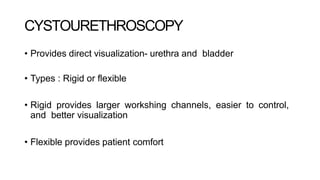
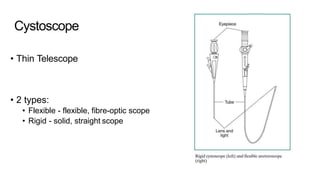
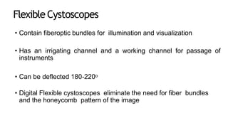
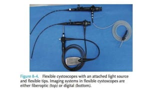
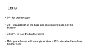
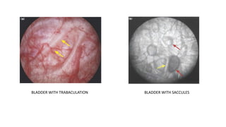
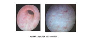
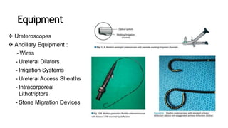
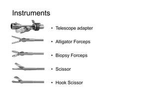
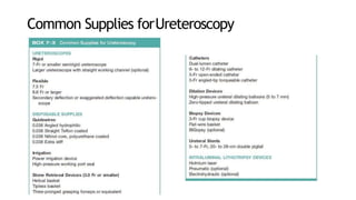
ENDOUROLOGY
Dr. Rachitha Radhakrishnan
Post graduate
Dept of General surgery
VMKVMCH
Various types
CYSTOURETHEROSCOPY
• Flexible
• Rigid
URETEROSCOPY
• Flexible
• Rigid
DIAGNOSTIC ENDOUROLOGY
 Follow up of TCC
 Biopsy from suspicious
sites
THERAPEUTIC ENDOUROLOGY
 Calculi – URSL
 Upper tract neoplasm
 Strictures
 Fistulas
 Benign lesions
CYSTOURETHROSCOPY
• Provides direct visualization- urethra and bladder
• Types : Rigid or flexible
• Rigid provides larger workshing channels, easier to control,
and better visualization
• Flexible provides patient comfort
Cystoscope
• Thin Telescope
• 2 types:
• Flexible - flexible, fibre-optic scope
• Rigid - solid, straight scope
Rigid Cystoscope
• Optical lenses come with tip angles ranging from 0 to 120 degrees.
• Visualization of the urethra is best performed with a 0- or 12-degree
lens.
• A 25- or 30-degree lens is commonly used for therapeutic purposes.
• A 70- or 120-degree lens may be required to completely inspect the
anterior and inferolateral walls, dome, and neck of the bladder.
Flexible Cystoscopes
• Contain fiberoptic bundles for illumination and visualization
• Has an irrigating channel and a working channel for passage of
instruments
• Can be deflected 180-220o
• Digital Flexible cystoscopes eliminate the need for fiber bundles
and the honeycomb pattern of the image
Advantages of rigid endoscope:
• Better optics
• Larger working channel allows
greater versatility in passage of
instruments
• Larger lumen for water flow, thus
improving visualization
• Ease of manipulation and
maintaining orientation during
inspection within the bladder
• Better for evacuating clots
Advantages of flexible endoscope:
• Greater comfort for the patient
• Ability to perform the procedure with the
patient in the supine position
• Ease of passing the instrument
over an elevated bladder neck
• Ability to inspect at any angle with
deflection of the tip of the instrumen
Lens
• 0O – for urethroscopy
• 30O - visualization of the base and anterolateral aspect of the
Bladder
• 70-90O - to view the bladder dome
• Retrograde lenses with an angle of view > 90O - visualize the anterior
bladder neck
Basiccomponents
ens
• Cystoscope bridge
• Sheath & obturator
BLADDER WITH TRABACULATION BLADDER WITH SACCULES
CALCULI SEEN ON CYSTOSCOPY
PAPPILLARY CA IN THE BLADDER
BLADDER DIVERTICULUM
NORMAL URETER ON URETEROSCOPY
URETHRAL STRICTURE
URETHROTOMY BEING PERFORMED
UPPER URINARY TRACT ENDOSCOPY
 Direct visualization of the upper urinary tract, facilitating
both diagnostic and therapeutic interventions
 Indication :
• Urolithiasis
• Upper urinary tract Transitional Cell Carcinoma
• Uretreropelvic junction obstruction
• Urethral stricture
Equipment
 Ureteroscopes
 Ancillary Equipment :
- Wires
- Ureteral Dilators
- Irrigation Systems
- Ureteral Access Sheaths
- Intracorporeal
Lithotriptors
- Stone Migration Devices
Instruments
• Telescope adapter
• Alligator Forceps
• Biopsy Forceps
• Scissor
• Hook Scissor
Common Supplies forUreteroscopy
PatientPreparation
• Informed consent must be obtained
• A urinalysis and urine culture, if indicated,
should be completed before cystoscopy  UTI
must be treated
• AUA : prophylaxis antibiotic is not recommended for
routine diagnostic procedure.
• For therapeutic  Chlorhexidine gluconate and
alcohol-based solutions are not recommended for use
on the genitalia
PatientPreparation
• In women, rigid cystourethroscope insertion is safest
using the sheath obturator
• In men, the penis should be angled 45 - 90° relative to
the abdominal wall while the scope is passed through
the anterior urethra.
• Visualization of the lateral walls is accomplished by
rotating the cystoscope while keeping the camera
orientation fixed.
• If a Foley catheter is to be placed after the procedure, it is
best to leave the bladder at least partially full before
removing the cystoscope.
Ureteroscopy Technique
• Upper tract imaging  IVP or helical CT scan
• UTI are treated preoperatively
• Routine preoperative antibiotic is give to all
patients fluoroquinolones or based on antibiotic
sensitivity test
• Patient in cystolithotomy position
Semirigid UreteroscopyTechnique
• Maneuvering the tip of the ureteroscope next to the
guidewire posterolaterally and elevate the wire 
propping the ureteral orifice open to allow scope
passage.
• If the intramural ureter is too tight to allow safe
passage of the ureteroscope, a dilating balloon
catheter can be used.
• Normal saline should be used for
irrigation to prevent accumulation and
absorption of hypotonic solution and
resultant TUR syndrome.
• Do not activate the laser unless the tip of
the fiber is seen to be contact with the
stone
Flexible UreteroscopyTechnique
Care andSterilization
• Scraped inner working channel  deflection of
flexible ureteroscope when the fiber is introduced
• Fired fiber within the working channel  to prevent the
tip must be seen in the central portion of the field of
view
• Should be cleased with warm water and a non
abrasive detergent after each use
THANKYOU

Endourology

  • 1.
    ENDOUROLOGY Dr. Rachitha Radhakrishnan Postgraduate Dept of General surgery VMKVMCH
  • 2.
    Various types CYSTOURETHEROSCOPY • Flexible •Rigid URETEROSCOPY • Flexible • Rigid
  • 3.
    DIAGNOSTIC ENDOUROLOGY  Followup of TCC  Biopsy from suspicious sites THERAPEUTIC ENDOUROLOGY  Calculi – URSL  Upper tract neoplasm  Strictures  Fistulas  Benign lesions
  • 4.
    CYSTOURETHROSCOPY • Provides directvisualization- urethra and bladder • Types : Rigid or flexible • Rigid provides larger workshing channels, easier to control, and better visualization • Flexible provides patient comfort
  • 6.
    Cystoscope • Thin Telescope •2 types: • Flexible - flexible, fibre-optic scope • Rigid - solid, straight scope
  • 7.
    Rigid Cystoscope • Opticallenses come with tip angles ranging from 0 to 120 degrees. • Visualization of the urethra is best performed with a 0- or 12-degree lens. • A 25- or 30-degree lens is commonly used for therapeutic purposes. • A 70- or 120-degree lens may be required to completely inspect the anterior and inferolateral walls, dome, and neck of the bladder.
  • 9.
    Flexible Cystoscopes • Containfiberoptic bundles for illumination and visualization • Has an irrigating channel and a working channel for passage of instruments • Can be deflected 180-220o • Digital Flexible cystoscopes eliminate the need for fiber bundles and the honeycomb pattern of the image
  • 11.
    Advantages of rigidendoscope: • Better optics • Larger working channel allows greater versatility in passage of instruments • Larger lumen for water flow, thus improving visualization • Ease of manipulation and maintaining orientation during inspection within the bladder • Better for evacuating clots Advantages of flexible endoscope: • Greater comfort for the patient • Ability to perform the procedure with the patient in the supine position • Ease of passing the instrument over an elevated bladder neck • Ability to inspect at any angle with deflection of the tip of the instrumen
  • 12.
    Lens • 0O –for urethroscopy • 30O - visualization of the base and anterolateral aspect of the Bladder • 70-90O - to view the bladder dome • Retrograde lenses with an angle of view > 90O - visualize the anterior bladder neck
  • 13.
  • 15.
    BLADDER WITH TRABACULATIONBLADDER WITH SACCULES
  • 16.
    CALCULI SEEN ONCYSTOSCOPY
  • 17.
    PAPPILLARY CA INTHE BLADDER
  • 18.
  • 19.
    NORMAL URETER ONURETEROSCOPY
  • 20.
  • 21.
    UPPER URINARY TRACTENDOSCOPY  Direct visualization of the upper urinary tract, facilitating both diagnostic and therapeutic interventions  Indication : • Urolithiasis • Upper urinary tract Transitional Cell Carcinoma • Uretreropelvic junction obstruction • Urethral stricture
  • 22.
    Equipment  Ureteroscopes  AncillaryEquipment : - Wires - Ureteral Dilators - Irrigation Systems - Ureteral Access Sheaths - Intracorporeal Lithotriptors - Stone Migration Devices
  • 23.
    Instruments • Telescope adapter •Alligator Forceps • Biopsy Forceps • Scissor • Hook Scissor
  • 24.
  • 27.
    PatientPreparation • Informed consentmust be obtained • A urinalysis and urine culture, if indicated, should be completed before cystoscopy  UTI must be treated • AUA : prophylaxis antibiotic is not recommended for routine diagnostic procedure. • For therapeutic  Chlorhexidine gluconate and alcohol-based solutions are not recommended for use on the genitalia
  • 28.
  • 29.
    • In women,rigid cystourethroscope insertion is safest using the sheath obturator • In men, the penis should be angled 45 - 90° relative to the abdominal wall while the scope is passed through the anterior urethra. • Visualization of the lateral walls is accomplished by rotating the cystoscope while keeping the camera orientation fixed. • If a Foley catheter is to be placed after the procedure, it is best to leave the bladder at least partially full before removing the cystoscope. Ureteroscopy Technique
  • 30.
    Ureteroscopy Technique • Uppertract imaging  IVP or helical CT scan • UTI are treated preoperatively • Routine preoperative antibiotic is give to all patients fluoroquinolones or based on antibiotic sensitivity test • Patient in cystolithotomy position
  • 31.
    Semirigid UreteroscopyTechnique • Maneuveringthe tip of the ureteroscope next to the guidewire posterolaterally and elevate the wire  propping the ureteral orifice open to allow scope passage. • If the intramural ureter is too tight to allow safe passage of the ureteroscope, a dilating balloon catheter can be used.
  • 34.
    • Normal salineshould be used for irrigation to prevent accumulation and absorption of hypotonic solution and resultant TUR syndrome. • Do not activate the laser unless the tip of the fiber is seen to be contact with the stone Flexible UreteroscopyTechnique
  • 35.
    Care andSterilization • Scrapedinner working channel  deflection of flexible ureteroscope when the fiber is introduced • Fired fiber within the working channel  to prevent the tip must be seen in the central portion of the field of view • Should be cleased with warm water and a non abrasive detergent after each use
  • 36.