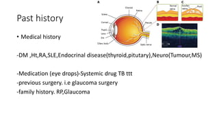
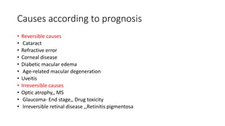
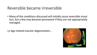
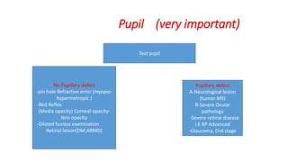
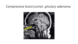
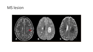
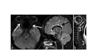
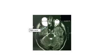
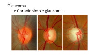
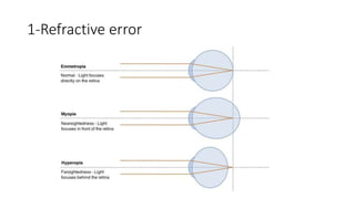
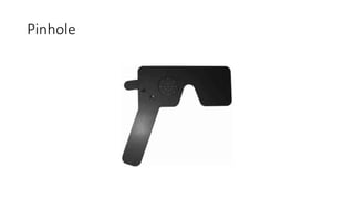
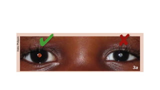
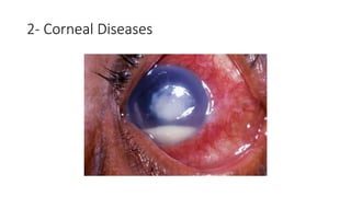
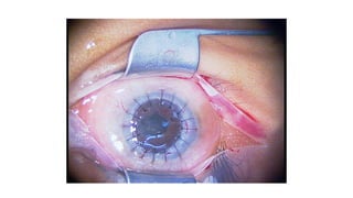
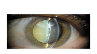
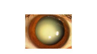
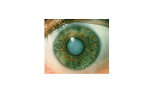
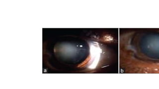
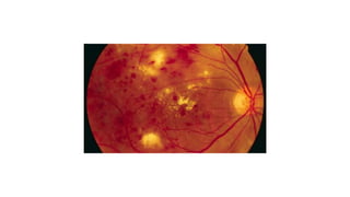
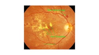
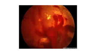
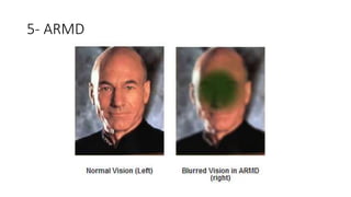
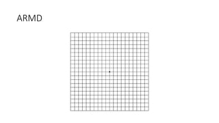
Gradual Causes of Visual
Loss
Ahmed Osama Hashem
FRCS ophth
PHD,Msc Ain Shams University
Lecturer , consultant of ophthalmology KFS
Causes according to prognosis
• Reversible causes
• Cataract
• Refractive error
• Corneal disease
• Diabetic macular oedema,Hypertensive retinopathy
• Age-related macular degeneration
• Uveitis
• Irreversible causes
• Optic atrophy,, MS
• Glaucoma- End stage
• Irreversible retinal disease ,,Retinitis pigmentosa
History
• Age Young (error of refraction) –Middle age MS -Old(Senile Cataract)
• A history of the type of visual loss, e.g. central or peripheral.
-Central loss is found with lesions of the macula
- peripheral loss is found in glaucoma.
• Patients with myopia and certain retinal degeneration may
experience poor vision with dim light, and those with a cataract may
find bright light to affect the vision more, although this symptom
depends on the morphology of the lens opacity.
Myopia,,,,,,History
Pin hole ,,,, same idea
Pupil size and effect on Vision.
Cataract,,,History - location of opacity
(Nodal point)
Central visual field loss,,,History
Peripheral loss of visual field
Lesion in visual pathway,,corresponding VF
loss…
Past history
• Medical history
-DM ,Ht,RA,SLE,Endocrinal disease(thyroid,pitutary),Neuro(Tumour,MS)
-Medication (eye drops)-Systemic drug TB ttt
-previous surgery. i.e glaucoma surgery
-family history. RP,Glaucoma
Causes according to prognosis
• Reversible causes
• Cataract
• Refractive error
• Corneal disease
• Diabetic macular edema
• Age-related macular degeneration
• Uveitis
• Irreversible causes
• Optic atrophy,, MS
• Glaucoma- End stage,, Drug toxicity
• Irreversible retinal disease ,,Retinitis pigmentosa
Reversible became irreversible
• Many of the conditions discussed will initially cause reversible visual
loss, but a few may become permanent if they are not appropriately
managed.
i,e Age related macular degeneration…
Examination (Clinical)
• The examination should focus on :
• visual acuity,
•pupil testing for the presence of an afferent pupil defect, and
assessment of the red reflex and fundoscopy.
• visual field testing,
• IOP, Slit lamp ,Fundus(Direct,Indirect)
Pupil (very important)
Test pupil
No Pupillary defect
-pin hole Refractive error (myopia-
hypermetropic )
-Red Reflex
(Media opacity) Corneal opacity-
lens opacity
-Dilated fundus examination
Retinal lesion(DM,ARMD)
Pupillary defect
A-Neurological lesion
(tumor-MS)
B-Severe Ocular
pathology
-Severe retinal disease
i.E RP Advanced
-Glaucoma, End stage
Compressive lesion,tumor ,pitutary adenoma
MS lesion
Glaucoma
i,e Chronic simple glaucoma….
Slit lamp + Gonio lens
Slit lamp + applanation tonometry
Applanation + FL
Digital method
Tono pen
Drug toxicity Ethambutol,,,
Fundus ,,, RP
Tubular vision
Confrontation test
Visual field by projection
Reversible causes
1-Refractive error
Pinhole
Phoropter
Not refractive error ?!!!
• What should we do next?
By torch ,, dilate the patient ,, observe Red
Reflex
2- Corneal Diseases
Pterygium
Corneal dystrophy
3-Cataract
Types of cataract
Iris shadow,,,Cataract
Iris shadow
4-Diabetic maculopathy
Hypertensive retinopathy
5- ARMD
ARMD
Drug preparation
Anti Vascular Endothelial Growth Factor
OCT
6-Complication of certain Diseases:
I,E Uveitis (Intraocular inflammation)
What is the most probable diagnosis?
Mention lines of treatment?
What is your diagnosis ?
Mention the ttt briefly ?
What you can see at the bottom of the cornea ?
What is the treatment ?
Mention 3 abnormal findings ?
Mention the most common cause?
Thank You

Gradual Visual loss.pptx