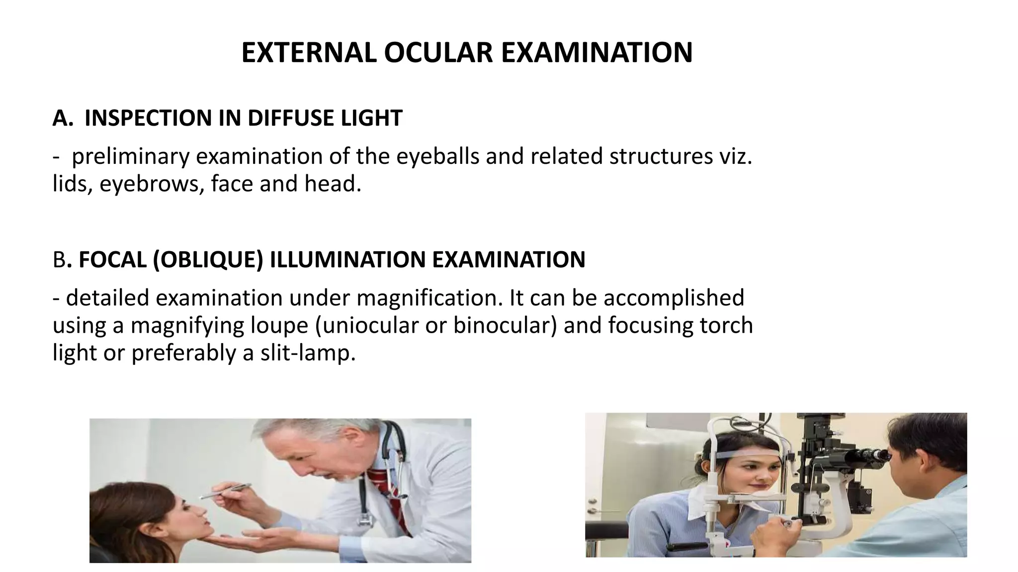
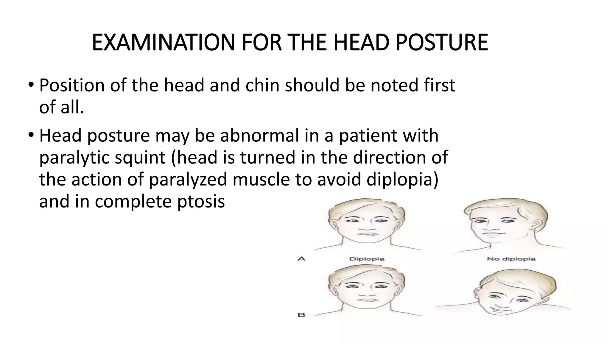
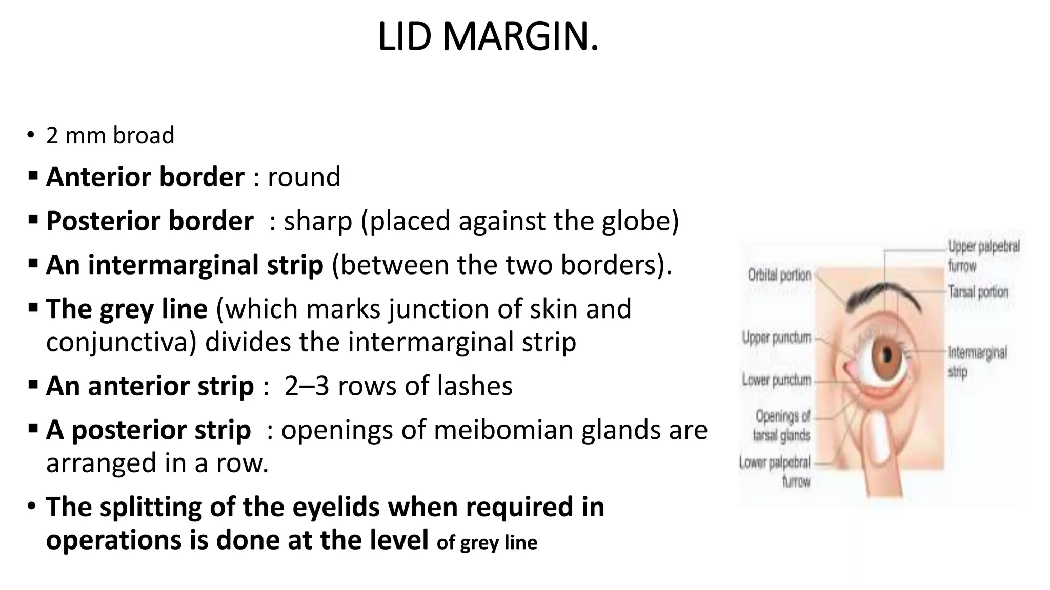
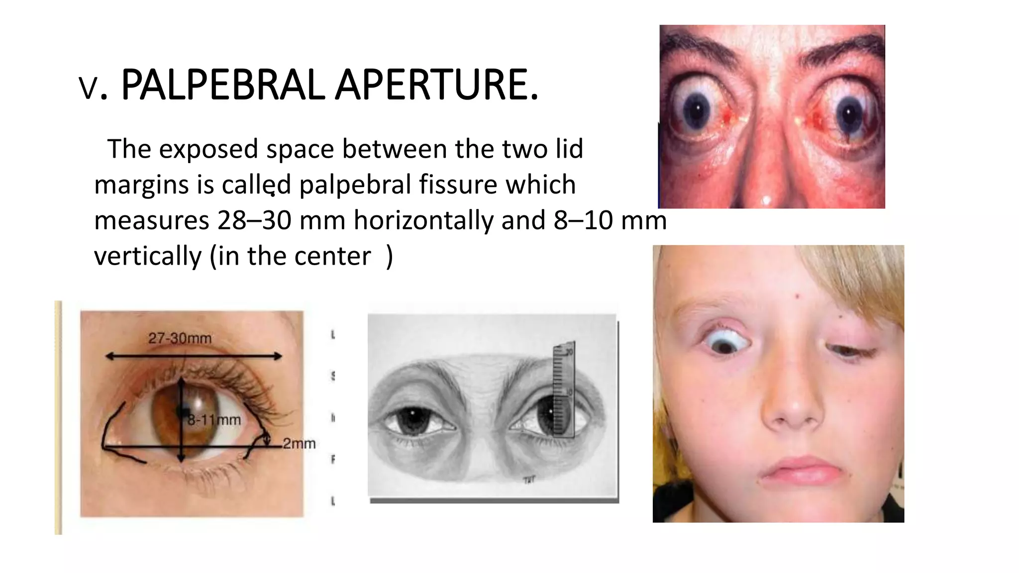
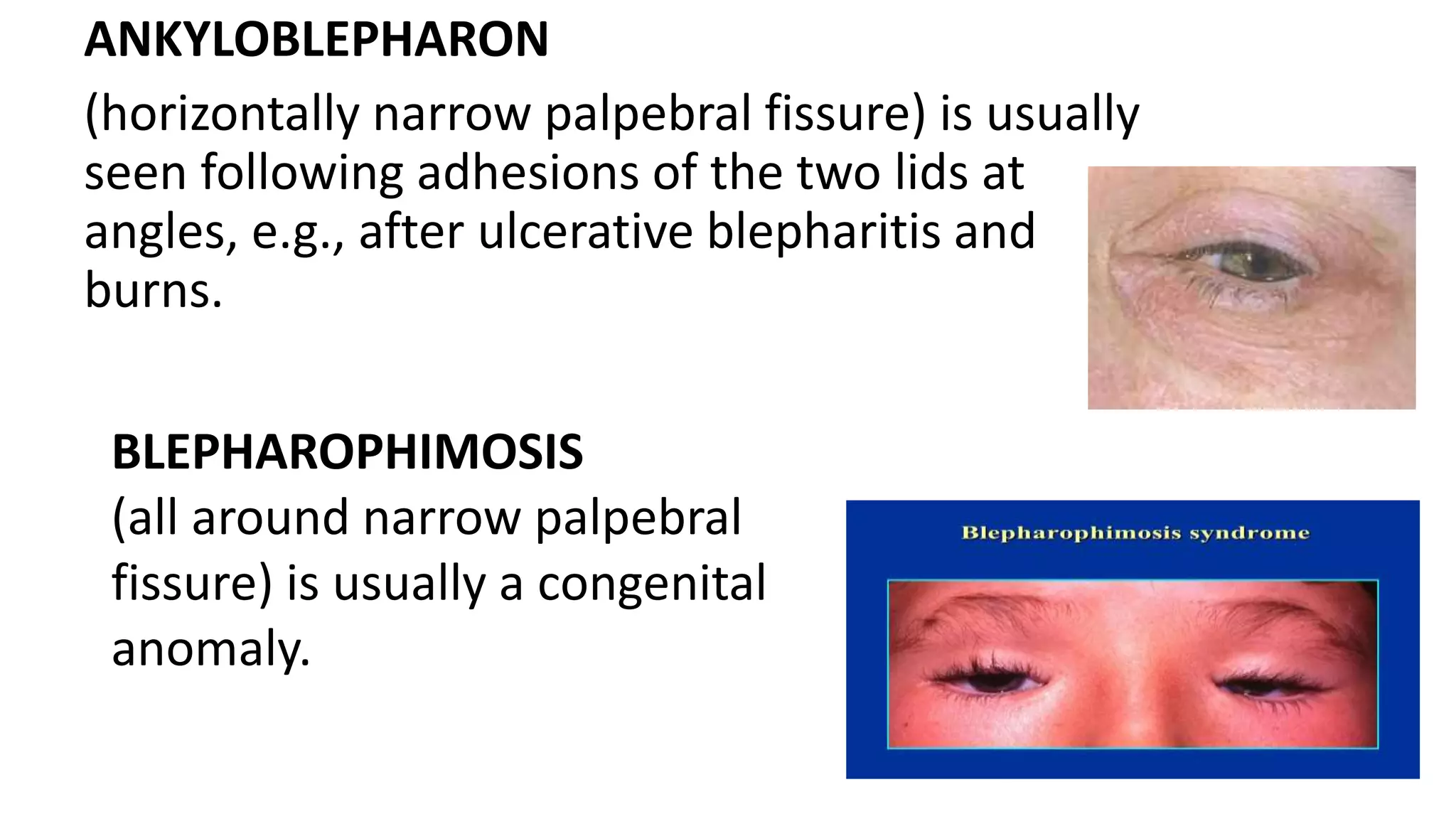
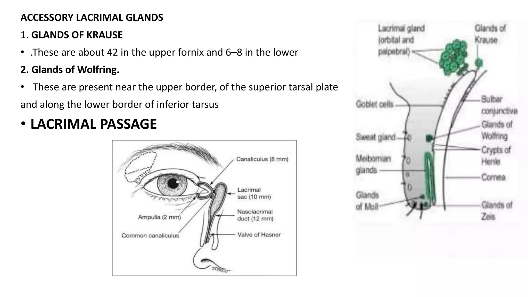
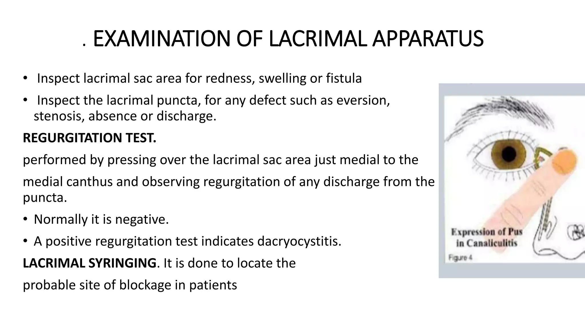
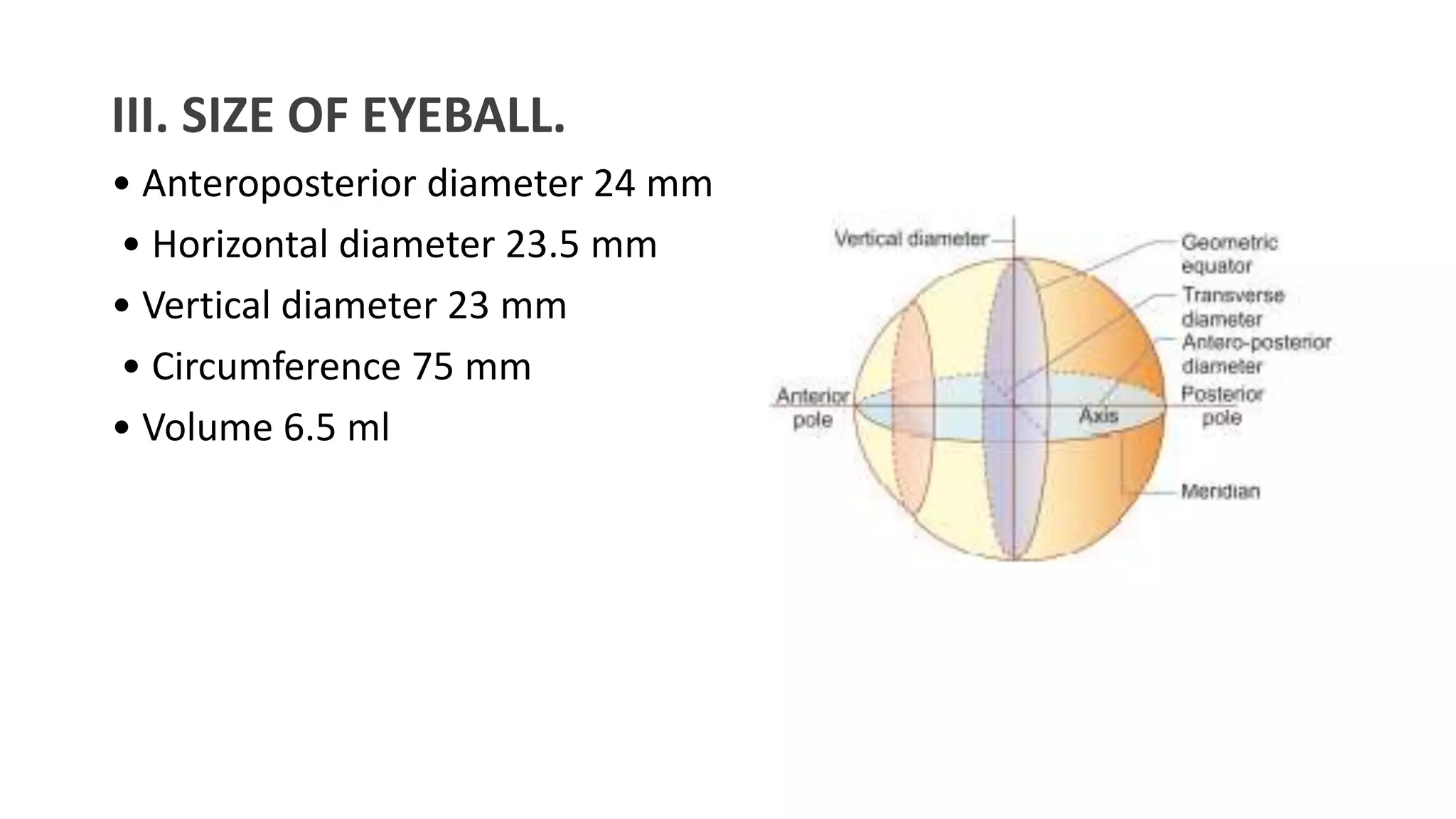
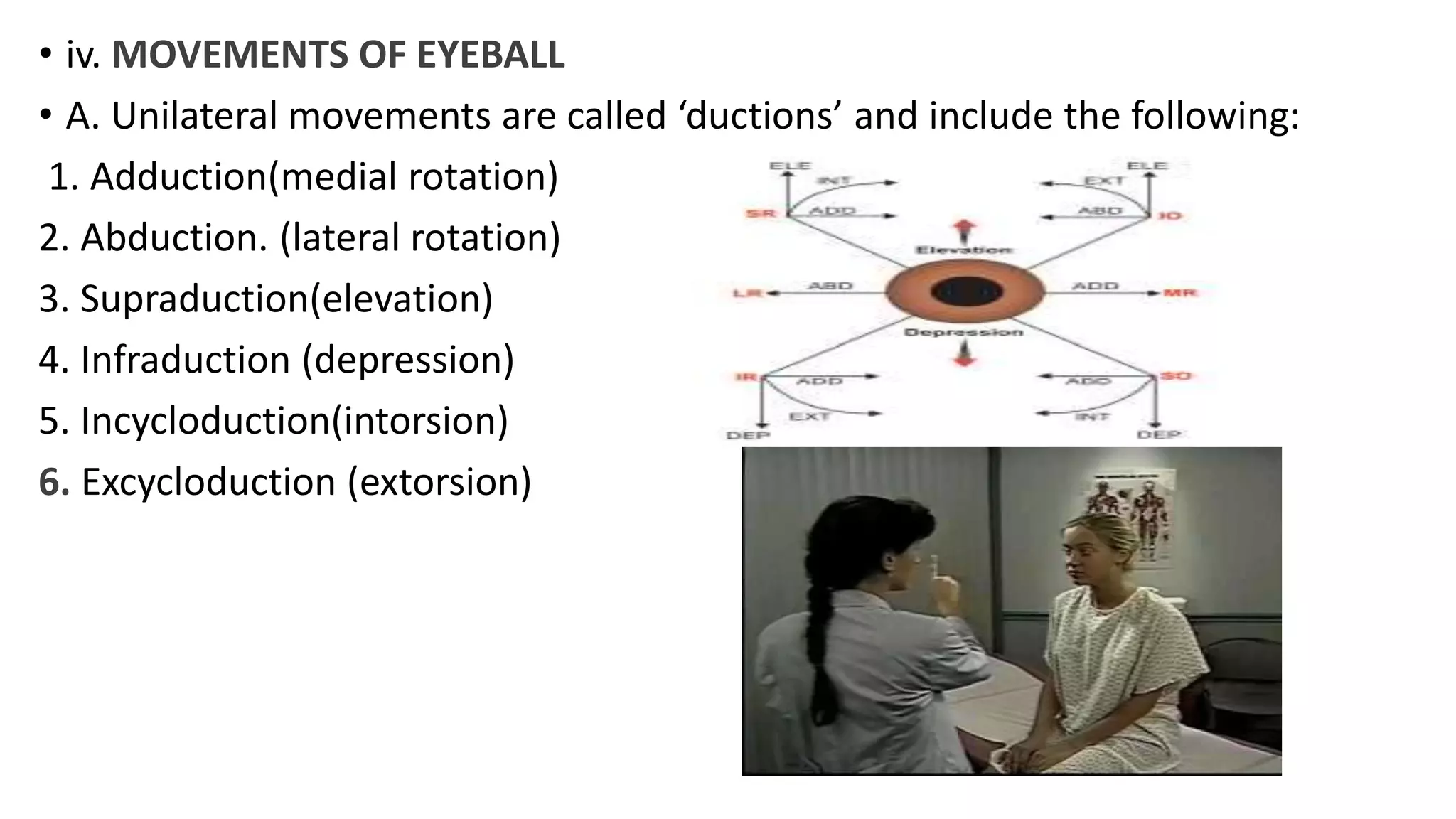
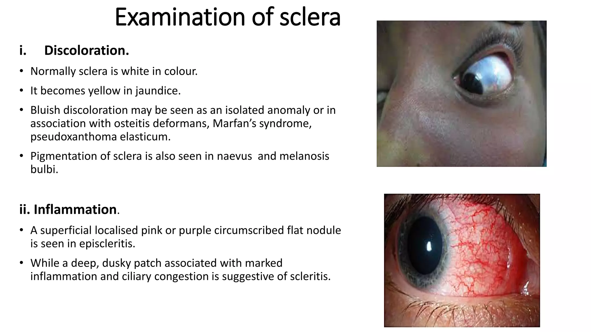
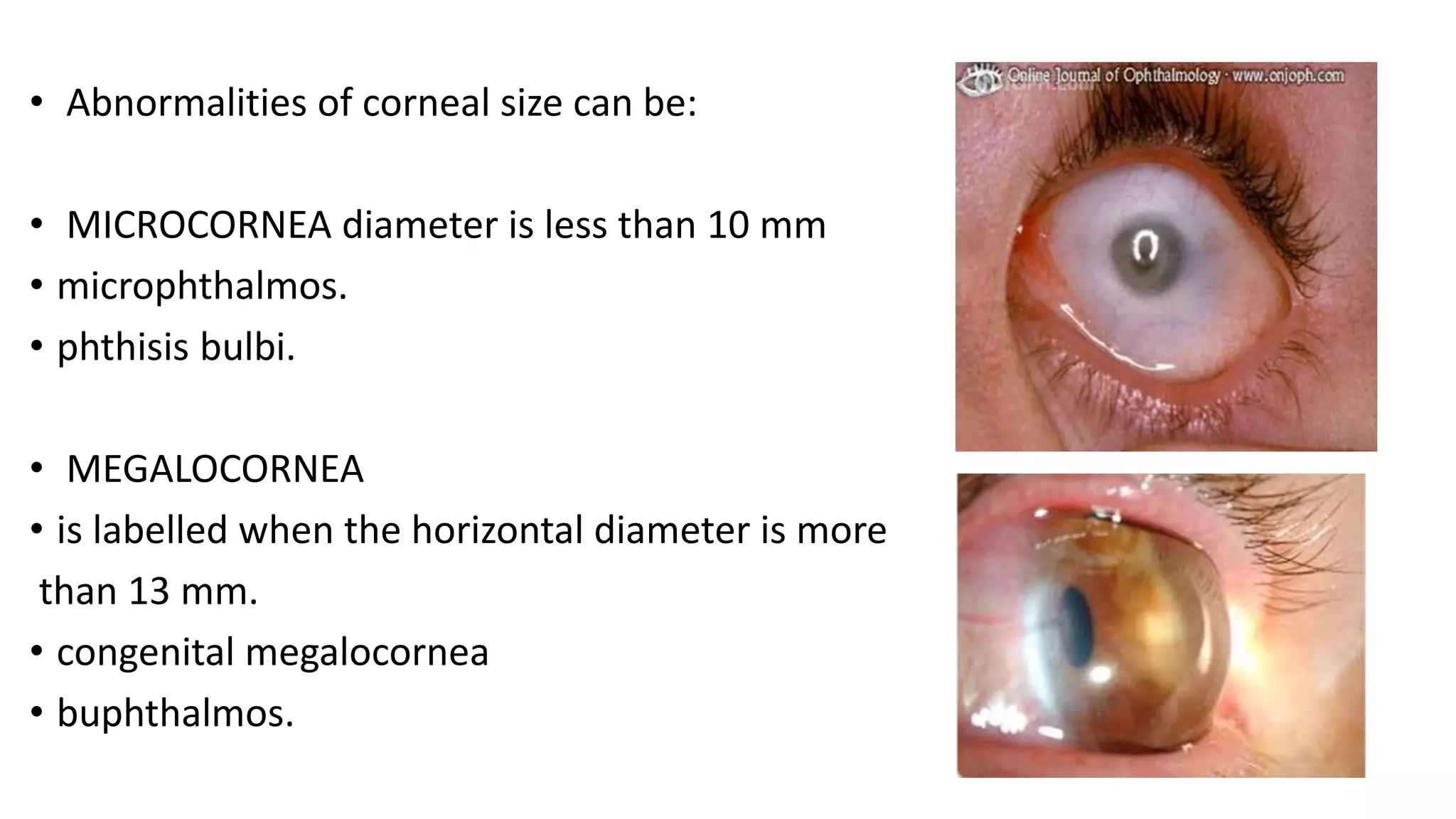
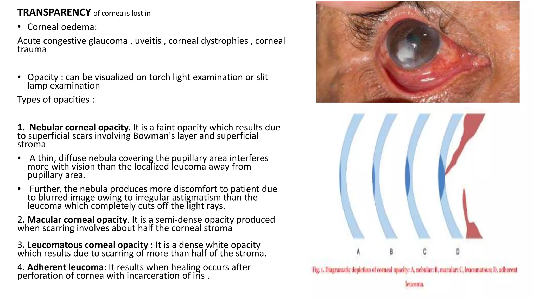
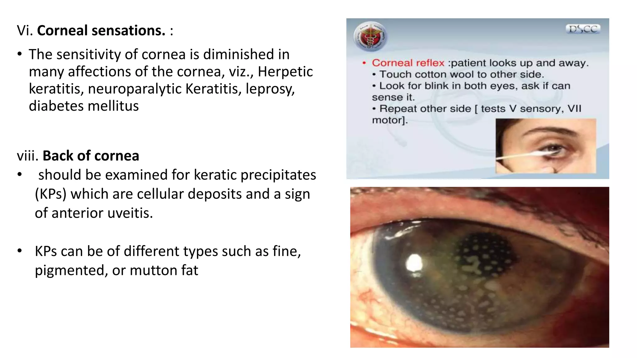
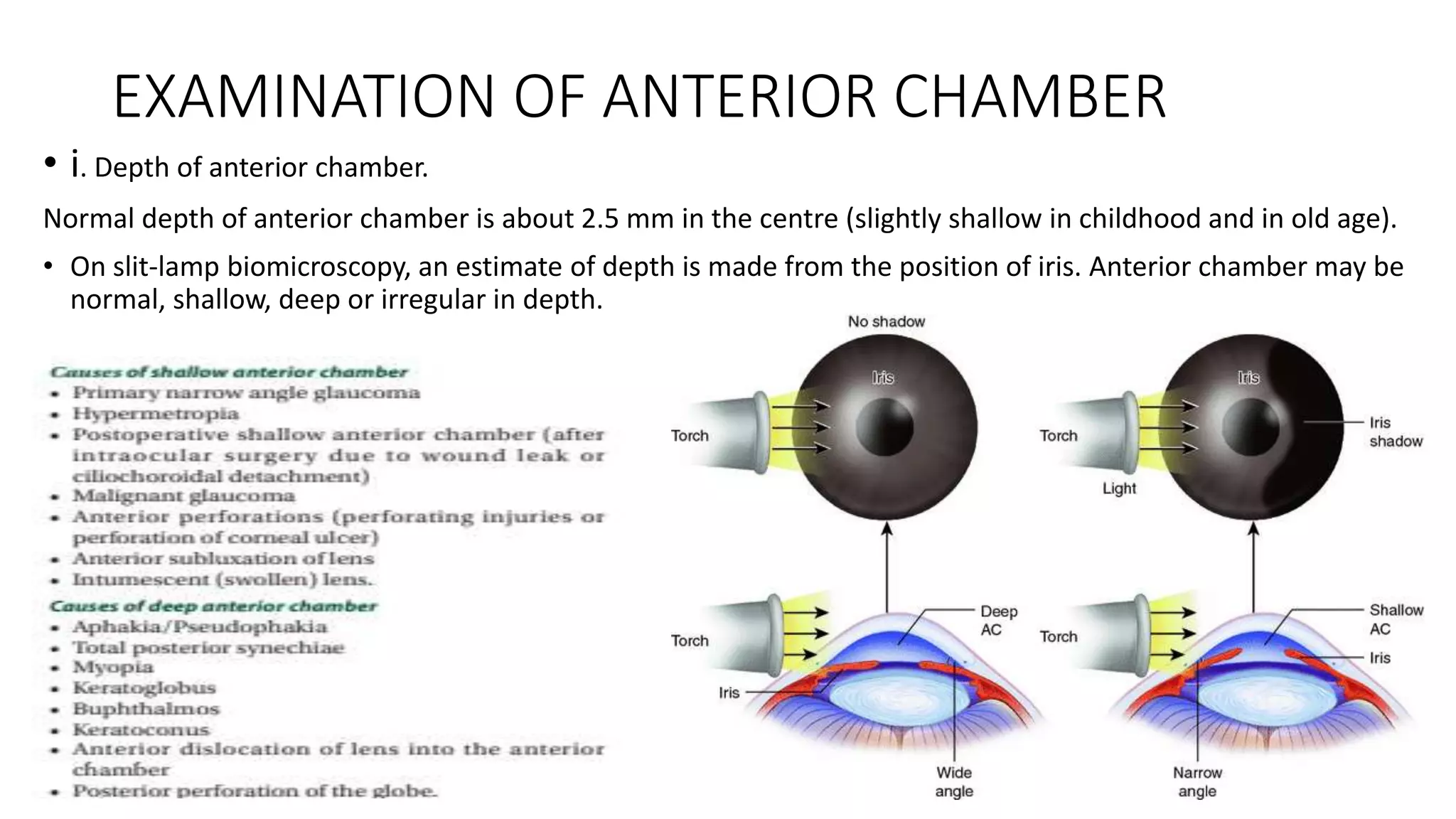
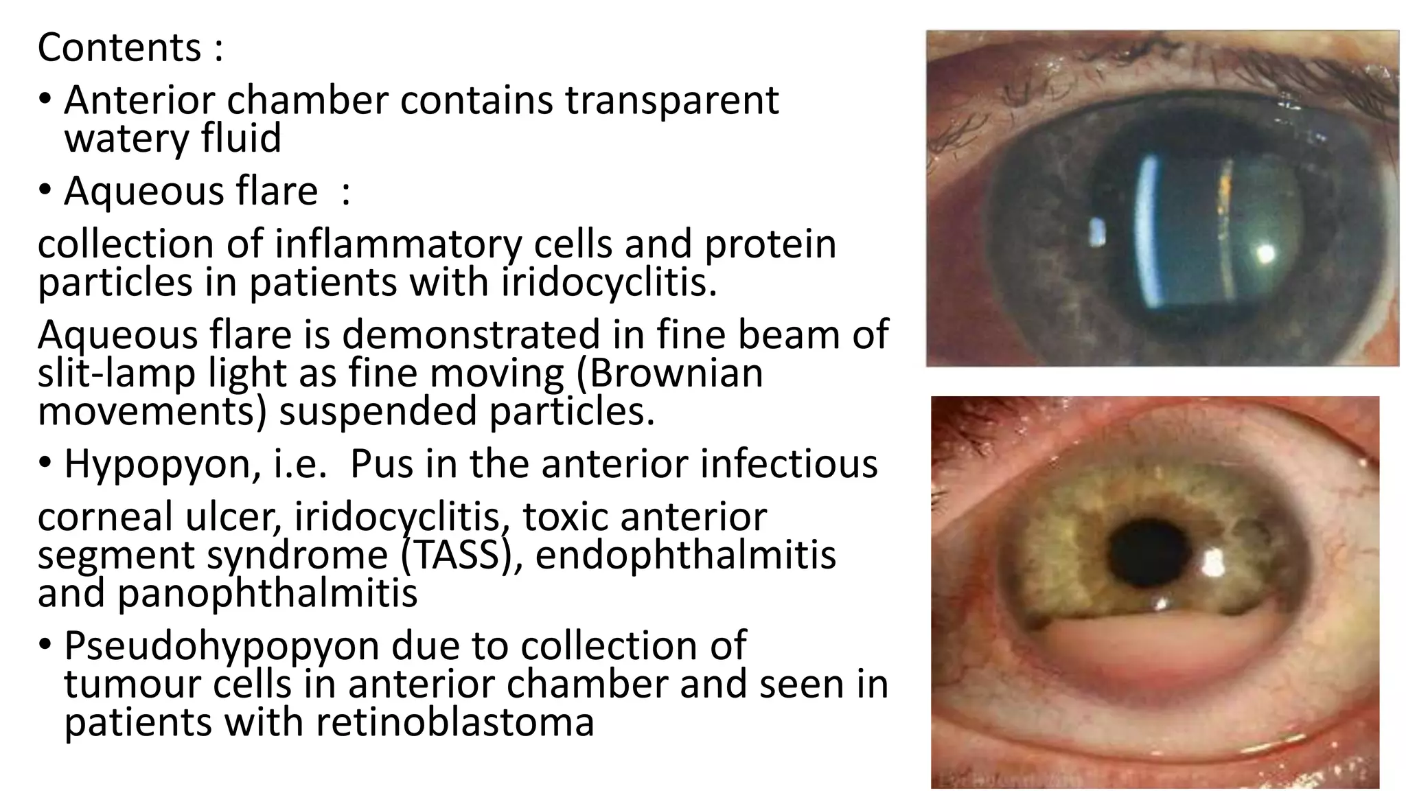
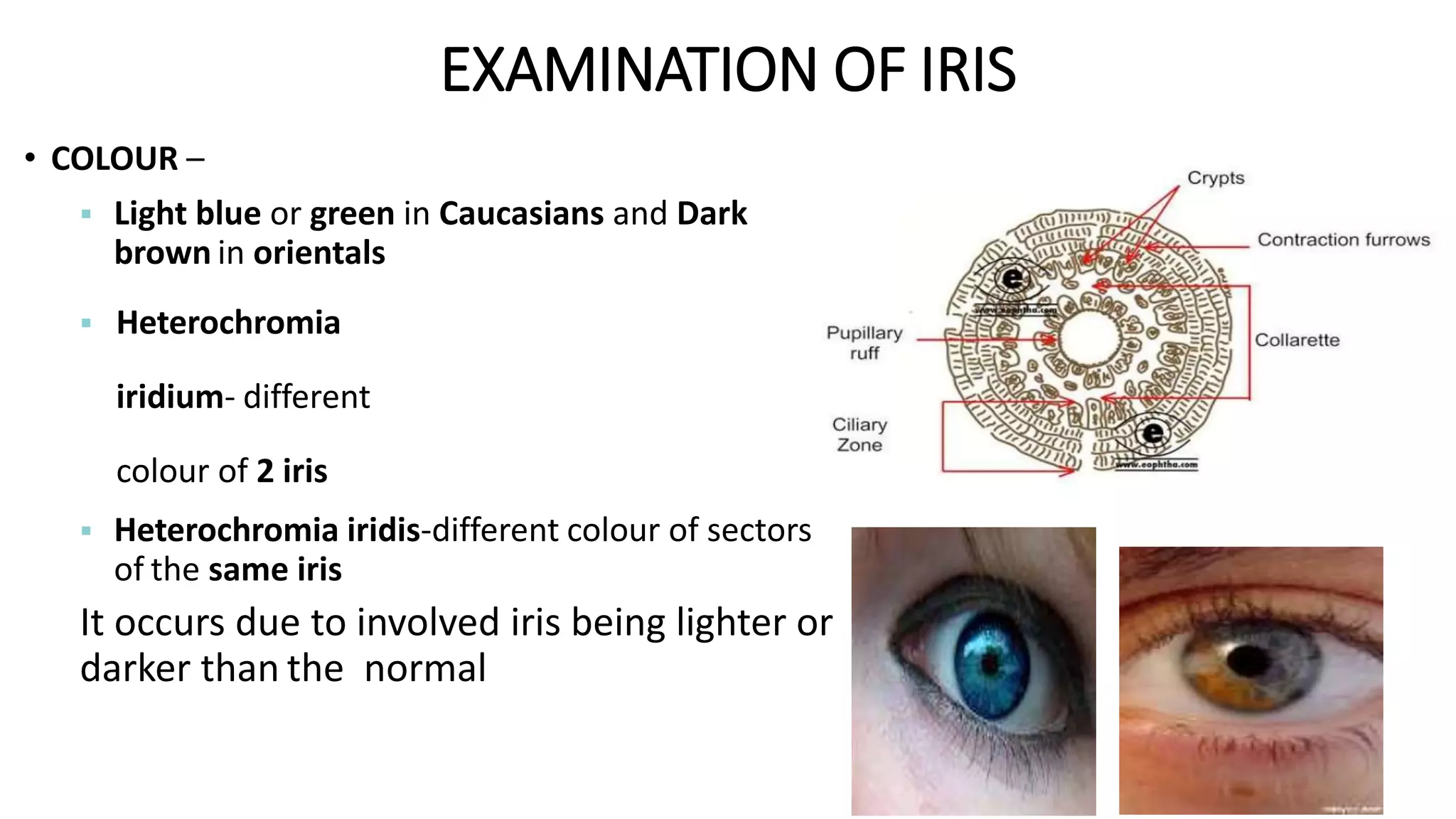
EXAMINATION OF EYE
GENERAL PHYSICAL AND
SYSTEMIC
EXAMINATION
• General physical and systemic examination should
be carried out in each case.
• Sometimes it may help in establishing the etiological
diagnosis, e.g., ankylosing spondylitis may be associated with
uveitis.
OCULAR EXAMINATION
• Testing of visual acuity
• Extra ocular examination
• Intra ocular examination :
1. Anterior segment examination
2. Posterior segment examination ( Fundus examination)
ASSESSMENT OF VISUAL FUNCTION
Forms of visual perception are
-form sense
-the field of vision
-The light sense
-The colour sense
-Visual acuity
Visual field examination with confrontation test, perimetry (kinetic and static)
Dark adaptation – measurement of least luminance required to produce a visual
sensation
Contrast sensitivity – is measurement of the smallest distinguishable contrast ,it is
assessment of quality of vision
Colour vision –with lantern test (edridge green lantern) and Isochromatic charts
VISUAL ACUITY
The principal of assessment is measurement of spatial
resolution of the eye i.e. an estimation of ability of eye to
discriminate between two points.
DISTANCE VISION
Two distance point can be visible as separate only when they subtend an angle of 1
minute at the nodal point of eye.
5
A. INTERPRETATION OF SNELLEN’S CHART FOR
VISUAL ACUITY
B. INTERPRETATION OF PINHOLE
EXTERNAL OCULAR EXAMINATION
A. INSPECTION IN DIFFUSE LIGHT
- preliminary examination of the eyeballs and related structures viz.
lids, eyebrows, face and head.
B. FOCAL (OBLIQUE) ILLUMINATION EXAMINATION
- detailed examination under magnification. It can be accomplished
using a magnifying loupe (uniocular or binocular) and focusing torch
light or preferably a slit-lamp.
EXAMINATION FOR THE HEAD POSTURE
• Position of the head and chin should be noted first
of all.
• Head posture may be abnormal in a patient with
paralytic squint (head is turned in the direction of
the action of paralyzed muscle to avoid diplopia)
and in complete ptosis
EXAMINATION OF FOREHEAD
• Forehead may show increased wrinkling (due to
overaction of frontalis muscle) in patient with ptosis.
• Complete loss of wrinkling in one-half of the forehead is
observed in patients with lower motor neuron facial
palsy.
• Facial asymmetry may be noted in patient with bell’s
palsy
EXAMINATION OF EYEBROWS
 Level of the two eyebrows may be changed in a patient
with ptosis (due to overaction of frontalis).
 Cilia of lateral one-third of the eyebrows may be
Absent (madarosis) in patients with leprosy or
Myxoedema.
4. EXAMINATION OF THE EYELIDS
• i. POSITION. :
Normally the lower lid just touches the limbus while
the upper lid covers about 1/6th (2 mm) of cornea.
• In ptosis, upper lid covers more than 1/6th of
cornea.
• Upper limbus is visible due to lid retraction as in
thyrotoxicosis and sympathetic overactivity
ii. MOVEMENTS OF LIDS :
• Normally the upper lid follows the eyeball in downward movement.
• Blinking is involuntary movement of eyelids.
• Normal rate is 12-16 blinks per minute
Layers of eyelid
Lagophthalmos
– Facial nerve palsy
– Extreme degree of proptosis
– Symblepharon
LID MARGIN.
• 2 mm broad
 Anterior border : round
 Posterior border : sharp (placed against the globe)
 An intermarginal strip (between the two borders).
 The grey line (which marks junction of skin and
conjunctiva) divides the intermarginal strip
 An anterior strip : 2–3 rows of lashes
 A posterior strip : openings of meibomian glands are
arranged in a row.
• The splitting of the eyelids when required in
operations is done at the level of grey line
Abnormalities :
•Entropion
• Ectropion
•Trichiasis
•Distichiasis
•Poliosis
•Madarosis
ECTROPION ENTROPION
TRICHIASIS MADROSIS
v. PALPEBRAL APERTURE.
•
The exposed space between the two lid
margins is called palpebral fissure which
measures 28–30 mm horizontally and 8–10 mm
vertically (in the center )
ANKYLOBLEPHARON
(horizontally narrow palpebral fissure) is usually
seen following adhesions of the two lids at
angles, e.g., after ulcerative blepharitis and
burns.
BLEPHAROPHIMOSIS
(all around narrow palpebral
fissure) is usually a congenital
anomaly.
VERTICALLY NARROW PALPEBRAL FISSURE is seen in:
– Inflammatory conditions of conjunctiva, cornea and uvea due to
blepharospasm
– Ptosis (drooping) of upper eyelid
– Enophthalmos (sunken eyeball)
– Anophthalmos (absent eyeball)
– Microphthalmos (congenital small eyeball)
– Phthisis bulbi
– Atrophic bulbi
VERTICALLY WIDE PALPEBRAL FISSURE may be noted in patients
with:
– Proptosis
– Retraction of upper lid
– Facial nerve palsy
LACRIMAL APPARATUS
LACRIMAL GLANDS
• Lacrimal glands includes main and accessory.
Main lacrimal gland It consists of an upper orbital and a lower palpebral part.
1. ORBITAL PART
• larger, about the size and shape of a small almond
2. PALPEBRAL PART
• small and consists of only one or two lobules
3. DUCTS OF LACRIMAL GLAND
• 10–12 ducts pass downward to open in the lateral part of superior fornix.
• One or two ducts also open in the lateral part of inferior fornix.
ACCESSORY LACRIMAL GLANDS
1. GLANDS OF KRAUSE
• .These are about 42 in the upper fornix and 6–8 in the lower
2. Glands of Wolfring.
• These are present near the upper border, of the superior tarsal plate
and along the lower border of inferior tarsus
• LACRIMAL PASSAGE
. EXAMINATION OF LACRIMAL APPARATUS
• Inspect lacrimal sac area for redness, swelling or fistula
• Inspect the lacrimal puncta, for any defect such as eversion,
stenosis, absence or discharge.
REGURGITATION TEST.
performed by pressing over the lacrimal sac area just medial to the
medial canthus and observing regurgitation of any discharge from the
puncta.
• Normally it is negative.
• A positive regurgitation test indicates dacryocystitis.
LACRIMAL SYRINGING. It is done to locate the
probable site of blockage in patients
6. EXAMINATION OF EYEBALL
I. POSITION.
Normally, the two eyeballs are symmetrically placed in the orbits in such a
way that a line joining the central points of superior and inferior orbital
margins just touches the cornea.
• Abnormalities of the position of eyeball can be:
(a)Proptosis/exophthalmos
(b) Enophthalmos (sunken eyeball)
ii. VISUAL AXES OF EYEBALLS.
Normally the visual axes of the two eyes are simultaneously
directed at the same object which is maintained in all the
directions of gaze.
III. SIZE OF EYEBALL.
• Anteroposterior diameter 24 mm
• Horizontal diameter 23.5 mm
• Vertical diameter 23 mm
• Circumference 75 mm
• Volume 6.5 ml
THE SIZE OF EYEBALL IS INCREASED IN CONDITIONS
• buphthalmos
• unilateral high myopia.
THE CAUSES OF SMALL-SIZED EYEBALL :
• Microphthalmos
• Phthisis bulbi
• And atrophic bulbi.
Buphthalmos
• iv. MOVEMENTS OF EYEBALL
• A. Unilateral movements are called ‘ductions’ and include the following:
1. Adduction(medial rotation)
2. Abduction. (lateral rotation)
3. Supraduction(elevation)
4. Infraduction (depression)
5. Incycloduction(intorsion)
6. Excycloduction (extorsion)
Binocular movements.
Versions : conjugate movements , are synchronous (simultaneous) symmetric
movements of both eyes in the same direction.
These include:
1. Dextroversion
2. Levoversion.
3. Supraversion .
4. Infraversion.
5. Dextroelevation.
6. Dextrodepression
7. Levoelevation
8. Levodepression
9. Dextrocycloversion
10. Levocycloversion
b. Vergences : disconjugate movements
are synchronous and symmetric movements
of both eyes in opposite directions e.g.:
1. Convergence
2. Divergence.
EXAMINATION OF CONJUNCTIVA
BULBAR CONJUNCTIVA can be examined by simply
retracting the upper lid with index finger and lower lid
with thumb of the left hand.
Lower palpebral conjunctiva and lower fornix can be examined by
just pulling down the lower lid and instructing the patient to look up
Upper palpebral conjunctiva can be examined
only after everting the upper eyelid. Eversion of upper lid can be
carried out by one-hand or two-hand technique.
CONGESTION :
• SUPERFICIAL (in conjunctivitis)
• CILIARY/CIRCUMCORNEAL /DEEP
(in iridocyclitis, and keratitis)
REACTION :
• Follicular reaction :
greyish white raised areas (mimicking boiled sago-grains)
on fornices and palpebral conjunctiva.
Follicles represent areas of aggregation of lymphocytes.
may be seen in following conditions:
• Trachoma
• Acute follicular conjunctivitis
• Chronic follicular conjunctivitis
PAPILLAE
• reddish raised areas with flat tops and
velvety appearance.
• These represent areas of vascular and
epithelial hyperplasia.
• Trachoma
• Spring catarrh
• Allergic conjunctivitis
• Giant papillary conjunctivitis
DISCHARGE :
• Serous : watery exudate in acute viral and acute allergic conjunctivitis.
• Mucoid : mucus discharge in VKC and KCS (dry eyes).
• Purulent : puss in severe acute bacterial conjunctivitis.
• Mucopurulent : puss plus mucus in mild
bacterial conjunctivitis and Chlamydial conjunctivitis.
Examination of sclera
i. Discoloration.
• Normally sclera is white in colour.
• It becomes yellow in jaundice.
• Bluish discoloration may be seen as an isolated anomaly or in
association with osteitis deformans, Marfan’s syndrome,
pseudoxanthoma elasticum.
• Pigmentation of sclera is also seen in naevus and melanosis
bulbi.
ii. Inflammation.
• A superficial localised pink or purple circumscribed flat nodule
is seen in episcleritis.
• While a deep, dusky patch associated with marked
inflammation and ciliary congestion is suggestive of scleritis.
iii. Staphyloma
• is a thinned out bulging area of sclera
which is lined by the uveal tissue.
• Types : Depending upon its location, scleral
staphylomas may be intercalary, ciliary,
equatorial and posterior
EXAMINATION OF CORNEA
i. SIZE.
• The anterior surface of normal cornea is elliptical
• average horizontal diameter of 11.7
• mm and vertical diameter of 11 mm.
Layers of cornea :
• Abnormalities of corneal size can be:
• MICROCORNEA diameter is less than 10 mm
• microphthalmos.
• phthisis bulbi.
• MEGALOCORNEA
• is labelled when the horizontal diameter is more
than 13 mm.
• congenital megalocornea
• buphthalmos.
II SHAPE (CURVATURE).
Normal cornea is like a watchglass with a
uniform posterior curve in its central area
• KERATOGLOBUS. It is an ectatic condition in
which cornea becomes thin and bulges out
like a globe.
• KERATOCONUS. It is an ectatic condition in
which cornea becomes cone shaped.
• CORNEA PLANA I.e., flat curvature of cornea
which may occur in patients with severe
hypotony and phthisis bulbi and rarely as a
congenital anomaly.
III. SURFACE
• abrasions, ulceration, ectatic scars and facets.
• Changes in smoothness of surface can be detected by slit-lamp biomicroscopy,
window reflex test and Placido’s disc.
• Placido’s keratoscopic disc
• It is a disc painted with alternating black and white circles.
• It may be used to assess the smoothness and curvature of corneal surface.
• Normally, on looking through thehole in the centre of disc a uniform sharp image of
the circles is seen on the cornea .
• Irregularities in the corneal surface cause distortion of the circles
Placido’s disc reflex from normal cornea. Placido’s disc reflex from irregular
corneal surface.
TRANSPARENCY of cornea is lost in
• Corneal oedema:
Acute congestive glaucoma , uveitis , corneal dystrophies , corneal
trauma
• Opacity : can be visualized on torch light examination or slit
lamp examination
Types of opacities :
1. Nebular corneal opacity. It is a faint opacity which results due
to superficial scars involving Bowman's layer and superficial
stroma
• A thin, diffuse nebula covering the pupillary area interferes
more with vision than the localized leucoma away from
pupillary area.
• Further, the nebula produces more discomfort to patient due
to blurred image owing to irregular astigmatism than the
leucoma which completely cuts off the light rays.
2. Macular corneal opacity. It is a semi-dense opacity produced
when scarring involves about half the corneal stroma
3. Leucomatous corneal opacity : It is a dense white opacity
which results due to scarring of more than half of the stroma.
4. Adherent leucoma: It results when healing occurs after
perforation of cornea with incarceration of iris .
• ULCERATION : bacterial, fungal , viral
• Examination for corneal ulcer. Once corneal ulcer is suspected, a
thorough biomicroscopic examination before and after fluorescein
staining should be performed to note the site, size, shape, depth,
floor and edges of the corneal ulcer.
• VASCULARIZATION :
EXAMINATION FOR CORNEAL ULCER
site , size, shape, depth, floor
And edges of the corneal ulcer.
EXAMINATION FOR CORNEAL OPACITY
Note the number, site, size,
Shape, density (nebular, macular or leucomatous) and
Surface of the opacity.
Vi. Corneal sensations. :
• The sensitivity of cornea is diminished in
many affections of the cornea, viz., Herpetic
keratitis, neuroparalytic Keratitis, leprosy,
diabetes mellitus
viii. Back of cornea
• should be examined for keratic precipitates
(KPs) which are cellular deposits and a sign
of anterior uveitis.
• KPs can be of different types such as fine,
pigmented, or mutton fat
EXAMINATION OF ANTERIOR CHAMBER
• i. Depth of anterior chamber.
Normal depth of anterior chamber is about 2.5 mm in the centre (slightly shallow in childhood and in old age).
• On slit-lamp biomicroscopy, an estimate of depth is made from the position of iris. Anterior chamber may be
normal, shallow, deep or irregular in depth.
Contents :
• Anterior chamber contains transparent
watery fluid
• Aqueous flare :
collection of inflammatory cells and protein
particles in patients with iridocyclitis.
Aqueous flare is demonstrated in fine beam of
slit-lamp light as fine moving (Brownian
movements) suspended particles.
• Hypopyon, i.e. Pus in the anterior infectious
corneal ulcer, iridocyclitis, toxic anterior
segment syndrome (TASS), endophthalmitis
and panophthalmitis
• Pseudohypopyon due to collection of
tumour cells in anterior chamber and seen in
patients with retinoblastoma
• Foreign bodies , crystalline lens may be
observed in anterior chamber after anterior
dislocation of lens , lens particles in anterior
chamber after trauma
• Blood in the anterior chamber is called
hyphaema
ocular trauma, surgery, herpes zoster and
gonococcal iridocyclitis, blood dyscrasias,
clotting disorder and intraocular tumours (e.g.
retinoblastoma, angioma).
• Parasitic cyst, e.g. cysticercus cellulosae has
been demonstrated in anterior chamber.
• Artificial lens. Anterior chamber intraocular
lens may be observed in patients with
pseudophakia
• Angle of anterior chamber :
• Examination of angle of anterior chamber is performed with the help
of a gonioscope and slitlamp. Gonioscopy is a specialized examination
required in patients with glaucoma
EXAMINATION OF IRIS
• COLOUR –
 Light blue or green in Caucasians and Dark
brown in orientals
 Heterochromia
iridium- different
colour of 2 iris
 Heterochromia iridis-different colour of sectors
of the same iris
It occurs due to involved iris being lighter or
darker than the normal
• Iris lighter than normal-
• congenital heterochromia,
• atrophic patches in chronic uveitis
• metastatic carcinoma
• Iris darker than normal-
Iris naevi-appear as freckles ocular melanocytosis,
haemosiderosis, siderosis bulbi, retained iris
foreign body, malignant melanoma of iris
lymphoma
PATTERN OF NORMAL IRIS
o Pattern occurs due to presence of collarette, crypts and radial striations on its
anterior surface
o It is disturbed in
i. Acute iridocyclitis-muddy iris
ii. Healed iridocyclitis-atrophy of iris
 PERSISTENT PUPILLARYMEMBRANE
oIt is seen as abnormal congenital tags of iris tissue
adherent to the collarette area
 SYNECHIAE
o It is the adhesion of iris to other intraocular
structures
o Types-
anterior (in adherent leucoma) or posterior (in
iridocyclitis).
Posterior synechiae may be total, annular
(ring), or segmental
IRIDODONESIS
o It is the tremulousness(trembling) of the iris
o Seen in aphakia , psudophakia
and subluxation of lens (since
posterior support is lost)
 NODULES ON THE IRIS SURFACE
• granulomatous uveitis, melanoma,
tuberculoma and gumma of iris
• Koeppe's nodules are small nodules seen at the
inner margin of the iris in patients with
granulomatous anterior uveitis, which occurs in
conditions such as sarcoidosis and tuberculosis.
• Busacca nodules inflammatory, granulomatous
nodules located away from the pupillary margin
of the iris.
• Lisch nodules are melanocytic hamartomas that
appear as well-defined, dome-shaped elevations
projecting from the surface of the iris and are
clear to yellow or brown
 RUBEOSIS IRIDIS
o New vessel formation on the iris
o Seen in diabetic retinopathy, central retinal vein occlusion, chronic uveitis, chronic retinal
detachment, retinoblastoma
GAP OR HOLE IN THE IRIS
o May be due to congenital coboloma or due to
iridectomy(surgical coboloma)
o Iridodialysis-separation of iris from ciliary
body , leads a D - shaped pupil
 ANIRIDIA ORIRIDEREMIA
o Complete absence of iris
o Rare congenital condition
 IRIS CYST
o In patients using strong
miotic drops, it may be
seen in the pupillary
margin
Examination of PUPIL
NUMBER
o Normal: 1 pupil
o Rarely: more than 1 pupil (polycoria)
 LOCATION
o Normal: almost centre of the iris, slightly nasal
o Rarely: congenitally eccentric (corectopia)
 SIZE
o Normal: 3-4mm depending upon illumination
o It may be abnormally small (miosis) or
large(mydriasis)
o Anisocoria- It is a condition where there is difference
between the size of two pupils
o Causes of Miosis
• Effect of local miotic drugs-
Parasympathomimetic drugs
• Iridocyclitis-narrow, irregular,
non-reacting pupil
• Head injury-pontine
haemorrhage
• Senile rigid miotic pupil
• Due to effect of strong light
• During sleep pupil is pinpoint
o Causes of Mydriasis
• Effect of topical
sympathomimetic drugs-
Adrenaline, phenylephrine
• Effect of topical
parasympatholytic drugs-
Atropine, homatropine,
tropicamide, cyclopentolate
• Acute congestive glaucoma
• Absolute glaucoma
 SHAPE
o Normal: circular
o Irregular narrow- iridocyclitis
o Festooned- effect of mydriatics
on posterior segment synechiae
o Vertically oval/pear
shaped/updrawn-
postoperatively (due to
incarceration of iris or
vitreous in the wound at 12
o’clock postion)
 COLOUR
o Normal: greyish black
o Aphakia-jet black
o Immature senile cortical cataract-greyish white
o Mature cortical cataract-pearly white
o Hypermature cataract-milky white
o Cataracta brunescens-brown
o Cataracta nigra-brownish black
o Leucocoria-white reflex in pupil
o Glaucoma-greenish hue
o Iridocyclitis-dirty white exudates
PUPILLARY REACTIONS
o Direct light reflex-
Normal: pupil reacts briskly and constricts
o Consensual light reflex
Normal: contralateral pupil should also
constrict
o Swinging flash light test-(when RAPD is
suspected)
• Normal: both pupils constrict equally
and the pupil to which
light is transferred remains tightly
constricted
• RAPD present: affected pupil will
dilate when flash light is moved from
normal eye to abnormal eye. This is
called Marcus Gunn Pupil
Abnormal pupillary reactions are seen in
i. Amaurotic pupil
ii. Efferent pathway defect
iii. Wernicke’s hemianopic pupil
iv. Marcus Gunn pupil
v. Argyll Robertson pupil
vi. Tonic pupil
Examination of LENS
• Can be examined using oblique illumination,
slit lamp biomicroscopy and distant direct
ophthalmoscopy with FULLY DILATED PUPILS
• POSITION
o Normal: patellar fossa by the zonules
o Dislocation of lens: lens not present in its
normal position
i. Anterior dislocation-present in anterior
chamber
ii. Posterior dislocation-present in
vitreous cavity either floating(LENSA
NUTANS) or fixed to theretina(LENSA
FIXATE)
o Subluxation of lens-lens is partially displaced from its
position
• Causes-trauma, marfan’s syndrome,
homocystinuria
o Aphakia-absence of lens
• It is diagnosed by
i. jet black pupil, deep anterior chamber, empty
patellar fossa by slit lamp biomicroscopy
ii. hypermetropic eye on ophthalmoscopy,
retinoscopy
iii. ABSENCE of 3rd and 4th purkinjeimages
o Pseudophakia-
• When posterior chamber IOL is present, it is
diagnosed by black pupil, deep anterior chamber,
shining reflexes (from anterior surface of IOL) and
PRESENCE of all the four Purkinje images
 SHAPE
o Normal: biconvex structure, on slit lamp-optical section shows
embryonic, foetal, infantile and adult nuclei, cortex and
capsule
o Spherophakia-spherical
o Lenticonus anterior-anterior cone shaped bulge (Alport
syndrome)
o Lenticonus posterior-posterior cone shaped bulge
o Coloboma of lens-Notch in the lens
 Colour
o Normal: In young age, it is almost clear or gives a faint blue hue
o Old age-greyish white (mistaken to be cataract)
o CORTICAL cataract- greyish white (immature), pearlywhite
(mature), milky white (hypermature)
o NUCLEAR cataract-amber, brown or black
o Cataractous lens with siderosis bulbi-rusty (orange)
• Thank you

Examination of eye

  • 1.
  • 2.
    GENERAL PHYSICAL AND SYSTEMIC EXAMINATION •General physical and systemic examination should be carried out in each case. • Sometimes it may help in establishing the etiological diagnosis, e.g., ankylosing spondylitis may be associated with uveitis.
  • 3.
    OCULAR EXAMINATION • Testingof visual acuity • Extra ocular examination • Intra ocular examination : 1. Anterior segment examination 2. Posterior segment examination ( Fundus examination)
  • 4.
    ASSESSMENT OF VISUALFUNCTION Forms of visual perception are -form sense -the field of vision -The light sense -The colour sense -Visual acuity Visual field examination with confrontation test, perimetry (kinetic and static) Dark adaptation – measurement of least luminance required to produce a visual sensation Contrast sensitivity – is measurement of the smallest distinguishable contrast ,it is assessment of quality of vision Colour vision –with lantern test (edridge green lantern) and Isochromatic charts
  • 5.
    VISUAL ACUITY The principalof assessment is measurement of spatial resolution of the eye i.e. an estimation of ability of eye to discriminate between two points. DISTANCE VISION Two distance point can be visible as separate only when they subtend an angle of 1 minute at the nodal point of eye. 5
  • 6.
    A. INTERPRETATION OFSNELLEN’S CHART FOR VISUAL ACUITY B. INTERPRETATION OF PINHOLE
  • 7.
    EXTERNAL OCULAR EXAMINATION A.INSPECTION IN DIFFUSE LIGHT - preliminary examination of the eyeballs and related structures viz. lids, eyebrows, face and head. B. FOCAL (OBLIQUE) ILLUMINATION EXAMINATION - detailed examination under magnification. It can be accomplished using a magnifying loupe (uniocular or binocular) and focusing torch light or preferably a slit-lamp.
  • 8.
    EXAMINATION FOR THEHEAD POSTURE • Position of the head and chin should be noted first of all. • Head posture may be abnormal in a patient with paralytic squint (head is turned in the direction of the action of paralyzed muscle to avoid diplopia) and in complete ptosis
  • 9.
    EXAMINATION OF FOREHEAD •Forehead may show increased wrinkling (due to overaction of frontalis muscle) in patient with ptosis. • Complete loss of wrinkling in one-half of the forehead is observed in patients with lower motor neuron facial palsy. • Facial asymmetry may be noted in patient with bell’s palsy
  • 10.
    EXAMINATION OF EYEBROWS Level of the two eyebrows may be changed in a patient with ptosis (due to overaction of frontalis).  Cilia of lateral one-third of the eyebrows may be Absent (madarosis) in patients with leprosy or Myxoedema.
  • 11.
    4. EXAMINATION OFTHE EYELIDS • i. POSITION. : Normally the lower lid just touches the limbus while the upper lid covers about 1/6th (2 mm) of cornea. • In ptosis, upper lid covers more than 1/6th of cornea. • Upper limbus is visible due to lid retraction as in thyrotoxicosis and sympathetic overactivity ii. MOVEMENTS OF LIDS : • Normally the upper lid follows the eyeball in downward movement. • Blinking is involuntary movement of eyelids. • Normal rate is 12-16 blinks per minute
  • 12.
    Layers of eyelid Lagophthalmos –Facial nerve palsy – Extreme degree of proptosis – Symblepharon
  • 13.
    LID MARGIN. • 2mm broad  Anterior border : round  Posterior border : sharp (placed against the globe)  An intermarginal strip (between the two borders).  The grey line (which marks junction of skin and conjunctiva) divides the intermarginal strip  An anterior strip : 2–3 rows of lashes  A posterior strip : openings of meibomian glands are arranged in a row. • The splitting of the eyelids when required in operations is done at the level of grey line
  • 14.
  • 15.
    v. PALPEBRAL APERTURE. • Theexposed space between the two lid margins is called palpebral fissure which measures 28–30 mm horizontally and 8–10 mm vertically (in the center )
  • 16.
    ANKYLOBLEPHARON (horizontally narrow palpebralfissure) is usually seen following adhesions of the two lids at angles, e.g., after ulcerative blepharitis and burns. BLEPHAROPHIMOSIS (all around narrow palpebral fissure) is usually a congenital anomaly.
  • 17.
    VERTICALLY NARROW PALPEBRALFISSURE is seen in: – Inflammatory conditions of conjunctiva, cornea and uvea due to blepharospasm – Ptosis (drooping) of upper eyelid – Enophthalmos (sunken eyeball) – Anophthalmos (absent eyeball) – Microphthalmos (congenital small eyeball) – Phthisis bulbi – Atrophic bulbi VERTICALLY WIDE PALPEBRAL FISSURE may be noted in patients with: – Proptosis – Retraction of upper lid – Facial nerve palsy
  • 18.
    LACRIMAL APPARATUS LACRIMAL GLANDS •Lacrimal glands includes main and accessory. Main lacrimal gland It consists of an upper orbital and a lower palpebral part. 1. ORBITAL PART • larger, about the size and shape of a small almond 2. PALPEBRAL PART • small and consists of only one or two lobules 3. DUCTS OF LACRIMAL GLAND • 10–12 ducts pass downward to open in the lateral part of superior fornix. • One or two ducts also open in the lateral part of inferior fornix.
  • 19.
    ACCESSORY LACRIMAL GLANDS 1.GLANDS OF KRAUSE • .These are about 42 in the upper fornix and 6–8 in the lower 2. Glands of Wolfring. • These are present near the upper border, of the superior tarsal plate and along the lower border of inferior tarsus • LACRIMAL PASSAGE
  • 20.
    . EXAMINATION OFLACRIMAL APPARATUS • Inspect lacrimal sac area for redness, swelling or fistula • Inspect the lacrimal puncta, for any defect such as eversion, stenosis, absence or discharge. REGURGITATION TEST. performed by pressing over the lacrimal sac area just medial to the medial canthus and observing regurgitation of any discharge from the puncta. • Normally it is negative. • A positive regurgitation test indicates dacryocystitis. LACRIMAL SYRINGING. It is done to locate the probable site of blockage in patients
  • 21.
    6. EXAMINATION OFEYEBALL I. POSITION. Normally, the two eyeballs are symmetrically placed in the orbits in such a way that a line joining the central points of superior and inferior orbital margins just touches the cornea. • Abnormalities of the position of eyeball can be: (a)Proptosis/exophthalmos (b) Enophthalmos (sunken eyeball)
  • 22.
    ii. VISUAL AXESOF EYEBALLS. Normally the visual axes of the two eyes are simultaneously directed at the same object which is maintained in all the directions of gaze.
  • 23.
    III. SIZE OFEYEBALL. • Anteroposterior diameter 24 mm • Horizontal diameter 23.5 mm • Vertical diameter 23 mm • Circumference 75 mm • Volume 6.5 ml
  • 24.
    THE SIZE OFEYEBALL IS INCREASED IN CONDITIONS • buphthalmos • unilateral high myopia. THE CAUSES OF SMALL-SIZED EYEBALL : • Microphthalmos • Phthisis bulbi • And atrophic bulbi. Buphthalmos
  • 25.
    • iv. MOVEMENTSOF EYEBALL • A. Unilateral movements are called ‘ductions’ and include the following: 1. Adduction(medial rotation) 2. Abduction. (lateral rotation) 3. Supraduction(elevation) 4. Infraduction (depression) 5. Incycloduction(intorsion) 6. Excycloduction (extorsion)
  • 26.
    Binocular movements. Versions :conjugate movements , are synchronous (simultaneous) symmetric movements of both eyes in the same direction. These include: 1. Dextroversion 2. Levoversion. 3. Supraversion . 4. Infraversion. 5. Dextroelevation. 6. Dextrodepression 7. Levoelevation 8. Levodepression 9. Dextrocycloversion 10. Levocycloversion b. Vergences : disconjugate movements are synchronous and symmetric movements of both eyes in opposite directions e.g.: 1. Convergence 2. Divergence.
  • 27.
    EXAMINATION OF CONJUNCTIVA BULBARCONJUNCTIVA can be examined by simply retracting the upper lid with index finger and lower lid with thumb of the left hand. Lower palpebral conjunctiva and lower fornix can be examined by just pulling down the lower lid and instructing the patient to look up
  • 28.
    Upper palpebral conjunctivacan be examined only after everting the upper eyelid. Eversion of upper lid can be carried out by one-hand or two-hand technique.
  • 29.
    CONGESTION : • SUPERFICIAL(in conjunctivitis) • CILIARY/CIRCUMCORNEAL /DEEP (in iridocyclitis, and keratitis) REACTION : • Follicular reaction : greyish white raised areas (mimicking boiled sago-grains) on fornices and palpebral conjunctiva. Follicles represent areas of aggregation of lymphocytes. may be seen in following conditions: • Trachoma • Acute follicular conjunctivitis • Chronic follicular conjunctivitis
  • 30.
    PAPILLAE • reddish raisedareas with flat tops and velvety appearance. • These represent areas of vascular and epithelial hyperplasia. • Trachoma • Spring catarrh • Allergic conjunctivitis • Giant papillary conjunctivitis
  • 31.
    DISCHARGE : • Serous: watery exudate in acute viral and acute allergic conjunctivitis. • Mucoid : mucus discharge in VKC and KCS (dry eyes). • Purulent : puss in severe acute bacterial conjunctivitis. • Mucopurulent : puss plus mucus in mild bacterial conjunctivitis and Chlamydial conjunctivitis.
  • 32.
    Examination of sclera i.Discoloration. • Normally sclera is white in colour. • It becomes yellow in jaundice. • Bluish discoloration may be seen as an isolated anomaly or in association with osteitis deformans, Marfan’s syndrome, pseudoxanthoma elasticum. • Pigmentation of sclera is also seen in naevus and melanosis bulbi. ii. Inflammation. • A superficial localised pink or purple circumscribed flat nodule is seen in episcleritis. • While a deep, dusky patch associated with marked inflammation and ciliary congestion is suggestive of scleritis.
  • 33.
    iii. Staphyloma • isa thinned out bulging area of sclera which is lined by the uveal tissue. • Types : Depending upon its location, scleral staphylomas may be intercalary, ciliary, equatorial and posterior
  • 34.
    EXAMINATION OF CORNEA i.SIZE. • The anterior surface of normal cornea is elliptical • average horizontal diameter of 11.7 • mm and vertical diameter of 11 mm. Layers of cornea :
  • 35.
    • Abnormalities ofcorneal size can be: • MICROCORNEA diameter is less than 10 mm • microphthalmos. • phthisis bulbi. • MEGALOCORNEA • is labelled when the horizontal diameter is more than 13 mm. • congenital megalocornea • buphthalmos.
  • 36.
    II SHAPE (CURVATURE). Normalcornea is like a watchglass with a uniform posterior curve in its central area • KERATOGLOBUS. It is an ectatic condition in which cornea becomes thin and bulges out like a globe. • KERATOCONUS. It is an ectatic condition in which cornea becomes cone shaped. • CORNEA PLANA I.e., flat curvature of cornea which may occur in patients with severe hypotony and phthisis bulbi and rarely as a congenital anomaly.
  • 37.
    III. SURFACE • abrasions,ulceration, ectatic scars and facets. • Changes in smoothness of surface can be detected by slit-lamp biomicroscopy, window reflex test and Placido’s disc. • Placido’s keratoscopic disc • It is a disc painted with alternating black and white circles. • It may be used to assess the smoothness and curvature of corneal surface. • Normally, on looking through thehole in the centre of disc a uniform sharp image of the circles is seen on the cornea . • Irregularities in the corneal surface cause distortion of the circles Placido’s disc reflex from normal cornea. Placido’s disc reflex from irregular corneal surface.
  • 38.
    TRANSPARENCY of corneais lost in • Corneal oedema: Acute congestive glaucoma , uveitis , corneal dystrophies , corneal trauma • Opacity : can be visualized on torch light examination or slit lamp examination Types of opacities : 1. Nebular corneal opacity. It is a faint opacity which results due to superficial scars involving Bowman's layer and superficial stroma • A thin, diffuse nebula covering the pupillary area interferes more with vision than the localized leucoma away from pupillary area. • Further, the nebula produces more discomfort to patient due to blurred image owing to irregular astigmatism than the leucoma which completely cuts off the light rays. 2. Macular corneal opacity. It is a semi-dense opacity produced when scarring involves about half the corneal stroma 3. Leucomatous corneal opacity : It is a dense white opacity which results due to scarring of more than half of the stroma. 4. Adherent leucoma: It results when healing occurs after perforation of cornea with incarceration of iris .
  • 39.
    • ULCERATION :bacterial, fungal , viral • Examination for corneal ulcer. Once corneal ulcer is suspected, a thorough biomicroscopic examination before and after fluorescein staining should be performed to note the site, size, shape, depth, floor and edges of the corneal ulcer. • VASCULARIZATION :
  • 40.
    EXAMINATION FOR CORNEALULCER site , size, shape, depth, floor And edges of the corneal ulcer. EXAMINATION FOR CORNEAL OPACITY Note the number, site, size, Shape, density (nebular, macular or leucomatous) and Surface of the opacity.
  • 41.
    Vi. Corneal sensations.: • The sensitivity of cornea is diminished in many affections of the cornea, viz., Herpetic keratitis, neuroparalytic Keratitis, leprosy, diabetes mellitus viii. Back of cornea • should be examined for keratic precipitates (KPs) which are cellular deposits and a sign of anterior uveitis. • KPs can be of different types such as fine, pigmented, or mutton fat
  • 42.
    EXAMINATION OF ANTERIORCHAMBER • i. Depth of anterior chamber. Normal depth of anterior chamber is about 2.5 mm in the centre (slightly shallow in childhood and in old age). • On slit-lamp biomicroscopy, an estimate of depth is made from the position of iris. Anterior chamber may be normal, shallow, deep or irregular in depth.
  • 43.
    Contents : • Anteriorchamber contains transparent watery fluid • Aqueous flare : collection of inflammatory cells and protein particles in patients with iridocyclitis. Aqueous flare is demonstrated in fine beam of slit-lamp light as fine moving (Brownian movements) suspended particles. • Hypopyon, i.e. Pus in the anterior infectious corneal ulcer, iridocyclitis, toxic anterior segment syndrome (TASS), endophthalmitis and panophthalmitis • Pseudohypopyon due to collection of tumour cells in anterior chamber and seen in patients with retinoblastoma
  • 44.
    • Foreign bodies, crystalline lens may be observed in anterior chamber after anterior dislocation of lens , lens particles in anterior chamber after trauma • Blood in the anterior chamber is called hyphaema ocular trauma, surgery, herpes zoster and gonococcal iridocyclitis, blood dyscrasias, clotting disorder and intraocular tumours (e.g. retinoblastoma, angioma). • Parasitic cyst, e.g. cysticercus cellulosae has been demonstrated in anterior chamber. • Artificial lens. Anterior chamber intraocular lens may be observed in patients with pseudophakia
  • 45.
    • Angle ofanterior chamber : • Examination of angle of anterior chamber is performed with the help of a gonioscope and slitlamp. Gonioscopy is a specialized examination required in patients with glaucoma
  • 46.
    EXAMINATION OF IRIS •COLOUR –  Light blue or green in Caucasians and Dark brown in orientals  Heterochromia iridium- different colour of 2 iris  Heterochromia iridis-different colour of sectors of the same iris It occurs due to involved iris being lighter or darker than the normal
  • 47.
    • Iris lighterthan normal- • congenital heterochromia, • atrophic patches in chronic uveitis • metastatic carcinoma • Iris darker than normal- Iris naevi-appear as freckles ocular melanocytosis, haemosiderosis, siderosis bulbi, retained iris foreign body, malignant melanoma of iris lymphoma
  • 48.
    PATTERN OF NORMALIRIS o Pattern occurs due to presence of collarette, crypts and radial striations on its anterior surface o It is disturbed in i. Acute iridocyclitis-muddy iris ii. Healed iridocyclitis-atrophy of iris  PERSISTENT PUPILLARYMEMBRANE oIt is seen as abnormal congenital tags of iris tissue adherent to the collarette area
  • 49.
     SYNECHIAE o Itis the adhesion of iris to other intraocular structures o Types- anterior (in adherent leucoma) or posterior (in iridocyclitis). Posterior synechiae may be total, annular (ring), or segmental IRIDODONESIS o It is the tremulousness(trembling) of the iris o Seen in aphakia , psudophakia and subluxation of lens (since posterior support is lost)
  • 50.
     NODULES ONTHE IRIS SURFACE • granulomatous uveitis, melanoma, tuberculoma and gumma of iris • Koeppe's nodules are small nodules seen at the inner margin of the iris in patients with granulomatous anterior uveitis, which occurs in conditions such as sarcoidosis and tuberculosis. • Busacca nodules inflammatory, granulomatous nodules located away from the pupillary margin of the iris. • Lisch nodules are melanocytic hamartomas that appear as well-defined, dome-shaped elevations projecting from the surface of the iris and are clear to yellow or brown
  • 51.
     RUBEOSIS IRIDIS oNew vessel formation on the iris o Seen in diabetic retinopathy, central retinal vein occlusion, chronic uveitis, chronic retinal detachment, retinoblastoma
  • 52.
    GAP OR HOLEIN THE IRIS o May be due to congenital coboloma or due to iridectomy(surgical coboloma) o Iridodialysis-separation of iris from ciliary body , leads a D - shaped pupil  ANIRIDIA ORIRIDEREMIA o Complete absence of iris o Rare congenital condition  IRIS CYST o In patients using strong miotic drops, it may be seen in the pupillary margin
  • 53.
    Examination of PUPIL NUMBER oNormal: 1 pupil o Rarely: more than 1 pupil (polycoria)  LOCATION o Normal: almost centre of the iris, slightly nasal o Rarely: congenitally eccentric (corectopia)  SIZE o Normal: 3-4mm depending upon illumination o It may be abnormally small (miosis) or large(mydriasis) o Anisocoria- It is a condition where there is difference between the size of two pupils
  • 54.
    o Causes ofMiosis • Effect of local miotic drugs- Parasympathomimetic drugs • Iridocyclitis-narrow, irregular, non-reacting pupil • Head injury-pontine haemorrhage • Senile rigid miotic pupil • Due to effect of strong light • During sleep pupil is pinpoint
  • 55.
    o Causes ofMydriasis • Effect of topical sympathomimetic drugs- Adrenaline, phenylephrine • Effect of topical parasympatholytic drugs- Atropine, homatropine, tropicamide, cyclopentolate • Acute congestive glaucoma • Absolute glaucoma
  • 56.
     SHAPE o Normal:circular o Irregular narrow- iridocyclitis o Festooned- effect of mydriatics on posterior segment synechiae o Vertically oval/pear shaped/updrawn- postoperatively (due to incarceration of iris or vitreous in the wound at 12 o’clock postion)
  • 57.
     COLOUR o Normal:greyish black o Aphakia-jet black o Immature senile cortical cataract-greyish white o Mature cortical cataract-pearly white o Hypermature cataract-milky white o Cataracta brunescens-brown o Cataracta nigra-brownish black o Leucocoria-white reflex in pupil o Glaucoma-greenish hue o Iridocyclitis-dirty white exudates
  • 58.
    PUPILLARY REACTIONS o Directlight reflex- Normal: pupil reacts briskly and constricts o Consensual light reflex Normal: contralateral pupil should also constrict o Swinging flash light test-(when RAPD is suspected) • Normal: both pupils constrict equally and the pupil to which light is transferred remains tightly constricted • RAPD present: affected pupil will dilate when flash light is moved from normal eye to abnormal eye. This is called Marcus Gunn Pupil
  • 59.
    Abnormal pupillary reactionsare seen in i. Amaurotic pupil ii. Efferent pathway defect iii. Wernicke’s hemianopic pupil iv. Marcus Gunn pupil v. Argyll Robertson pupil vi. Tonic pupil
  • 60.
    Examination of LENS •Can be examined using oblique illumination, slit lamp biomicroscopy and distant direct ophthalmoscopy with FULLY DILATED PUPILS • POSITION o Normal: patellar fossa by the zonules o Dislocation of lens: lens not present in its normal position i. Anterior dislocation-present in anterior chamber ii. Posterior dislocation-present in vitreous cavity either floating(LENSA NUTANS) or fixed to theretina(LENSA FIXATE)
  • 61.
    o Subluxation oflens-lens is partially displaced from its position • Causes-trauma, marfan’s syndrome, homocystinuria o Aphakia-absence of lens • It is diagnosed by i. jet black pupil, deep anterior chamber, empty patellar fossa by slit lamp biomicroscopy ii. hypermetropic eye on ophthalmoscopy, retinoscopy iii. ABSENCE of 3rd and 4th purkinjeimages o Pseudophakia- • When posterior chamber IOL is present, it is diagnosed by black pupil, deep anterior chamber, shining reflexes (from anterior surface of IOL) and PRESENCE of all the four Purkinje images
  • 62.
     SHAPE o Normal:biconvex structure, on slit lamp-optical section shows embryonic, foetal, infantile and adult nuclei, cortex and capsule o Spherophakia-spherical o Lenticonus anterior-anterior cone shaped bulge (Alport syndrome) o Lenticonus posterior-posterior cone shaped bulge o Coloboma of lens-Notch in the lens
  • 63.
     Colour o Normal:In young age, it is almost clear or gives a faint blue hue o Old age-greyish white (mistaken to be cataract) o CORTICAL cataract- greyish white (immature), pearlywhite (mature), milky white (hypermature) o NUCLEAR cataract-amber, brown or black o Cataractous lens with siderosis bulbi-rusty (orange)
  • 65.