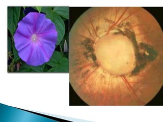
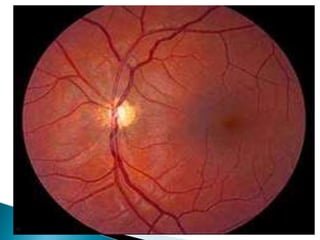
Dr. sidesh Hendavitharana(Registrar-
ophthalmology)
 Means swelling of the optic disc.
 It includes all causes of active or passive
oedematous swelling of the optic disc other
than the causes of papillioedema
 Disc hyperemia
 Blurred disc margins
 Nerve fiber layer swelling
 Disc elevation
 Tortuous vessels
 Causes for pseudodisc edema,
1) Optic nerve head drusen
2) Myelinated nerve fibres
3) Morning glory syndrome
4) Tilted disc
5) Small hypermetropic disc
6) Optic disc dysplasia
7) Bergmeister’s papilla
 Composed of hyaline like calcific material within the
substance of the optic nerve head.
 Present in 0.3% of population
 Are often bilateral
 Associations include,
◦ Retinitis pigmentosa
◦ Angioid streak
◦ Alagille syndrome
 Complications-usually rare,
◦ include CNV,CRVO,CRAO
 Imaging,
 FA-shows progressive hyperfluorescence due to staining
but absence of leakage
 US-most reliable method because of its ability to detect
calcific deposits that show high acoustic reflectivity
 In normal eyes ,optic nerve myelination stops
at the cribriform plate.
 In eyes with myelinated nerve fibres the
ganglion cells retain a myelin sheath.
 Most often unilateral.
 Ocular associations include high
myopia,anisometropia and amblyopia.
 Systemic associations include NF1 and Gorlin
syndrome
 Very rare usually unilateral sporadic condition.
 Signs,
◦ VA-may be normal or impaired to a variable extent.
◦ Large disc with funnel shaped excavation surrounded by
an annulus of chorioretinal disturbance.
◦ Blood vessels emerge from the rim of the excavation in a
radial pattern like the spokes of a wheel.
 Complications,
◦ Serous retinal detachment develops in about 30% of
cases.
◦ CNV is less common and develop adjecent to the lesion
 Frontonasal dysplasia-mid facial anomalies
consisting of hypertelorism,flat nasal bridge and
occasionally a mid line notch in the upper lip and
a midline cleft in the soft palate.
 Midline brain malformations such as absent
corpus callosum,hypoplastic cerebellar
vermis,small optic chiasm,malformed occipital
lobe,pituitary deficiency.
 NF2 less common.
 PHASE syndrome-posterior fossa brain
malformations,large facial haemangiomas and
cardiovascular anomalies.
 Is common usually bilateral anomaly caused by an
oblique entry of the optic nerve into the globe.
 Signs,
◦ Small oval or D shaped disc
◦ Disc margin is indistinct where the retinal nerve fibres are
elevated.
◦ Associated finding includes inferonasal chorioretinal thining
and myopic astigmatic refractive error.
 Perimetry-
◦ superotemporal defects that do not respect vertical midline.
 Complications-
◦ uncommon,include CNV and sensory macular detachment.
 Unilateral or bilateral
 Characterized by a diminished number of
nerve fibres.
 Commonly involving the midline structures of
the brain.
 Predisposition,
◦ Agents taken by mother during gestation.
◦ LSD,quinine,anticonvulsants,excess alcohol
 Superior segmental hypoplasia may be
associated with maternal diabetes.
 Severe bilateral cases present with blindness in early
infancy.
 Less severe bilateral involvement may cause minor minor
visual defects or squint at any time in childhood.
 Unilateral cases usually present with squint.
 Signs,
◦ VA may be normal or impaired to a variable degree,even to NPL.
◦ Double ring sign-small gray disc surrounded by a yellow halo of
hypopigmentation caused by concentric chorioretinal atrophy.
◦ Distance from the fovea to the temporal border of the optic disc
often equals or exceeds 3 times the disc diameter
◦ Retinal vessels are normal in caliber but tortous
 Depends on the severity
 Includes astigmatism,field defect ,
dyschromatopsia , APD,foveal
hypoplasia,aniridia,microphthalmos,strabismus,
nystagmus
 Systemic associations,
 Wide variety of midline developmental brain
defects-most common De Morsier
syndrome(septo-optic dysplasia)
 Characterized by absence of septum pellucidum
agenesis of corpus callosum,hypopituitarism
 Bilateral optic nerve hypoplasia may be a marker
for potential endocrine dysfunction.
 Inflammatory-papillitis, neuroretinitis,
papillophlebitis and uveitis
 Ocular hypotony-due to any cause
 Vascular-CRVO,diabetic papillopathy ,
uremia, AION
 Orbital causes-tumors,Graves’ orbitopathy,
orbital cellulitis
 Infiltrative conditions-leukaemias,lymphomas
 Is hydrostatic non inflammatory edema of
optic disc due to raised intracranial pressure.
 Is almost always bilateral although it may be
asymetrical.
 Etiology-
 Intracranial causes,
◦ congenital-
 Aqueductal stenosis
 craniosynostosis
◦ Brain tumors-commonly tumors arising in posterior
fossa-cerebellum,midbrain,parieto-ocipital region
which obstruct aqueduct of sylvius.
◦ Abscess,tuberculoma,gumma,subdural
hematoma,aneurysms,hydrocephalus
 Intracranial infections-meningitis,encephalitis
 IC h’age-SAH,SDH
◦ Direct blockage of villi by blood and protein
◦ Cavernous sinus thrombosis
◦ Damaged arachnoid villi
 Hypersecretion of CSF
 Tumors of spinal cord
 Idiopathic intracranial hypertension-
◦ pseudotumor cerebri,due to drugs like
tetracycline,vit.A,N.acid,contraceptives,
corticosteroids
 Systemic conditions
◦ Malignant hypertension
◦ Pregnancy induced hypertension
◦ Cardiopulmonary insufficiency
◦ Blood dyscrasias
◦ Nephritis
 Diffuse cerebral edema-due to blunt trauma
to head.
 Metabolic disorders
◦ Addison’s diseases,diabetic ketoaccidosis,
hypoparathyroidism
 Tumors
 Orbital abscess
 Orbital cellulitis
 Aneurysm of ophthalmic artery
 Pseudotumor
 Endocrinal exophthalmos
 Marked ocular hypotony
 Acutely raised intraocular tension
 CRVO
 AION
 uveitis
Increased IC pressure,malignant hypertension and orbital lesions
Inceased in tissue pressure in retrolaminar region
Alteration in pressure gradient across lamina cribrosa
Stasis of axoplasm in prelaminar region of optic disc
Axonal swelling in prelaminar region
Extracellular edema
papilledema
 Usually bilateral(in case of raised ICP) but may
be unilateral(in case oforbital or ocular
causes)
 General features,
 Features of raised ICP
◦ Headache,worsen on coughing,sneezing or
straining
◦ Projectile vomiting(without nausea)
◦ Diplopia
 Focal neurological deficit with changes in
level of consciousness.
 Early(incipient)papilledema
 Symptoms
◦ Usually absent
 Normal VA
 Signs
◦ Normal pupillary reactions
 Fundus picture
◦ Obscuration of disc margin-1st sign
◦ Absence of spontaneous venous pulsation at disc
◦ Mild hyperemia of disc dilated retinal capillaries splinter
h’ages in peripapillary region
 Visual field-normal
 Symptoms,
◦ Amaurosis fugax-recurrent attacks of transient
blackout of vision in one or both eyes lasting few
seconds,after standing
◦ Normal VA-until optic atrophy sets in,
◦ Painless progressive gradual loss of vision
 Signs,
◦ Normal pupillary reactions(until optic atrophy sets
in)
 Blurring of disc margins-1st sign,
 Markedly hyperemic disc
 Obliteration of physiological cup
 Bending of vessels over elevated optic disc
 Apparent optic disc edema evident by forward
elevation up to 1-2mm of disc above the
retina.
 Visual field,
 Enlargement of blind spot
 Progressive contraction of visual field
 Symptoms,
◦ Variably reduced VA
 Signs,
◦ Normal pupillary reactions
 Fundus picture
◦ Resolving acute h’age and exudates
◦ Resorption of peripapillary edema
◦ Disc appears like dome of champagne cork
◦ Obliteration of central cup
◦ Deposition of small drusen like crystal(corpora
amylacea)and opto-ciliary shunts on disc surface
 Visual fields,
◦ Enlarged blind spot
◦ Constriction of peripheral visual field.
 Papillitis,
◦ Unilateral
◦ Sudden diminution of vision
◦ Marcus Gunn pupil
◦ Ocular tenderness at insertion of superior and
medial rectus
◦ Pain on ocular movements
◦ Pulfrich phenomenon
◦ Hazy media
◦ Central or centrocaecal scotoma
 Hypermetropia,
◦ Small porus opticus
◦ Blurring of disc margins(due to heaped up optic
nerve fibers)
◦ Swelling never more than 2D
◦ Absence of venous engorgement,h’ages and
exudates
◦ No enlargement of blindspot
◦ No leakage on FFA
 Neurological emergency warranting urgent
hospitalization
 Treatment of underlying cause
 Surgical decompression of optic nerve before
field changes occur.
Disc oedema

Disc oedema

  • 1.
  • 2.
     Means swellingof the optic disc.  It includes all causes of active or passive oedematous swelling of the optic disc other than the causes of papillioedema
  • 3.
     Disc hyperemia Blurred disc margins  Nerve fiber layer swelling  Disc elevation  Tortuous vessels
  • 4.
     Causes forpseudodisc edema, 1) Optic nerve head drusen 2) Myelinated nerve fibres 3) Morning glory syndrome 4) Tilted disc 5) Small hypermetropic disc 6) Optic disc dysplasia 7) Bergmeister’s papilla
  • 5.
     Composed ofhyaline like calcific material within the substance of the optic nerve head.  Present in 0.3% of population  Are often bilateral  Associations include, ◦ Retinitis pigmentosa ◦ Angioid streak ◦ Alagille syndrome  Complications-usually rare, ◦ include CNV,CRVO,CRAO  Imaging,  FA-shows progressive hyperfluorescence due to staining but absence of leakage  US-most reliable method because of its ability to detect calcific deposits that show high acoustic reflectivity
  • 7.
     In normaleyes ,optic nerve myelination stops at the cribriform plate.  In eyes with myelinated nerve fibres the ganglion cells retain a myelin sheath.  Most often unilateral.  Ocular associations include high myopia,anisometropia and amblyopia.  Systemic associations include NF1 and Gorlin syndrome
  • 9.
     Very rareusually unilateral sporadic condition.  Signs, ◦ VA-may be normal or impaired to a variable extent. ◦ Large disc with funnel shaped excavation surrounded by an annulus of chorioretinal disturbance. ◦ Blood vessels emerge from the rim of the excavation in a radial pattern like the spokes of a wheel.  Complications, ◦ Serous retinal detachment develops in about 30% of cases. ◦ CNV is less common and develop adjecent to the lesion
  • 11.
     Frontonasal dysplasia-midfacial anomalies consisting of hypertelorism,flat nasal bridge and occasionally a mid line notch in the upper lip and a midline cleft in the soft palate.  Midline brain malformations such as absent corpus callosum,hypoplastic cerebellar vermis,small optic chiasm,malformed occipital lobe,pituitary deficiency.  NF2 less common.  PHASE syndrome-posterior fossa brain malformations,large facial haemangiomas and cardiovascular anomalies.
  • 13.
     Is commonusually bilateral anomaly caused by an oblique entry of the optic nerve into the globe.  Signs, ◦ Small oval or D shaped disc ◦ Disc margin is indistinct where the retinal nerve fibres are elevated. ◦ Associated finding includes inferonasal chorioretinal thining and myopic astigmatic refractive error.  Perimetry- ◦ superotemporal defects that do not respect vertical midline.  Complications- ◦ uncommon,include CNV and sensory macular detachment.
  • 15.
     Unilateral orbilateral  Characterized by a diminished number of nerve fibres.  Commonly involving the midline structures of the brain.  Predisposition, ◦ Agents taken by mother during gestation. ◦ LSD,quinine,anticonvulsants,excess alcohol  Superior segmental hypoplasia may be associated with maternal diabetes.
  • 16.
     Severe bilateralcases present with blindness in early infancy.  Less severe bilateral involvement may cause minor minor visual defects or squint at any time in childhood.  Unilateral cases usually present with squint.  Signs, ◦ VA may be normal or impaired to a variable degree,even to NPL. ◦ Double ring sign-small gray disc surrounded by a yellow halo of hypopigmentation caused by concentric chorioretinal atrophy. ◦ Distance from the fovea to the temporal border of the optic disc often equals or exceeds 3 times the disc diameter ◦ Retinal vessels are normal in caliber but tortous
  • 18.
     Depends onthe severity  Includes astigmatism,field defect , dyschromatopsia , APD,foveal hypoplasia,aniridia,microphthalmos,strabismus, nystagmus  Systemic associations,  Wide variety of midline developmental brain defects-most common De Morsier syndrome(septo-optic dysplasia)  Characterized by absence of septum pellucidum agenesis of corpus callosum,hypopituitarism  Bilateral optic nerve hypoplasia may be a marker for potential endocrine dysfunction.
  • 19.
     Inflammatory-papillitis, neuroretinitis, papillophlebitisand uveitis  Ocular hypotony-due to any cause  Vascular-CRVO,diabetic papillopathy , uremia, AION  Orbital causes-tumors,Graves’ orbitopathy, orbital cellulitis  Infiltrative conditions-leukaemias,lymphomas
  • 20.
     Is hydrostaticnon inflammatory edema of optic disc due to raised intracranial pressure.  Is almost always bilateral although it may be asymetrical.  Etiology-  Intracranial causes, ◦ congenital-  Aqueductal stenosis  craniosynostosis
  • 21.
    ◦ Brain tumors-commonlytumors arising in posterior fossa-cerebellum,midbrain,parieto-ocipital region which obstruct aqueduct of sylvius. ◦ Abscess,tuberculoma,gumma,subdural hematoma,aneurysms,hydrocephalus  Intracranial infections-meningitis,encephalitis  IC h’age-SAH,SDH
  • 22.
    ◦ Direct blockageof villi by blood and protein ◦ Cavernous sinus thrombosis ◦ Damaged arachnoid villi  Hypersecretion of CSF  Tumors of spinal cord  Idiopathic intracranial hypertension- ◦ pseudotumor cerebri,due to drugs like tetracycline,vit.A,N.acid,contraceptives, corticosteroids
  • 23.
     Systemic conditions ◦Malignant hypertension ◦ Pregnancy induced hypertension ◦ Cardiopulmonary insufficiency ◦ Blood dyscrasias ◦ Nephritis  Diffuse cerebral edema-due to blunt trauma to head.  Metabolic disorders ◦ Addison’s diseases,diabetic ketoaccidosis, hypoparathyroidism
  • 24.
     Tumors  Orbitalabscess  Orbital cellulitis  Aneurysm of ophthalmic artery  Pseudotumor  Endocrinal exophthalmos
  • 25.
     Marked ocularhypotony  Acutely raised intraocular tension  CRVO  AION  uveitis
  • 26.
    Increased IC pressure,malignanthypertension and orbital lesions Inceased in tissue pressure in retrolaminar region Alteration in pressure gradient across lamina cribrosa Stasis of axoplasm in prelaminar region of optic disc Axonal swelling in prelaminar region Extracellular edema papilledema
  • 27.
     Usually bilateral(incase of raised ICP) but may be unilateral(in case oforbital or ocular causes)  General features,  Features of raised ICP ◦ Headache,worsen on coughing,sneezing or straining ◦ Projectile vomiting(without nausea) ◦ Diplopia  Focal neurological deficit with changes in level of consciousness.
  • 28.
     Early(incipient)papilledema  Symptoms ◦Usually absent  Normal VA  Signs ◦ Normal pupillary reactions  Fundus picture ◦ Obscuration of disc margin-1st sign ◦ Absence of spontaneous venous pulsation at disc ◦ Mild hyperemia of disc dilated retinal capillaries splinter h’ages in peripapillary region  Visual field-normal
  • 30.
     Symptoms, ◦ Amaurosisfugax-recurrent attacks of transient blackout of vision in one or both eyes lasting few seconds,after standing ◦ Normal VA-until optic atrophy sets in, ◦ Painless progressive gradual loss of vision  Signs, ◦ Normal pupillary reactions(until optic atrophy sets in)
  • 31.
     Blurring ofdisc margins-1st sign,  Markedly hyperemic disc  Obliteration of physiological cup  Bending of vessels over elevated optic disc  Apparent optic disc edema evident by forward elevation up to 1-2mm of disc above the retina.  Visual field,  Enlargement of blind spot  Progressive contraction of visual field
  • 33.
     Symptoms, ◦ Variablyreduced VA  Signs, ◦ Normal pupillary reactions  Fundus picture ◦ Resolving acute h’age and exudates ◦ Resorption of peripapillary edema ◦ Disc appears like dome of champagne cork ◦ Obliteration of central cup ◦ Deposition of small drusen like crystal(corpora amylacea)and opto-ciliary shunts on disc surface  Visual fields, ◦ Enlarged blind spot ◦ Constriction of peripheral visual field.
  • 35.
     Papillitis, ◦ Unilateral ◦Sudden diminution of vision ◦ Marcus Gunn pupil ◦ Ocular tenderness at insertion of superior and medial rectus ◦ Pain on ocular movements ◦ Pulfrich phenomenon ◦ Hazy media ◦ Central or centrocaecal scotoma
  • 36.
     Hypermetropia, ◦ Smallporus opticus ◦ Blurring of disc margins(due to heaped up optic nerve fibers) ◦ Swelling never more than 2D ◦ Absence of venous engorgement,h’ages and exudates ◦ No enlargement of blindspot ◦ No leakage on FFA
  • 37.
     Neurological emergencywarranting urgent hospitalization  Treatment of underlying cause  Surgical decompression of optic nerve before field changes occur.