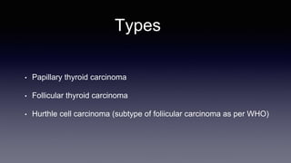
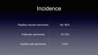
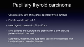
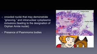
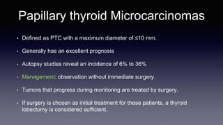
DIFFERENTIATED THYROID
CANCER
Dr. Ankur
Deptt. of General Surgery
PGIMS, Rohtak
Introduction
• Thyroid carcinoma that derived from thyroid follicular cells are
included in differentiated thyroid carcinoma.
• Most common presentation is solitary thyroid nodule.
Types
• Papillary thyroid carcinoma
• Follicular thyroid carcinoma
• Hurthle cell carcinoma (subtype of foliicular carcinoma as per WHO)
Incidence
Papillary thyroid carcinoma 80- 85%
Follicular carcinoma 10-15%
Hurthle cell carcinoma 3-5%
Molecular genetics
Cancer Oncogenes Tumor supressor genes
PTC RET, MET, RAS, BRAF p53
FTC RAS, PAX8/PPAR p53, PTEN
Papillary thyroid carcinoma
• Constitutes 80-85% of malignant epithelial thyroid tumours
• Female to male ratio is 2:1
• mean age at presentation 30 to 40 yrs
• Most patients are euthyroid and present with a slow-growing
painless mass in the neck.
• Dysphagia, dyspnea, and dysphonia usually are associated with
locally advanced invasive disease
• Diagnosis is established by FNAC of the thyroid mass or lymph
node.
• On gross examination, PTCs generally are hard and whitish and
remain flat on sectioning, in contrast to normal tissue or benign
nodular lesions that tend to bulge.
• Macroscopic calcification, necrosis, or cystic change may be
apparent.
• Histologically types:
• Papillary
• Mixed papillary and follicular
• Pure follicular ( follicular variant of PTC)
• diagnosis is established by characteristic nuclear cellular features.
• crowded nuclei that may demonstrate
“grooving,” and intranuclear cytoplasmic
inclusions (leading to the designation of
Orphan Annie nuclei)
• Presence of Psammoma bodies
• Mixed follicular & pure follicular variant are classified as papillary
carcinomas because they behave biologically as PTC.
• Follicular variant are of two types
• Encapsulated
• Non encapsulated
• Encapsulated tumours have an indolent behaviour and are
genetically distinct from their infiltrative counterparts.
• These tumors are now designated noninvasive follicular thyroid
neoplasm with papillary-like nuclear features (NIFTP).
• Multifocality is common in papillary carcinoma around (85% cases)
• PTC rarely invade adjacent structures
• Uncommon variants of PTC are about 1% of all papillary carcinomas and are
generally associated with a worse prognosis. These are
• tall cell
• insular
• columnar
• diffuse sclerosing
• clear cell
• trabecular
• poorly differentiated types
Prognostic Indicators
• PTC have an excellent prognosis with a >95% 10-year survival rate
• Several prognostic indicators have been incorporated into various staging systems. The
most widely used systems include
• AGES (age, tumor grade, tumor extent, tumor size)
• AMES (age, metastatic disease, extrathyroidal extension, size)
• MACIS (metastasis, patient age, completeness of resection, local invasion, and
tumor size)
• TNM
• all of these classification systems rely on data that are not available preoperatively.
Prognostic Risk Classification
Low risk High risk
Age <40 years >40 years
Sex Female Male
Extent
No local extension, intrathyroidal,
no capsular invasion
Capsular invasion, extrathyroidal
extension
Metastasis None Regional or distant
Size <2 cm >4 cm
Grade Well differentiated Poorly differentiated
Surgical Treatment
• Total or near total thyroidectomy for high risk and bilateral tumours
• Multifocal disease
• Familial disease
• Extra thyroid extension
• Lymph node positive
• Distant metastasis
• History of radiation exposure
• ATA recommended a near-total or total thyroidectomy for primary cancers >1 cm.
• Prophylactic lateral neck node dissection is not necessary in patients with
PTC because these cancers do not appear to metastasize systemically
from lymph nodes.
• Prophylactic (ipsilateral or bilateral) dissection may be performed in
patients with advanced (T3 or T4) PTC, or if the lateral neck nodes are
involved with tumor (N1b)
• According to the European Thyroid Association, ATA & National
Comprehensive Cancer Network, therapeutic neck dissection is performed
with initial thyroidectomy for patients with PTC with NNM.
• Prophylactic lateral neck dissection is not recommended; however,
prophylactic central neck dissection remains controversial.
Papillary thyroid Microcarcinomas
• Defined as PTC with a maximum diameter of ≤10 mm.
• Generally has an excellent prognosis
• Autopsy studies reveal an incidence of 6% to 36%
• Management: observation without immediate surgery.
• Tumors that progress during monitoring are treated by surgery.
• If surgery is chosen as initial treatment for these patients, a thyroid
lobectomy is considered sufficient.
Follicular cell carcinoma
• 5-10% of thyroid malignancy
• Occur more commonly in iodine deficient areas.
• Most common malignancy seen in patients having long standing goiter
• overall incidence is declining probably due to iodine supplementation and
improved histologic classification.
• female-to-male ratio of 3:1
• mean age at presentation of 50 years
• usually present as solitary thyroid nodules, occasionally with a history
of rapid size increase, and long-standing goiter.
• In <1% of cases, follicular cancers may be hyperfunctioning, leading
patients to present with signs and symptoms of thyrotoxicosis
• FNAC is unable to distinguish benign follicular lesions from follicular
carcinomas
• Large follicular tumors (>4 cm) in older men are more likely to be
malignant.
Pathology
• usually are solitary lesions.
• the majority are surrounded by a capsule.
• Histologically, follicles are present, but the
lumen may be devoid of colloid.
• Malignancy is defined by the presence of
capsular and vascular invasion
Surgical Treatment and
Prognosis
• Patients diagnosed by FNAC as having a follicular lesion should undergo thyroid
lobectomy because 70% to 80% of these patients will have benign adenomas.
• Total thyroidectomy is recommended in
• older patients with lesions >4 cm
• atypia on FNA
• family history of thyroid cancer
• history of radiation exposure.
• Prophylactic nodal dissection is not needed because nodal
involvement is infrequent
• Mortality from follicular thyroid cancer is approximately 5% at 10
years and 30% at 20 years.
Hürthle Cell Carcinoma
• approximately 3% of all thyroid malignancies.
• According to WHO classification, are considered to be a subtype of
follicular thyroid cancer.
• Hürthle cell cancers also are characterized by vascular or capsular
invasion and, therefore, cannot be diagnosed by FNAC.
• Microscopically contain sheets of eosinophilic cells packed with
mitochondria
• HCC differ from follicular carcinoma in that they are
• More often multifocal & bilateral (30%)
• Usually don’t take up RAI
• More likely to metastasize local nodes (25%), & distant site
• Higher mortality rate ( 20% at 10 yr)
• Management
• similar to that of follicular neoplasms
• lobectomy and isthmusectomy for unilateral Hürthle cell adenomas.
• For invasive HCC, Total thyroidectomy should be performed
• Routine central neck node removal should be performed, and
modified radical neck dissection when lateral neck nodes are
palpable or identified by ultrasonography.
Postoperative Management of
Differentiated Thyroid Cancer
Radio iodine Therapy
• RAI therapy benefit in differentiated thyroid cancer remains
controversial.
• Screening with RAI is more sensitive than chest X-ray or CT
scanning for detecting metastases.
• however, it is less sensitive than Tg measurements for detecting
metastatic disease in most differentiated thyroid cancers except
Hürthle cell tumors.
• The ATA recommend RRA in
• stage II disease older than 45 years of age
• stage III and IV disease.
• Only selected patients with stage I disease should be considered for RRA
• aggressive histologies
• multifocal disease
• nodal metastases
• extra-thyroidal or vascular invasion
• Postoperative RRA is typically performed approximately 6 weeks
after neartotal or total thyroidectomy.
• Most centers perform a pretherapy whole-body iodine scan. If
performed, a pretherapy scan should use a low dose of I131(1 to 3
mCi) or I123
• TSH must be >30 mU/L to obtain optimal uptake of radioiodine.
• Post therapy whole-body iodine scanning is typically performed 1
week after I131 treatment to identify metastases.
Complications of RAI therapy
• ACUTE
• Neck pain, swelling, sialadenitis (50-450 mCi)
• Taste dysfunction, cerebral edema ( 200 mCi)
• Vocal cord paralysis, nausea & vomiting ( 50-450 mCi)
• Bone marrow supression (200 mCi)
• LONG-TERM
• Leukemia, breast cancer (>1000 mCi)
• Infertility, pulmonary fibrosis, chronic sialadenitis
• Anaplastic thyroid cancer
• Gastric cancer, hepatocellular cancer
Thyroid Hormone
• T4 is necessary as
• replacement therapy
• suppressing TSH (TSH suppression reduces tumor recurrence
rates)
• Current guidelines advise maintaining initial TSH levels
• <0.1 mU/mL in patients with high-risk thyroid cancer
• 0.1 to 0.5 mU/mL in patients with intermediate-risk disease.
• 0.5–2 mU/L For low-risk patients (with or without remnant
ablation) with undetectable serum Tg levels.
• The risk of tumor recurrence must be balanced with the side effects
associated with prolonged TSH suppression, including osteopenia
and cardiac problems, particularly in older patients.
Follow-Up of Patients With Differentiated
Thyroid Cancer
• Thyroglobulin
• Ultrasound
• CXR
• FDG- PET
Thyroglobulin Measurement
• Tg and anti-Tg antibody levels should be measured initially at 6 to
12 month intervals and more frequently in patients with high-risk
tumors.
• Response to treatment excellent if suppressed Tg is <0.2 ng/mL and
stimulated Tg is <1 ng/mL with negative imaging.
Imaging
• diagnostic whole-body scans 6 to 12 months after remnant ablation with
high- or intermediate-risk patients.
• Other indications for follow-up scans include
• patients with abnormal uptake outside the thyroid bed on
post therapy scan
• those with poorly informative Post ablation scans (e.g., due
to high thyroid bed uptake)
• patients with Tg antibodies.
• High-sensitivity ultrasonography -mainstay of imaging surveillance
with low-risk disease.
• Cervical ultrasound be performed
• at 6 and 12 months after thyroidectomy
• then annually for at least 3 to 5 years
Additional Treatment Modalities
Radiotherapy
• External-beam radiotherapy is occasionally required
• to control unresectable, locally invasive, or recurrent
disease
• to treat metastases in support bones to decrease the
risk of fractures.
• for the treatment and control of pain from bony
metastases when there is minimal or no RAIU.
Thermal Ablation
• Percutaneous thermal ablation either
• by increasing temperature - radiofrequency ablation
• By decreasing temperature - cryoablation
• Used to induce irreversible cellular damage has shown good results for
lung, bone, and liver lesions.
Chemotherapy
• Doxorubicin (Adriamycin) and Paclitaxel (Taxol) were previously the
most frequently used agents.
• Doxorubicin ( Adriamycin) also acts as a radiation sensitizer and
should be considered in patients undergoing external-beam
radiation.
Novel therapies
• Sorafenib and lenvatinib are US-FDA approved for advanced
differentiated thyroid cancer that is non-responsive to RAI.
• Both drugs are multikinase inhibitors and target RET kinase and the
vascular endothelial growth factor (VEGF)-receptor.
Management of Local Recurrence
and Distant Metastasis
Local recurrence
• Significant nodal locoregional recurrence in the neck - MRND or central
compartment (level VI) neck dissection (depending on the location)
• smaller regional lymph node metastasis
• patients who are not amenable to surgical therapy
• have distant metastasis, locoregional disease control can also be achieved
using ultrasound-guided percutaneous ethanol ablation.
Metastatic disease
• Metastatic disease that is detected with whole-body iodine scan, and
is considered radioiodine avid, is treated with 131I therapy.
• high dose ranging between 150 to 300 mCi.
• Pulmonary metastases are frequently detected exclusively on
radioiodine scanning and tend to respond to 131I treatment.
• Treatment can be performed every 6 to 12 months as long as the
disease continues to respond. It should be noted, however, that
pulmonary fibrosis may limit further 131I treatment.
• There has been a great deal of debate regarding the optimal
management of patients who are Tg positive with negative imaging
including negative whole body iodine scans and ultrasonography.
• Previous reports have indicated benefit from empiric RAI of 100 to
200 mCi 131I in these patients. If the post therapy scan is positive,
then repeated RAI is given until the scan is negative.
• FDG-PET has been used in re-staging of residual or recurrent
advanced disease in patients with negative WBS but with increased
Tg.
• FDG-PET is best performed with a high Tg.
THANK YOU

Differentiated thyroid carcinoma

  • 1.
    DIFFERENTIATED THYROID CANCER Dr. Ankur Deptt.of General Surgery PGIMS, Rohtak
  • 2.
    Introduction • Thyroid carcinomathat derived from thyroid follicular cells are included in differentiated thyroid carcinoma. • Most common presentation is solitary thyroid nodule.
  • 3.
    Types • Papillary thyroidcarcinoma • Follicular thyroid carcinoma • Hurthle cell carcinoma (subtype of foliicular carcinoma as per WHO)
  • 4.
    Incidence Papillary thyroid carcinoma80- 85% Follicular carcinoma 10-15% Hurthle cell carcinoma 3-5%
  • 5.
    Molecular genetics Cancer OncogenesTumor supressor genes PTC RET, MET, RAS, BRAF p53 FTC RAS, PAX8/PPAR p53, PTEN
  • 6.
    Papillary thyroid carcinoma •Constitutes 80-85% of malignant epithelial thyroid tumours • Female to male ratio is 2:1 • mean age at presentation 30 to 40 yrs • Most patients are euthyroid and present with a slow-growing painless mass in the neck. • Dysphagia, dyspnea, and dysphonia usually are associated with locally advanced invasive disease
  • 7.
    • Diagnosis isestablished by FNAC of the thyroid mass or lymph node. • On gross examination, PTCs generally are hard and whitish and remain flat on sectioning, in contrast to normal tissue or benign nodular lesions that tend to bulge. • Macroscopic calcification, necrosis, or cystic change may be apparent.
  • 8.
    • Histologically types: •Papillary • Mixed papillary and follicular • Pure follicular ( follicular variant of PTC) • diagnosis is established by characteristic nuclear cellular features.
  • 9.
    • crowded nucleithat may demonstrate “grooving,” and intranuclear cytoplasmic inclusions (leading to the designation of Orphan Annie nuclei) • Presence of Psammoma bodies
  • 10.
    • Mixed follicular& pure follicular variant are classified as papillary carcinomas because they behave biologically as PTC. • Follicular variant are of two types • Encapsulated • Non encapsulated • Encapsulated tumours have an indolent behaviour and are genetically distinct from their infiltrative counterparts. • These tumors are now designated noninvasive follicular thyroid neoplasm with papillary-like nuclear features (NIFTP).
  • 11.
    • Multifocality iscommon in papillary carcinoma around (85% cases) • PTC rarely invade adjacent structures • Uncommon variants of PTC are about 1% of all papillary carcinomas and are generally associated with a worse prognosis. These are • tall cell • insular • columnar • diffuse sclerosing • clear cell • trabecular • poorly differentiated types
  • 12.
    Prognostic Indicators • PTChave an excellent prognosis with a >95% 10-year survival rate • Several prognostic indicators have been incorporated into various staging systems. The most widely used systems include • AGES (age, tumor grade, tumor extent, tumor size) • AMES (age, metastatic disease, extrathyroidal extension, size) • MACIS (metastasis, patient age, completeness of resection, local invasion, and tumor size) • TNM • all of these classification systems rely on data that are not available preoperatively.
  • 13.
    Prognostic Risk Classification Lowrisk High risk Age <40 years >40 years Sex Female Male Extent No local extension, intrathyroidal, no capsular invasion Capsular invasion, extrathyroidal extension Metastasis None Regional or distant Size <2 cm >4 cm Grade Well differentiated Poorly differentiated
  • 14.
    Surgical Treatment • Totalor near total thyroidectomy for high risk and bilateral tumours • Multifocal disease • Familial disease • Extra thyroid extension • Lymph node positive • Distant metastasis • History of radiation exposure • ATA recommended a near-total or total thyroidectomy for primary cancers >1 cm.
  • 15.
    • Prophylactic lateralneck node dissection is not necessary in patients with PTC because these cancers do not appear to metastasize systemically from lymph nodes. • Prophylactic (ipsilateral or bilateral) dissection may be performed in patients with advanced (T3 or T4) PTC, or if the lateral neck nodes are involved with tumor (N1b) • According to the European Thyroid Association, ATA & National Comprehensive Cancer Network, therapeutic neck dissection is performed with initial thyroidectomy for patients with PTC with NNM. • Prophylactic lateral neck dissection is not recommended; however, prophylactic central neck dissection remains controversial.
  • 16.
    Papillary thyroid Microcarcinomas •Defined as PTC with a maximum diameter of ≤10 mm. • Generally has an excellent prognosis • Autopsy studies reveal an incidence of 6% to 36% • Management: observation without immediate surgery. • Tumors that progress during monitoring are treated by surgery. • If surgery is chosen as initial treatment for these patients, a thyroid lobectomy is considered sufficient.
  • 17.
    Follicular cell carcinoma •5-10% of thyroid malignancy • Occur more commonly in iodine deficient areas. • Most common malignancy seen in patients having long standing goiter • overall incidence is declining probably due to iodine supplementation and improved histologic classification. • female-to-male ratio of 3:1 • mean age at presentation of 50 years
  • 18.
    • usually presentas solitary thyroid nodules, occasionally with a history of rapid size increase, and long-standing goiter. • In <1% of cases, follicular cancers may be hyperfunctioning, leading patients to present with signs and symptoms of thyrotoxicosis • FNAC is unable to distinguish benign follicular lesions from follicular carcinomas • Large follicular tumors (>4 cm) in older men are more likely to be malignant.
  • 19.
    Pathology • usually aresolitary lesions. • the majority are surrounded by a capsule. • Histologically, follicles are present, but the lumen may be devoid of colloid. • Malignancy is defined by the presence of capsular and vascular invasion
  • 20.
    Surgical Treatment and Prognosis •Patients diagnosed by FNAC as having a follicular lesion should undergo thyroid lobectomy because 70% to 80% of these patients will have benign adenomas. • Total thyroidectomy is recommended in • older patients with lesions >4 cm • atypia on FNA • family history of thyroid cancer • history of radiation exposure.
  • 21.
    • Prophylactic nodaldissection is not needed because nodal involvement is infrequent • Mortality from follicular thyroid cancer is approximately 5% at 10 years and 30% at 20 years.
  • 22.
    Hürthle Cell Carcinoma •approximately 3% of all thyroid malignancies. • According to WHO classification, are considered to be a subtype of follicular thyroid cancer. • Hürthle cell cancers also are characterized by vascular or capsular invasion and, therefore, cannot be diagnosed by FNAC.
  • 23.
    • Microscopically containsheets of eosinophilic cells packed with mitochondria • HCC differ from follicular carcinoma in that they are • More often multifocal & bilateral (30%) • Usually don’t take up RAI • More likely to metastasize local nodes (25%), & distant site • Higher mortality rate ( 20% at 10 yr)
  • 24.
    • Management • similarto that of follicular neoplasms • lobectomy and isthmusectomy for unilateral Hürthle cell adenomas. • For invasive HCC, Total thyroidectomy should be performed • Routine central neck node removal should be performed, and modified radical neck dissection when lateral neck nodes are palpable or identified by ultrasonography.
  • 25.
  • 26.
    Radio iodine Therapy •RAI therapy benefit in differentiated thyroid cancer remains controversial. • Screening with RAI is more sensitive than chest X-ray or CT scanning for detecting metastases. • however, it is less sensitive than Tg measurements for detecting metastatic disease in most differentiated thyroid cancers except Hürthle cell tumors.
  • 27.
    • The ATArecommend RRA in • stage II disease older than 45 years of age • stage III and IV disease. • Only selected patients with stage I disease should be considered for RRA • aggressive histologies • multifocal disease • nodal metastases • extra-thyroidal or vascular invasion
  • 28.
    • Postoperative RRAis typically performed approximately 6 weeks after neartotal or total thyroidectomy. • Most centers perform a pretherapy whole-body iodine scan. If performed, a pretherapy scan should use a low dose of I131(1 to 3 mCi) or I123 • TSH must be >30 mU/L to obtain optimal uptake of radioiodine. • Post therapy whole-body iodine scanning is typically performed 1 week after I131 treatment to identify metastases.
  • 29.
    Complications of RAItherapy • ACUTE • Neck pain, swelling, sialadenitis (50-450 mCi) • Taste dysfunction, cerebral edema ( 200 mCi) • Vocal cord paralysis, nausea & vomiting ( 50-450 mCi) • Bone marrow supression (200 mCi)
  • 30.
    • LONG-TERM • Leukemia,breast cancer (>1000 mCi) • Infertility, pulmonary fibrosis, chronic sialadenitis • Anaplastic thyroid cancer • Gastric cancer, hepatocellular cancer
  • 31.
    Thyroid Hormone • T4is necessary as • replacement therapy • suppressing TSH (TSH suppression reduces tumor recurrence rates) • Current guidelines advise maintaining initial TSH levels • <0.1 mU/mL in patients with high-risk thyroid cancer • 0.1 to 0.5 mU/mL in patients with intermediate-risk disease. • 0.5–2 mU/L For low-risk patients (with or without remnant ablation) with undetectable serum Tg levels.
  • 32.
    • The riskof tumor recurrence must be balanced with the side effects associated with prolonged TSH suppression, including osteopenia and cardiac problems, particularly in older patients.
  • 33.
    Follow-Up of PatientsWith Differentiated Thyroid Cancer • Thyroglobulin • Ultrasound • CXR • FDG- PET
  • 34.
    Thyroglobulin Measurement • Tgand anti-Tg antibody levels should be measured initially at 6 to 12 month intervals and more frequently in patients with high-risk tumors. • Response to treatment excellent if suppressed Tg is <0.2 ng/mL and stimulated Tg is <1 ng/mL with negative imaging.
  • 35.
    Imaging • diagnostic whole-bodyscans 6 to 12 months after remnant ablation with high- or intermediate-risk patients. • Other indications for follow-up scans include • patients with abnormal uptake outside the thyroid bed on post therapy scan • those with poorly informative Post ablation scans (e.g., due to high thyroid bed uptake) • patients with Tg antibodies.
  • 36.
    • High-sensitivity ultrasonography-mainstay of imaging surveillance with low-risk disease. • Cervical ultrasound be performed • at 6 and 12 months after thyroidectomy • then annually for at least 3 to 5 years
  • 37.
  • 38.
    Radiotherapy • External-beam radiotherapyis occasionally required • to control unresectable, locally invasive, or recurrent disease • to treat metastases in support bones to decrease the risk of fractures. • for the treatment and control of pain from bony metastases when there is minimal or no RAIU.
  • 39.
    Thermal Ablation • Percutaneousthermal ablation either • by increasing temperature - radiofrequency ablation • By decreasing temperature - cryoablation • Used to induce irreversible cellular damage has shown good results for lung, bone, and liver lesions.
  • 40.
    Chemotherapy • Doxorubicin (Adriamycin)and Paclitaxel (Taxol) were previously the most frequently used agents. • Doxorubicin ( Adriamycin) also acts as a radiation sensitizer and should be considered in patients undergoing external-beam radiation.
  • 41.
    Novel therapies • Sorafeniband lenvatinib are US-FDA approved for advanced differentiated thyroid cancer that is non-responsive to RAI. • Both drugs are multikinase inhibitors and target RET kinase and the vascular endothelial growth factor (VEGF)-receptor.
  • 42.
    Management of LocalRecurrence and Distant Metastasis
  • 43.
    Local recurrence • Significantnodal locoregional recurrence in the neck - MRND or central compartment (level VI) neck dissection (depending on the location) • smaller regional lymph node metastasis • patients who are not amenable to surgical therapy • have distant metastasis, locoregional disease control can also be achieved using ultrasound-guided percutaneous ethanol ablation.
  • 44.
    Metastatic disease • Metastaticdisease that is detected with whole-body iodine scan, and is considered radioiodine avid, is treated with 131I therapy. • high dose ranging between 150 to 300 mCi. • Pulmonary metastases are frequently detected exclusively on radioiodine scanning and tend to respond to 131I treatment. • Treatment can be performed every 6 to 12 months as long as the disease continues to respond. It should be noted, however, that pulmonary fibrosis may limit further 131I treatment.
  • 45.
    • There hasbeen a great deal of debate regarding the optimal management of patients who are Tg positive with negative imaging including negative whole body iodine scans and ultrasonography. • Previous reports have indicated benefit from empiric RAI of 100 to 200 mCi 131I in these patients. If the post therapy scan is positive, then repeated RAI is given until the scan is negative. • FDG-PET has been used in re-staging of residual or recurrent advanced disease in patients with negative WBS but with increased Tg. • FDG-PET is best performed with a high Tg.
  • 46.