Downloaded 144 times


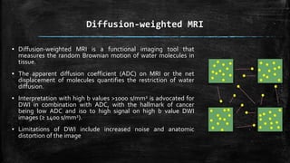
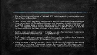
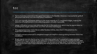
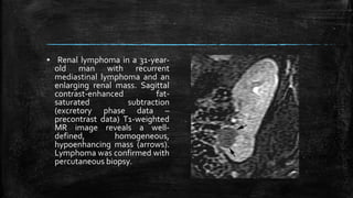
![▪ Studies have also shown an inverse correlation between quantitative ADC values and
Gleason score, and may therefore help in assigning accurate risk stratification for
selection of therapeutic options.
▪ DWI is a widely available technique and is considered to be the most important
functional imaging sequence in mp-MRI.
▪ Functional imaging (DWI, DCE and magnetic resonance spectroscopic imaging [MRSI]),
and in particular DWI, may help to differentiate cancer from benign abnormalities such
as prostatitis, fibrosis, scar tissue, post-biopsy hemorrhage or post-irradiation in the
peripheral zone](https://image.slidesharecdn.com/mriinurology-170810182854/85/Mri-in-urology-31-320.jpg)


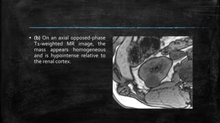
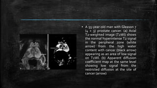
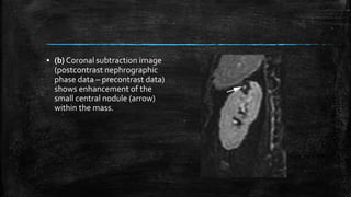
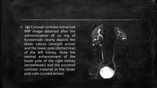
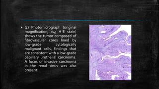
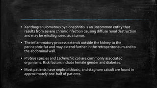
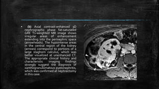
![▪ (c) Photomicrograph (original
magnification, ×10;
hematoxylin-eosin [H-E] stain)
helps confirm the presence of
conventional (clear cell) RCC
(Fuhrman grade 1–2). Clear cell
cytoplasm may contain lipid
that is responsible for the loss
of signal intensity on opposed-
phase MR images. Many tumor
cells contain finely granular
eosinophilic cytoplasm.](https://image.slidesharecdn.com/mriinurology-170810182854/85/Mri-in-urology-84-320.jpg)
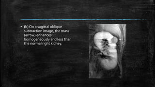


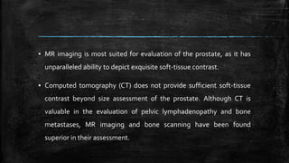
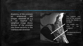
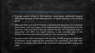
MRI has several applications in urology, including evaluating the prostate, penis, scrotum, kidneys, and adrenal glands. For the prostate, MRI is useful for detecting and localizing cancer, assessing tumor staging and extent, and guiding biopsies. It provides better soft tissue contrast than other modalities like CT or ultrasound. On T2-weighted MRI sequences, prostate cancer appears as an area of low signal intensity in the peripheral zone compared to the normally high signal prostate tissue. Functional sequences like diffusion-weighted imaging can also identify cancers. MRI is particularly helpful for detecting multifocal tumors and assessing extracapsular extension and seminal vesicle invasion.


![PERI-PROSTHETIC FRACTURE NAIL-PLATE CONSTRUCT [NPC].pptx](https://cdn.slidesharecdn.com/ss_thumbnails/drarunkumardrmohamedashrafperiprostheticfrasturenail-plateconstructnpc-260209164459-7e9d15a1-thumbnail.jpg?width=640&height=640&fit=bounds)


