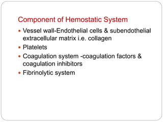
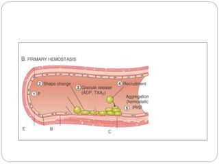
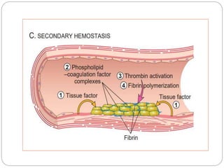
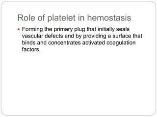
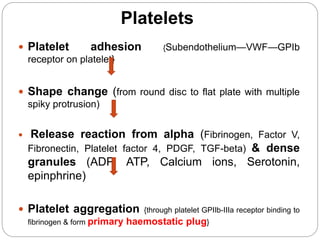
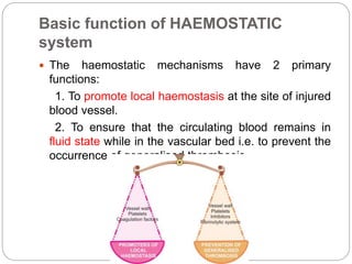
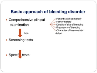
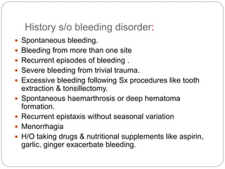
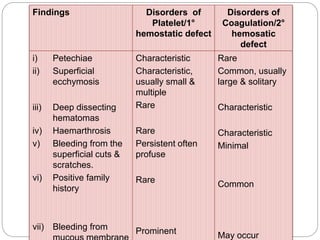
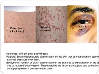
Dr sandeep singh
Haemostasis
Haemostasis
 Process of formation of hemosatic plug to halt
bleeding & restore normal blood flow.
 Hemostasis is a precisely orchestrated
process involving platelets, clotting factors,
and endothelium that occurs at the site of
vascular injury and culminates in the
formation of a blood clot, which serves to
prevent or limit the extent of bleeding.
Component of Hemostatic System
 Vessel wall-Endothelial cells & subendothelial
extracellular matrix i.e. collagen
 Platelets
 Coagulation system -coagulation factors &
coagulation inhibitors
 Fibrinolytic system
Sequence of events at site of
vascular injury during
hemostatsis
Clot stabilization and resorption.
 Polymerized fibrin and platelet aggregates
undergo contraction to form a solid, permanent
plug that prevents further hemorrhage.
 At this stage, counterregulatory mechanisms are
set into motion that limit clotting to the site of
injury and eventually lead to clot resorption and
tissue repair.
Role of platelet in hemostasis
 Forming the primary plug that initially seals
vascular defects and by providing a surface that
binds and concentrates activated coagulation
factors.
 Platelet adhesion {Subendothelium—VWF—GPIb
receptor on platelet}
 Shape change (from round disc to flat plate with multiple
spiky protrusion)
 Release reaction from alpha (Fibrinogen, Factor V,
Fibronectin, Platelet factor 4, PDGF, TGF-beta) & dense
granules (ADP, ATP, Calcium ions, Serotonin,
epinphrine)
 Platelet aggregation {through platelet GPIIb-IIIa receptor binding to
fibrinogen & form primary haemostatic plug}
Platelets
Coagulation cascade
 series of amplifying enzymatic reactions that lead
to the deposition of an insoluble fibrin clot –
secondary hemostatic plug
Common Pathway
Fibrinolytic system
Plasminogen
PAI-1
tPA
α2 antiplasmin Plasmin
Fibrin FDP (D dimer)
 Coagulation normally is restricted to sites of vascular
injury by:
 Coagulation factors present in inactivated forms require
enzymatic activation
 Circulating inhibitors of coagulation factors, such as
antithrombin III, whose activity is augmented by heparin-like
molecules expressed on endothelial cells
 Expression of thrombomodulin on normal endothelial cells,
which binds thrombin and converts it to an anticoagulant
 Activation of fibrinolytic pathways (e.g., by association of t-
PA with fibrin
Role of endothelium
 Has both prothrombotic and antithrombotic
properties.
 Antithrombotic properties are divided into :
 Platelet inhibitory effect: intact endothelium act as
a barrier prevents binding of platelet with vWF &
collagen. Release PGI2 & NO
 Anticoagulant effects: exert via expression of
various molecules like thrombomodulin, heparin
like molecule, TFPI
 Fibrinolytic effect-
Basic function of HAEMOSTATIC
system
 The haemostatic mechanisms have 2 primary
functions:
1. To promote local haemostasis at the site of injured
blood vessel.
2. To ensure that the circulating blood remains in
fluid state while in the vascular bed i.e. to prevent the
occurrence of generalised thrombosis.
Why Bleeding disorders?
 Failure of normal haemostatic mechanism.
o Injury to vascular endothelium
o Platelets defects
o Coagulation defects
o Fibrinolytic system defects
o Combination of all these
What are Bleeding disorders
• Group of disorders of diverse aetiology,
characterised by abnormal tendency to bleed
either spontaneously or after slight trauma.
 Accordingly, there are specific tests for
assessing each of these components:
A. Blood vessel wall: Tests for disordered vascular
haemostasis
B. Platelets: Tests for platelets and their functions
C. Plasma coagulation factors: Tests for
blood coagulation
D. Fibrinolytic system: Tests or fibrinolysis
E. Inhibitors: Tests for coagulation inhibitors
Basic approach of bleeding disorder
 Comprehensive clinical
examination
 Screening tests
 Specific tests
then
•Patient’s clinical history
•Family history
•Details of site of bleeding
•Frequency of bleeding
•Character of haemostatic
defect
History s/o bleeding disorder:
 Spontaneous bleeding.
 Bleeding from more than one site
 Recurrent episodes of bleeding .
 Severe bleeding from trivial trauma.
 Excessive bleeding following Sx procedures like tooth
extraction & tonsillectomy.
 Spontaneous haemarthrosis or deep hematoma
formation.
 Recurrent epistaxis without seasonal variation
 Menorrhagia
 H/O taking drugs & nutritional supplements like aspirin,
garlic, ginger exacerbate bleeding.
INHERITED DISORDERS ACQUIRED DISORDERS
 Early age of presentation
 Family history positive
 More severe
 Bleeding is the dominant
feature
 Single factor defect
 Later age of presentation
 Family history usually negative
 Less severe
 Clinical picture is dominated by
the underlying disorder e.g.DIC
 Multiple hemostatic defect
Findings Disorders of
Platelet/1°
hemostatic defect
Disorders of
Coagulation/2°
hemosatic
defect
i) Petechiae
ii) Superficial
ecchymosis
iii) Deep dissecting
hematomas
iv) Haemarthrosis
v) Bleeding from the
superficial cuts &
scratches.
vi) Positive family
history
vii) Bleeding from
Characteristic
Characteristic,
usually small &
multiple
Rare
Rare
Persistent often
profuse
Rare
Prominent
Rare
Common, usually
large & solitary
Characteristic
Characteristic
Minimal
Common
May occur
Petechiae
( 1 – 2 mm )
Purpura
(≥ 3 mm)
Ecchymosi
s
(> 1 – 2
cms)
Petechiae: Tiny pin point ecchymoses
Purpura: Small reddish-purple discoloration on the skin that do not blanch on applyin
external presssure over them.
Ecchymosis: reddish or bluish discoloration on the skin due to extravasation of the blo
due to ruptured blood vessels. These patches are larger than pupura and do not blan
on applying external presssure over them
Screening Test for Primary Haemostasis
 Hb,PCV-asses blood loss
 Hess test
 Bleeding time –platelet & vascular defects
 Platelet count-Quantitation of platelets
 PBS- 1) Quantitative & morphological
abnormalities of platelets 2)detection of
underlying hematological disorder
 PFA-100 system-platelet function
Screening Test For Secondary
Haemostasis
 CT- coagulation phase
 PT-Extrinsic & common pathway
 APTT-Intrinsic & common pathway
 TT-Fibrinogen function
Normal hemostasis.pptx

Normal hemostasis.pptx

  • 1.
  • 2.
    Haemostasis  Process offormation of hemosatic plug to halt bleeding & restore normal blood flow.  Hemostasis is a precisely orchestrated process involving platelets, clotting factors, and endothelium that occurs at the site of vascular injury and culminates in the formation of a blood clot, which serves to prevent or limit the extent of bleeding.
  • 3.
    Component of HemostaticSystem  Vessel wall-Endothelial cells & subendothelial extracellular matrix i.e. collagen  Platelets  Coagulation system -coagulation factors & coagulation inhibitors  Fibrinolytic system
  • 4.
    Sequence of eventsat site of vascular injury during hemostatsis
  • 8.
    Clot stabilization andresorption.  Polymerized fibrin and platelet aggregates undergo contraction to form a solid, permanent plug that prevents further hemorrhage.  At this stage, counterregulatory mechanisms are set into motion that limit clotting to the site of injury and eventually lead to clot resorption and tissue repair.
  • 10.
    Role of plateletin hemostasis  Forming the primary plug that initially seals vascular defects and by providing a surface that binds and concentrates activated coagulation factors.
  • 11.
     Platelet adhesion{Subendothelium—VWF—GPIb receptor on platelet}  Shape change (from round disc to flat plate with multiple spiky protrusion)  Release reaction from alpha (Fibrinogen, Factor V, Fibronectin, Platelet factor 4, PDGF, TGF-beta) & dense granules (ADP, ATP, Calcium ions, Serotonin, epinphrine)  Platelet aggregation {through platelet GPIIb-IIIa receptor binding to fibrinogen & form primary haemostatic plug} Platelets
  • 13.
    Coagulation cascade  seriesof amplifying enzymatic reactions that lead to the deposition of an insoluble fibrin clot – secondary hemostatic plug
  • 15.
  • 17.
  • 18.
     Coagulation normallyis restricted to sites of vascular injury by:  Coagulation factors present in inactivated forms require enzymatic activation  Circulating inhibitors of coagulation factors, such as antithrombin III, whose activity is augmented by heparin-like molecules expressed on endothelial cells  Expression of thrombomodulin on normal endothelial cells, which binds thrombin and converts it to an anticoagulant  Activation of fibrinolytic pathways (e.g., by association of t- PA with fibrin
  • 19.
    Role of endothelium Has both prothrombotic and antithrombotic properties.  Antithrombotic properties are divided into :  Platelet inhibitory effect: intact endothelium act as a barrier prevents binding of platelet with vWF & collagen. Release PGI2 & NO  Anticoagulant effects: exert via expression of various molecules like thrombomodulin, heparin like molecule, TFPI  Fibrinolytic effect-
  • 21.
    Basic function ofHAEMOSTATIC system  The haemostatic mechanisms have 2 primary functions: 1. To promote local haemostasis at the site of injured blood vessel. 2. To ensure that the circulating blood remains in fluid state while in the vascular bed i.e. to prevent the occurrence of generalised thrombosis.
  • 22.
    Why Bleeding disorders? Failure of normal haemostatic mechanism. o Injury to vascular endothelium o Platelets defects o Coagulation defects o Fibrinolytic system defects o Combination of all these
  • 23.
    What are Bleedingdisorders • Group of disorders of diverse aetiology, characterised by abnormal tendency to bleed either spontaneously or after slight trauma.
  • 24.
     Accordingly, thereare specific tests for assessing each of these components: A. Blood vessel wall: Tests for disordered vascular haemostasis B. Platelets: Tests for platelets and their functions C. Plasma coagulation factors: Tests for blood coagulation D. Fibrinolytic system: Tests or fibrinolysis E. Inhibitors: Tests for coagulation inhibitors
  • 25.
    Basic approach ofbleeding disorder  Comprehensive clinical examination  Screening tests  Specific tests then •Patient’s clinical history •Family history •Details of site of bleeding •Frequency of bleeding •Character of haemostatic defect
  • 26.
    History s/o bleedingdisorder:  Spontaneous bleeding.  Bleeding from more than one site  Recurrent episodes of bleeding .  Severe bleeding from trivial trauma.  Excessive bleeding following Sx procedures like tooth extraction & tonsillectomy.  Spontaneous haemarthrosis or deep hematoma formation.  Recurrent epistaxis without seasonal variation  Menorrhagia  H/O taking drugs & nutritional supplements like aspirin, garlic, ginger exacerbate bleeding.
  • 27.
    INHERITED DISORDERS ACQUIREDDISORDERS  Early age of presentation  Family history positive  More severe  Bleeding is the dominant feature  Single factor defect  Later age of presentation  Family history usually negative  Less severe  Clinical picture is dominated by the underlying disorder e.g.DIC  Multiple hemostatic defect
  • 28.
    Findings Disorders of Platelet/1° hemostaticdefect Disorders of Coagulation/2° hemosatic defect i) Petechiae ii) Superficial ecchymosis iii) Deep dissecting hematomas iv) Haemarthrosis v) Bleeding from the superficial cuts & scratches. vi) Positive family history vii) Bleeding from Characteristic Characteristic, usually small & multiple Rare Rare Persistent often profuse Rare Prominent Rare Common, usually large & solitary Characteristic Characteristic Minimal Common May occur
  • 29.
    Petechiae ( 1 –2 mm ) Purpura (≥ 3 mm) Ecchymosi s (> 1 – 2 cms) Petechiae: Tiny pin point ecchymoses Purpura: Small reddish-purple discoloration on the skin that do not blanch on applyin external presssure over them. Ecchymosis: reddish or bluish discoloration on the skin due to extravasation of the blo due to ruptured blood vessels. These patches are larger than pupura and do not blan on applying external presssure over them
  • 30.
    Screening Test forPrimary Haemostasis  Hb,PCV-asses blood loss  Hess test  Bleeding time –platelet & vascular defects  Platelet count-Quantitation of platelets  PBS- 1) Quantitative & morphological abnormalities of platelets 2)detection of underlying hematological disorder  PFA-100 system-platelet function
  • 31.
    Screening Test ForSecondary Haemostasis  CT- coagulation phase  PT-Extrinsic & common pathway  APTT-Intrinsic & common pathway  TT-Fibrinogen function

Editor's Notes

  • #12 Platelet contain two types of cytoplasmic granules: 1. Alpha granules have ptns involved in coagulation: Fibrinogen, Factor V, Fibronectin, Platelet factor 4, PDGF, TGF-beta. 2. Dense or delta granules have ADP, ATP, Calcium ions, Serotonin, epinphrine.
  • #16 The extrinsic pathway is activated by external trauma (tissue damge) that causes blood to escape from the vascular system. This pathway is quicker than the intrinsic pathway. The intrinsic pathway is activated by trauma inside the vascular system, and is activated by platelets, exposed endothelium, chemicals, or collagen.
  • #19 AAAAAWEEE468O;P[
  • #20 Anticoagulant : Normal endothelium shields coagulation factors from tissue factor in vessel wall, also release thrombomodulin, endothelial protein C receptor, heparin-like molecules, and tissue factor pathway inhibitor. Thrombomodulin bind thrombin , by this thrombin loses its ability to activate coagulation factors and platelets, and instead cleaves and activates protein C, a vitamin K–dependent protease that requires a cofactor, protein S. Activated protein C/protein S complex is a potent inhibitor of coagulation cofactors Va and VIIIa. Heparin-like molecules on the surface of endothelium bind and activate antithrombin III, which then inhibits thrombin and factors IXa, Xa, XIa, and XIIa. Tissue factor pathway inhibitor (TFPI), binds and inhibits tissue factor/factor VIIa complexes.