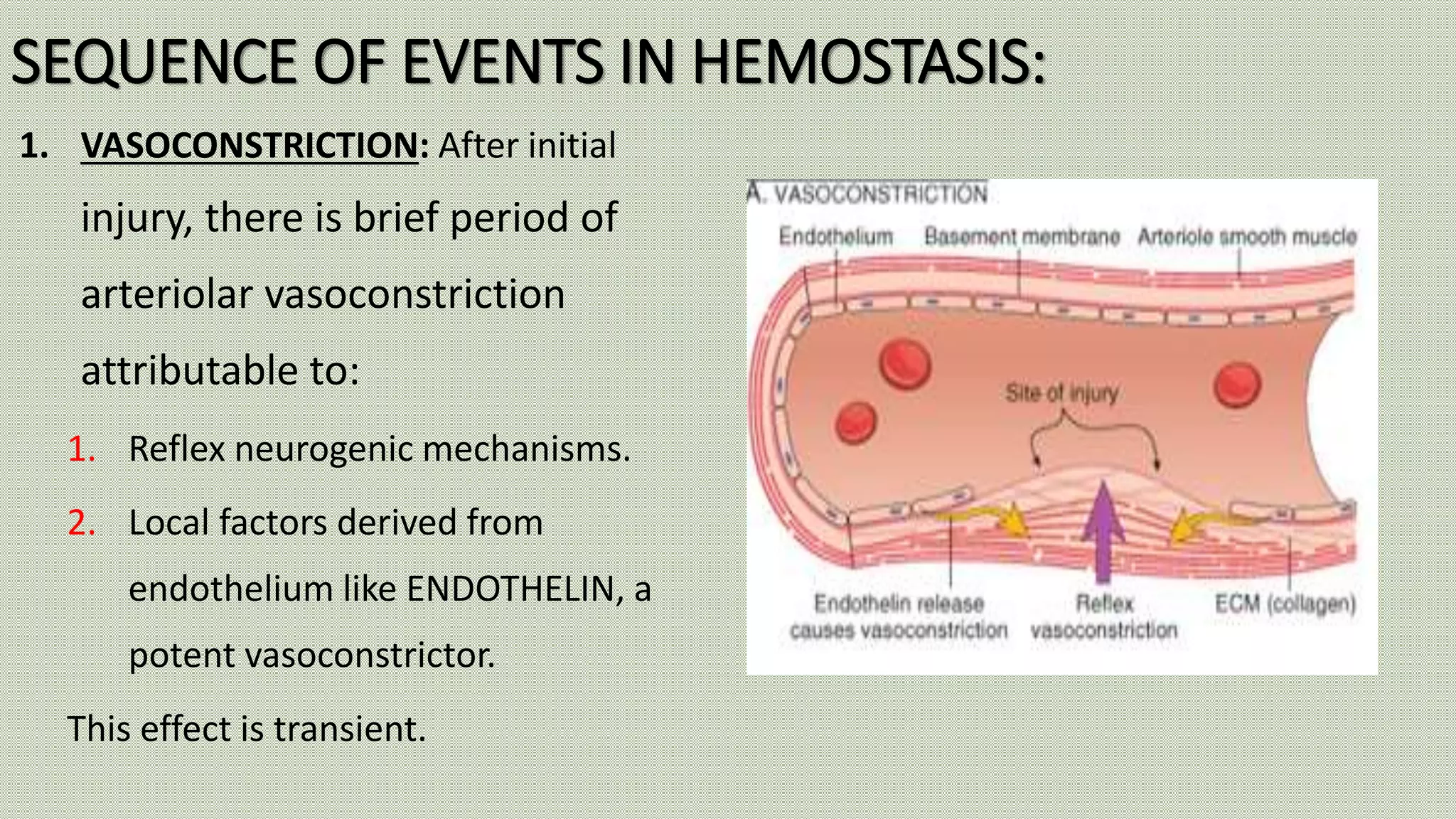
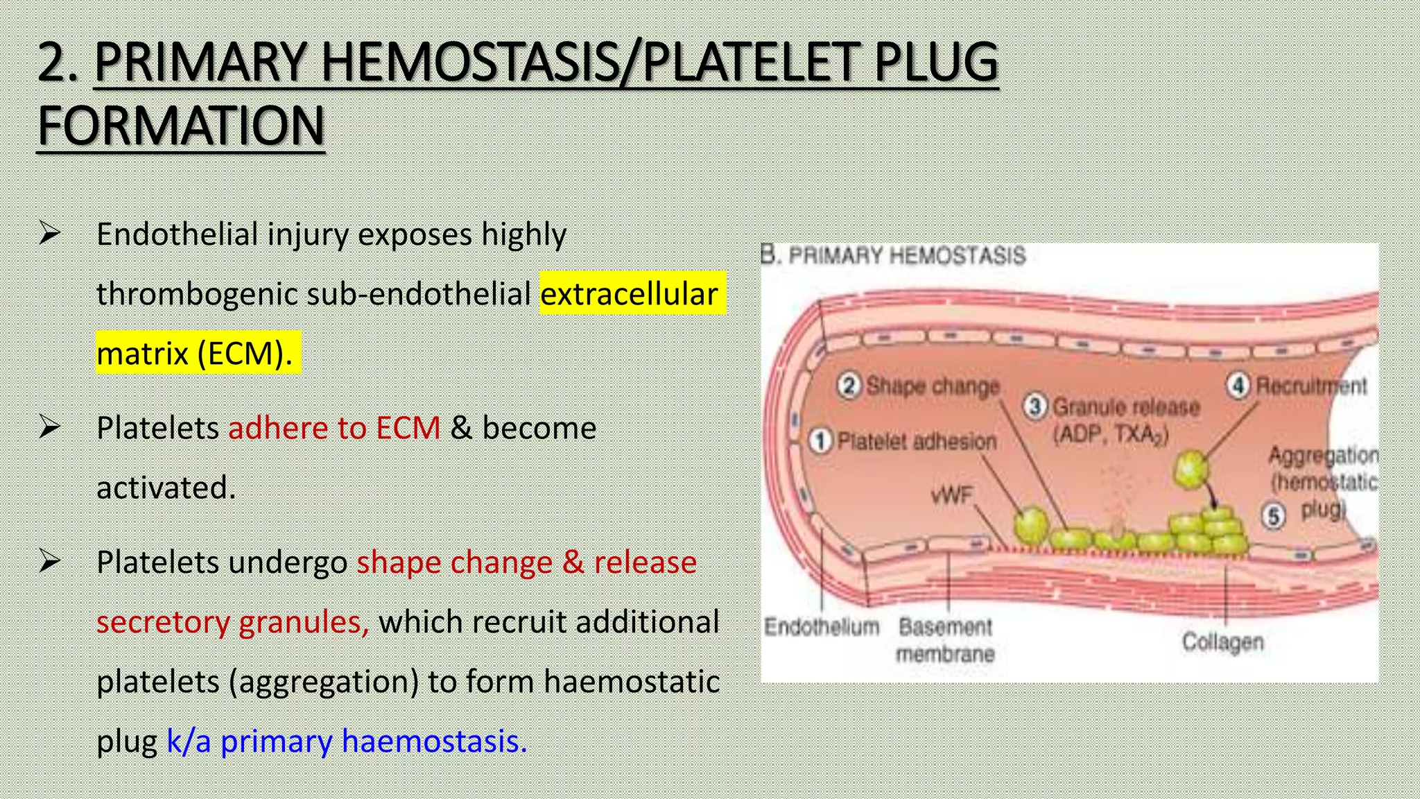
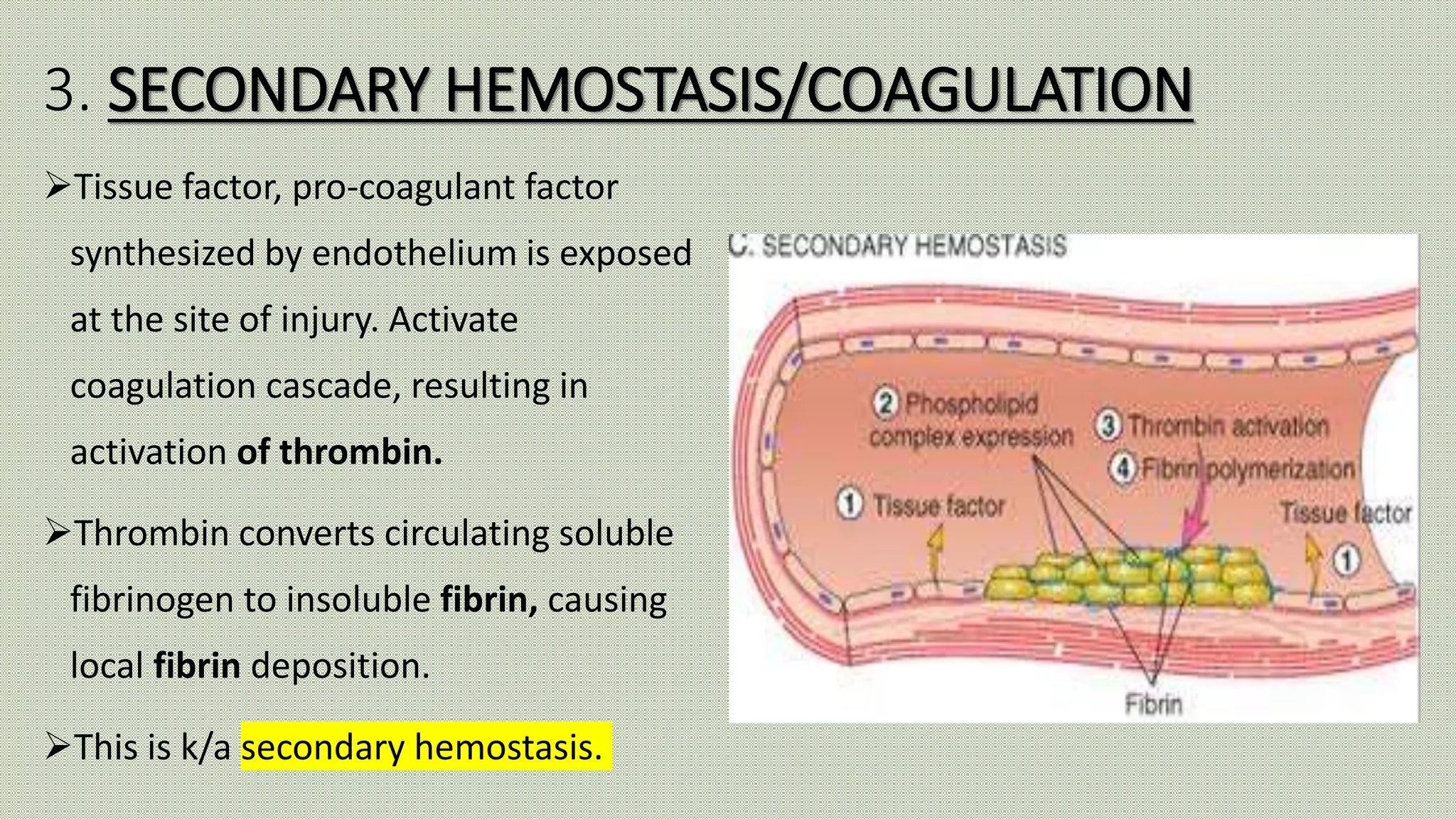
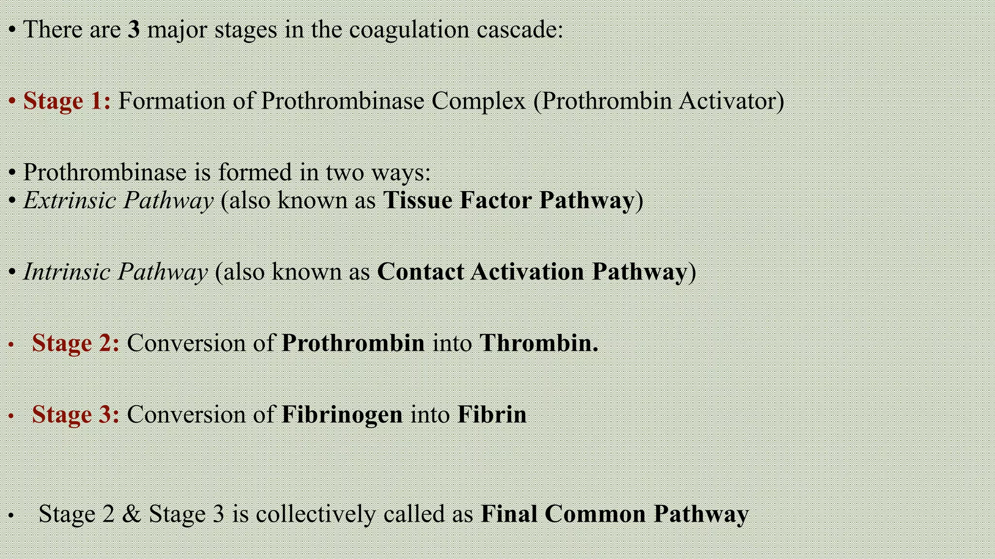
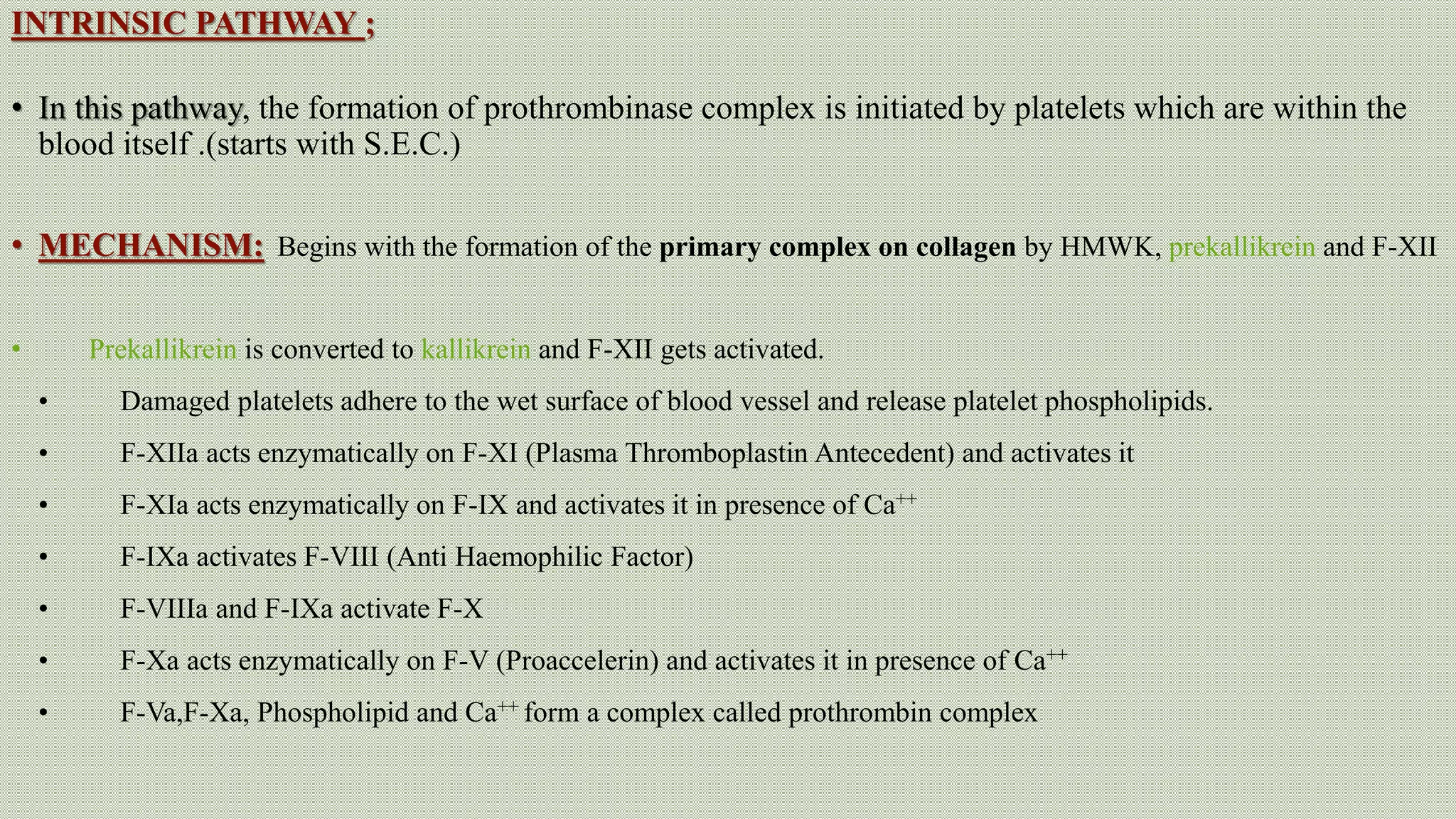
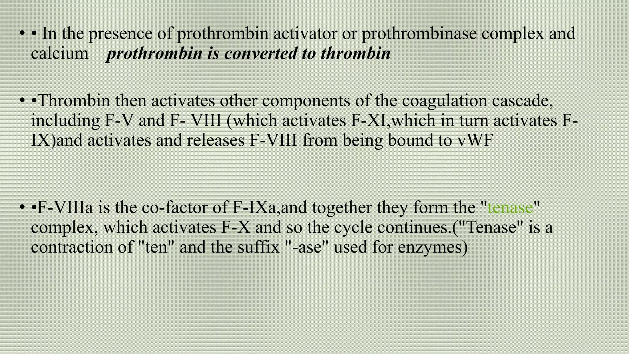
Normal haemostasis involves platelet plug formation, coagulation cascade, and fibrin clot formation to stop bleeding at the site of vascular injury. It is regulated by the endothelium, platelets, and coagulation factors. The coagulation cascade can be initiated through the intrinsic or extrinsic pathway, both leading to thrombin generation and fibrin clot formation. Counter-regulatory mechanisms like fibrinolysis and natural anticoagulants limit clot formation to the site of injury. Laboratory tests monitor components of the coagulation cascade like platelet count and function, prothrombin time, activated partial thromboplastin time, and fibrinogen levels.
This is the power point that explains about the blood and blood cells. Power point describes about the mechanism of coagulation and defense cells of our circulatory system.
This is the powerpoint for the students, faculties as well as any person who study medical and any life sciences subjects , the hemostasis portion is very comprehensively covered by diagrams and descriptions from standard books. Go through this, all the best.
Juxtaglomerular apparatus (The Guyton and Hall physiology)Maryam Fida
JUXTAGLOMERULAR APPARATUS
Juxtaglomerular apparatus is a specialized organ situated near the glomerulus of each nephron (juxta = near).
1..MACULA DENSA
Macula densa is the end portion of thick ascending segment. It is situated between afferent and efferent arterioles of the same nephron. It is very close to afferent arteriole.
Macula densa is formed by tightly packed cuboidal epithelial cells.
2..EXTRAGLOMERULAR MESANGIAL CELLS
Extraglomerular mesangial cells are situated in the triangular region bound by afferent arteriole, efferent arteriole and macula densa. These cells are also called agranular cells, lacis cells or Goormaghtigh cells.
3. Glomerular Mesangial Cell
Hemostasis and coagulation of blood For M.Sc & Basic Medical Students by Pand...Pandian M
Blood coagulation
Mechanism of coagulation
STAGES OF HEMOSTASIS
Coagulation of blood
Factors involved in blood clotting
Enzyme cascade theory
Mechanisms for formation of prothrombin activator
Fibrinolysis
Anticlotting mechanism in the body
Applied physiology
Indian Dental Academy: will be one of the most relevant and exciting training center with best faculty and flexible training programs for dental professionals who wish to advance in their dental practice,Offers certified courses in Dental implants,Orthodontics,Endodontics,Cosmetic Dentistry, Prosthetic Dentistry, Periodontics and General Dentistry.
Coagulation or clotting is defined as the process in which blood loses its fluidity and becomes a jelly-like mass few minutes after it is shed out or collected in a container
Hemostasis and coagulation of blood by Pandian M, Tutor, Dept of Physiology, ...Pandian M
DEFINITION Hemostasis
STAGES OF HEMOSTASIS
VASOCONSTRICTION
PLATELET PLUG FORMATION
COAGULATION OF BLOOD DEFINITION
FACTORS INVOLVED IN BLOOD CLOTTING
SEQUENCE OF CLOTTING MECHANISM
BLOOD CLOT
ANTICLOTTING MECHANISM IN THE BODY
ANTICOAGULANTS
PHYSICAL METHODS TO PREVENT BLOOD CLOTTING
PROCOAGULANTS
TESTS FOR BLOOD CLOTTING
APPLIED PHYSIOLOGY
Hemostasis definition, types and steps.
Hemostasis and coagulation physiology and pathology in steps and illustrated in simple way by diagrams.
Intrinsic and extrinsic pathways are mentioned in details.
Platelet function as a corner stone hemostasis in case of endothelial injury or another pathology taht affect endothelium or blood vessels.
Some pharmacological notes about drugs related to hemostasis and its clinical significance.
Hemostasis is the mechanism that leads to cessation of bleeding from a blood vessel. It is a process that involves multiple interlinked steps. This cascade culminates into the formation of a “plug” that closes up the damaged site of the blood vessel controlling the bleeding.
Hemostasis is the maintenance of blood flow is fluid state within the vascular system, the major components of hemostasis are vascular system, platelets, coagulation factors, inhibitors of coagulation and fibrinolytic system. details are given
Title: Sense of Taste
Presenter: Dr. Faiza, Assistant Professor of Physiology
Qualifications:
MBBS (Best Graduate, AIMC Lahore)
FCPS Physiology
ICMT, CHPE, DHPE (STMU)
MPH (GC University, Faisalabad)
MBA (Virtual University of Pakistan)
Learning Objectives:
Describe the structure and function of taste buds.
Describe the relationship between the taste threshold and taste index of common substances.
Explain the chemical basis and signal transduction of taste perception for each type of primary taste sensation.
Recognize different abnormalities of taste perception and their causes.
Key Topics:
Significance of Taste Sensation:
Differentiation between pleasant and harmful food
Influence on behavior
Selection of food based on metabolic needs
Receptors of Taste:
Taste buds on the tongue
Influence of sense of smell, texture of food, and pain stimulation (e.g., by pepper)
Primary and Secondary Taste Sensations:
Primary taste sensations: Sweet, Sour, Salty, Bitter, Umami
Chemical basis and signal transduction mechanisms for each taste
Taste Threshold and Index:
Taste threshold values for Sweet (sucrose), Salty (NaCl), Sour (HCl), and Bitter (Quinine)
Taste index relationship: Inversely proportional to taste threshold
Taste Blindness:
Inability to taste certain substances, particularly thiourea compounds
Example: Phenylthiocarbamide
Structure and Function of Taste Buds:
Composition: Epithelial cells, Sustentacular/Supporting cells, Taste cells, Basal cells
Features: Taste pores, Taste hairs/microvilli, and Taste nerve fibers
Location of Taste Buds:
Found in papillae of the tongue (Fungiform, Circumvallate, Foliate)
Also present on the palate, tonsillar pillars, epiglottis, and proximal esophagus
Mechanism of Taste Stimulation:
Interaction of taste substances with receptors on microvilli
Signal transduction pathways for Umami, Sweet, Bitter, Sour, and Salty tastes
Taste Sensitivity and Adaptation:
Decrease in sensitivity with age
Rapid adaptation of taste sensation
Role of Saliva in Taste:
Dissolution of tastants to reach receptors
Washing away the stimulus
Taste Preferences and Aversions:
Mechanisms behind taste preference and aversion
Influence of receptors and neural pathways
Impact of Sensory Nerve Damage:
Degeneration of taste buds if the sensory nerve fiber is cut
Abnormalities of Taste Detection:
Conditions: Ageusia, Hypogeusia, Dysgeusia (parageusia)
Causes: Nerve damage, neurological disorders, infections, poor oral hygiene, adverse drug effects, deficiencies, aging, tobacco use, altered neurotransmitter levels
Neurotransmitters and Taste Threshold:
Effects of serotonin (5-HT) and norepinephrine (NE) on taste sensitivity
Supertasters:
25% of the population with heightened sensitivity to taste, especially bitterness
Increased number of fungiform papillae
Ethanol (CH3CH2OH), or beverage alcohol, is a two-carbon alcohol
that is rapidly distributed in the body and brain. Ethanol alters many
neurochemical systems and has rewarding and addictive properties. It
is the oldest recreational drug and likely contributes to more morbidity,
mortality, and public health costs than all illicit drugs combined. The
5th edition of the Diagnostic and Statistical Manual of Mental Disorders
(DSM-5) integrates alcohol abuse and alcohol dependence into a single
disorder called alcohol use disorder (AUD), with mild, moderate,
and severe subclassifications (American Psychiatric Association, 2013).
In the DSM-5, all types of substance abuse and dependence have been
combined into a single substance use disorder (SUD) on a continuum
from mild to severe. A diagnosis of AUD requires that at least two of
the 11 DSM-5 behaviors be present within a 12-month period (mild
AUD: 2–3 criteria; moderate AUD: 4–5 criteria; severe AUD: 6–11 criteria).
The four main behavioral effects of AUD are impaired control over
drinking, negative social consequences, risky use, and altered physiological
effects (tolerance, withdrawal). This chapter presents an overview
of the prevalence and harmful consequences of AUD in the U.S.,
the systemic nature of the disease, neurocircuitry and stages of AUD,
comorbidities, fetal alcohol spectrum disorders, genetic risk factors, and
pharmacotherapies for AUD.
Ozempic: Preoperative Management of Patients on GLP-1 Receptor Agonists Saeid Safari
Preoperative Management of Patients on GLP-1 Receptor Agonists like Ozempic and Semiglutide
ASA GUIDELINE
NYSORA Guideline
2 Case Reports of Gastric Ultrasound
New Directions in Targeted Therapeutic Approaches for Older Adults With Mantl...i3 Health
i3 Health is pleased to make the speaker slides from this activity available for use as a non-accredited self-study or teaching resource.
This slide deck presented by Dr. Kami Maddocks, Professor-Clinical in the Division of Hematology and
Associate Division Director for Ambulatory Operations
The Ohio State University Comprehensive Cancer Center, will provide insight into new directions in targeted therapeutic approaches for older adults with mantle cell lymphoma.
STATEMENT OF NEED
Mantle cell lymphoma (MCL) is a rare, aggressive B-cell non-Hodgkin lymphoma (NHL) accounting for 5% to 7% of all lymphomas. Its prognosis ranges from indolent disease that does not require treatment for years to very aggressive disease, which is associated with poor survival (Silkenstedt et al, 2021). Typically, MCL is diagnosed at advanced stage and in older patients who cannot tolerate intensive therapy (NCCN, 2022). Although recent advances have slightly increased remission rates, recurrence and relapse remain very common, leading to a median overall survival between 3 and 6 years (LLS, 2021). Though there are several effective options, progress is still needed towards establishing an accepted frontline approach for MCL (Castellino et al, 2022). Treatment selection and management of MCL are complicated by the heterogeneity of prognosis, advanced age and comorbidities of patients, and lack of an established standard approach for treatment, making it vital that clinicians be familiar with the latest research and advances in this area. In this activity chaired by Michael Wang, MD, Professor in the Department of Lymphoma & Myeloma at MD Anderson Cancer Center, expert faculty will discuss prognostic factors informing treatment, the promising results of recent trials in new therapeutic approaches, and the implications of treatment resistance in therapeutic selection for MCL.
Target Audience
Hematology/oncology fellows, attending faculty, and other health care professionals involved in the treatment of patients with mantle cell lymphoma (MCL).
Learning Objectives
1.) Identify clinical and biological prognostic factors that can guide treatment decision making for older adults with MCL
2.) Evaluate emerging data on targeted therapeutic approaches for treatment-naive and relapsed/refractory MCL and their applicability to older adults
3.) Assess mechanisms of resistance to targeted therapies for MCL and their implications for treatment selection
TEST BANK for Operations Management, 14th Edition by William J. Stevenson, Ve...kevinkariuki227
TEST BANK for Operations Management, 14th Edition by William J. Stevenson, Verified Chapters 1 - 19, Complete Newest Version.pdf
NVBDCP.pptx Nation vector borne disease control programSapna Thakur
NVBDCP was launched in 2003-2004 . Vector-Borne Disease: Disease that results from an infection transmitted to humans and other animals by blood-feeding arthropods, such as mosquitoes, ticks, and fleas. Examples of vector-borne diseases include Dengue fever, West Nile Virus, Lyme disease, and malaria.
MANAGEMENT OF ATRIOVENTRICULAR CONDUCTION BLOCK.pdfJim Jacob Roy
Cardiac conduction defects can occur due to various causes.
Atrioventricular conduction blocks ( AV blocks ) are classified into 3 types.
This document describes the acute management of AV block.
Explore natural remedies for syphilis treatment in Singapore. Discover alternative therapies, herbal remedies, and lifestyle changes that may complement conventional treatments. Learn about holistic approaches to managing syphilis symptoms and supporting overall health.
ARTIFICIAL INTELLIGENCE IN HEALTHCARE.pdfAnujkumaranit
Artificial intelligence (AI) refers to the simulation of human intelligence processes by machines, especially computer systems. It encompasses tasks such as learning, reasoning, problem-solving, perception, and language understanding. AI technologies are revolutionizing various fields, from healthcare to finance, by enabling machines to perform tasks that typically require human intelligence.
263778731218 Abortion Clinic /Pills In Harare ,sisternakatoto
263778731218 Abortion Clinic /Pills In Harare ,ABORTION WOMEN’S CLINIC +27730423979 IN women clinic we believe that every woman should be able to make choices in her pregnancy. Our job is to provide compassionate care, safety,affordable and confidential services. That’s why we have won the trust from all generations of women all over the world. we use non surgical method(Abortion pills) to terminate…Dr.LISA +27730423979women Clinic is committed to providing the highest quality of obstetrical and gynecological care to women of all ages. Our dedicated staff aim to treat each patient and her health concerns with compassion and respect.Our dedicated group ABORTION WOMEN’S CLINIC +27730423979 IN women clinic we believe that every woman should be able to make choices in her pregnancy. Our job is to provide compassionate care, safety,affordable and confidential services. That’s why we have won the trust from all generations of women all over the world. we use non surgical method(Abortion pills) to terminate…Dr.LISA +27730423979women Clinic is committed to providing the highest quality of obstetrical and gynecological care to women of all ages. Our dedicated staff aim to treat each patient and her health concerns with compassion and respect.Our dedicated group of receptionists, nurses, and physicians have worked together as a teamof receptionists, nurses, and physicians have worked together as a team wwww.lisywomensclinic.co.za/
HOT NEW PRODUCT! BIG SALES FAST SHIPPING NOW FROM CHINA!! EU KU DB BK substit...GL Anaacs
Contact us if you are interested:
Email / Skype : kefaya1771@gmail.com
Threema: PXHY5PDH
New BATCH Ku !!! MUCH IN DEMAND FAST SALE EVERY BATCH HAPPY GOOD EFFECT BIG BATCH !
Contact me on Threema or skype to start big business!!
Hot-sale products:
NEW HOT EUTYLONE WHITE CRYSTAL!!
5cl-adba precursor (semi finished )
5cl-adba raw materials
ADBB precursor (semi finished )
ADBB raw materials
APVP powder
5fadb/4f-adb
Jwh018 / Jwh210
Eutylone crystal
Protonitazene (hydrochloride) CAS: 119276-01-6
Flubrotizolam CAS: 57801-95-3
Metonitazene CAS: 14680-51-4
Payment terms: Western Union,MoneyGram,Bitcoin or USDT.
Deliver Time: Usually 7-15days
Shipping method: FedEx, TNT, DHL,UPS etc.Our deliveries are 100% safe, fast, reliable and discreet.
Samples will be sent for your evaluation!If you are interested in, please contact me, let's talk details.
We specializes in exporting high quality Research chemical, medical intermediate, Pharmaceutical chemicals and so on. Products are exported to USA, Canada, France, Korea, Japan,Russia, Southeast Asia and other countries.
2. HAEMOSTASIS: (blood+ stable)
Haemostasis is defined as a set of well-regulated processes designed to
rapidly and specifically form a clot at sites of vascular injury.
It is the first stage of wound healing.
• Normal haemostasis accomplish two important functions:
• Maintenance of blood in a fluid, clot free state.
• A rapid & localized haemostatic plug formation at a site of vascular injury.
3. SEQUENCE OF EVENTS IN HEMOSTASIS:
1. VASOCONSTRICTION: After initial
injury, there is brief period of
arteriolar vasoconstriction
attributable to:
1. Reflex neurogenic mechanisms.
2. Local factors derived from
endothelium like ENDOTHELIN, a
potent vasoconstrictor.
This effect is transient.
4. 2. PRIMARY HEMOSTASIS/PLATELET PLUG
FORMATION
Endothelial injury exposes highly
thrombogenic sub-endothelial extracellular
matrix (ECM).
Platelets adhere to ECM & become
activated.
Platelets undergo shape change & release
secretory granules, which recruit additional
platelets (aggregation) to form haemostatic
plug k/a primary haemostasis.
5. 3. SECONDARY HEMOSTASIS/COAGULATION
Tissue factor, pro-coagulant factor
synthesized by endothelium is exposed
at the site of injury. Activate
coagulation cascade, resulting in
activation of thrombin.
Thrombin converts circulating soluble
fibrinogen to insoluble fibrin, causing
local fibrin deposition.
This is k/a secondary hemostasis.
6. 4. CLOT STABILIZATION AND RESORPTION
Polymerised fibrin & platelet
aggregates form solid permanent
plug to prevent any further
haemorrhage.
At this stage, counter-regulatory
mechanisms e.g. t-PA are set into
motion to limit the haemostatic plug
at the site of injury.
12. ANTI-COAGULANT EFFECTS CONTD..
• Endothelium is also a major source for tissue factor pathway
inhibitor, that complexes & inhibits activated tissue factor- factor
VIIa and factor Xa molecules.
15. ROLE OF PLATELETS:
•After vascular injury, platelets encounter ECM constituents &
undergo 3 general reactions:
1) Platelet adhesion
2) Secretion (Release Reaction)
3) Platelet aggregation
16. 1. PLATELET ADHESION
• Platelets adhere to extra-cellular
matrix (ECM) via vWF which acts as
bridge b/w plt. Surface receptors
(e.g. GP 1b) & exposed collagen.
17.
18. 2. SECRETION
Release of contents of α and δ-granules.
Dense body contents are especially important because:
I. Calcium is required in the coagulation Cascade.
II. ADP is a potent mediator of plt. Aggregation and
“Recruitment”
21. ROLE OF COAGULATION SYSTEM:
1. Constitutes 3rd component of the haemostatic process & is a major
contributor to thrombosis.
2. Results in formation of thrombin.
3. Each reaction step involves an enzyme (an activated coagulation factor), a
substrate (an inactive proenzyme form of a coagulation factor), and a
cofactor (a reaction accelerator).
4. Blood coagulation pathway has been divided into extrinsic & intrinsic
pathways, converging where factor X is activated.
22. There are major 13 factors which are involved in the coagulation
cascade. All these factors are blood proteins or their derivatives.
Even if one of the factor is defective, the whole clotting process is
impaired leading to hemorrhage. These factors are F-I to F-XIII.
23.
24. • There are 3 major stages in the coagulation cascade:
• Stage 1: Formation of Prothrombinase Complex (Prothrombin Activator)
• Prothrombinase is formed in two ways:
• Extrinsic Pathway (also known as Tissue Factor Pathway)
• Intrinsic Pathway (also known as Contact Activation Pathway)
• Stage 2: Conversion of Prothrombin into Thrombin.
• Stage 3: Conversion of Fibrinogen into Fibrin
• Stage 2 & Stage 3 is collectively called as Final Common Pathway
25.
26.
27. Extrinsic Pathway
• In this pathway, the formation of prothrombinase complex is initiated by the tissue thromboplastin
Mechanism:
•It begins with trauma to blood vessel or tissues outside the blood vessel. It releases F-VII and tissue
phosholipids.F-VII comes in contact with F- III (TF or Thromboplastin) expressed on TF-bearing cells
(stromal fibroblasts & leukocytes) forming an activated complex (TF-VIIa)
•TF-VIIa activates F-X in presence of Ca++ and tissue phospholipids
•F-Xa acts on F-V and activates it
•F-Xa complexes with tissue phospholipids, F-Va, Ca++ and forms a complex called prothrombinase
complex or prothrombin
28. INTRINSIC PATHWAY ;
• In this pathway, the formation of prothrombinase complex is initiated by platelets which are within the
blood itself .(starts with S.E.C.)
• MECHANISM: Begins with the formation of the primary complex on collagen by HMWK, prekallikrein and F-XII
• Prekallikrein is converted to kallikrein and F-XII gets activated.
• Damaged platelets adhere to the wet surface of blood vessel and release platelet phospholipids.
• F-XIIa acts enzymatically on F-XI (Plasma Thromboplastin Antecedent) and activates it
• F-XIa acts enzymatically on F-IX and activates it in presence of Ca++
• F-IXa activates F-VIII (Anti Haemophilic Factor)
• F-VIIIa and F-IXa activate F-X
• F-Xa acts enzymatically on F-V (Proaccelerin) and activates it in presence of Ca++
• F-Va,F-Xa, Phospholipid and Ca++ form a complex called prothrombin complex
29. • • In the presence of prothrombin activator or prothrombinase complex and
calcium prothrombin is converted to thrombin
• •Thrombin then activates other components of the coagulation cascade,
including F-V and F- VIII (which activates F-XI,which in turn activates F-
IX)and activates and releases F-VIII from being bound to vWF
• •F-VIIIa is the co-factor of F-IXa,and together they form the "tenase"
complex, which activates F-X and so the cycle continues.("Tenase" is a
contraction of "ten" and the suffix "-ase" used for enzymes)
30. • • Thrombin converts fibrinogen (plasma protein produced by the liver) to
fibrin
• • Thrombin also activates F-XIII (Fibrin Stabilizing Factor) which in
presence of Ca++ stabilizes the fibrin polymer through covalent bonding
of fibrin monomers
31. • Once activated, the coagulation cascade must be restricted to the local
site of vascular injury to prevent clotting of the entire vascular tree.
• Clotting is also regulated by 3 types of natural anti-coagulants:
1. Anti-thrombin
2. Protein C & protein S
3. Tissue factor pathway inhibitor (TFPI)
• Clotting cascade also sets into motion a fibrinolytic cascade that limits the
size of final clot. This is accomplished by the generation of plasmin from
plasminogen.
32. Factors limiting Coagulation
Once initiated, coagulation must be restricted to the site of vascular injury to
prevent deleterious consequences.
1. One limiting factor is simple dilution; blood flowing past the site of injury
washes out activated coagulation factors, which are rapidly removed by the
liver.
2. A second is the requirement for negatively charged phospholipids, which, as
mentioned, are mainly provided by platelets that have been activated by
contact with subendothelial matrix at sites of vascular injury.
However, the most important counterregulatory mechanisms involve factors that
are expressed by intact endothelium adjacent to the site of injury.
Activation of the coagulation cascade also sets into motion a fibrinolytic cascade
that limits the size of the clot and contributes to its later dissolution. Fibrinolysis is
largely accomplished through the enzymatic activity of plasmin, which breaks down
fibrin and interferes with its polymerization. An elevated level of breakdown
products of fibrinogen (often called fibrin split products), most notably fibrin-
derived D-dimers, are a useful clinical markers of several thrombotic states.
33. • Plasmin is generated by enzymatic catabolism of the inactive circulating
precursor plasminogen, either by a factor XII–dependent pathway (possibly
explaining the association of factor XII deficiency and thrombosis) or by
plasminogen activators. The most important plasminogen activator is t-PA; it is
synthesized principally by endothelium and is most active when bound to fibrin.
This characteristic makes t-PA a useful therapeutic agent, since its fibrinolytic
activity is largely confined to sites of recent thrombosis. Once activated, plasmin
is in turn tightly controlled by counterregulatory factors such as α2-plasmin
inhibitor, a plasma protein that binds and rapidly inhibits free plasmin.
34.
35. Coagulation Monitoring – Conventional Tests ( details in separate
session)
* Platelets – Number and Function
• Clotting studies= PT(PT-INR) ,APTT ,Thrombin time, Fibrinogen levels
etc.
Editor's Notes
A process involving platelets, clotting factors, and endothelium that occurs at the site of vascular injury and results in the formation of a blood clot, which serves to prevent or limit the extent of bleeding
Disruption of the endothelium exposes subendothelial ,von Willebrand factor (vWF) and collagen, which promote platelet adherence and activation. Activation of platelets results in a dramatic shape change (from small rounded discs to flat plates with spiky protrusions that markedly increased surface area), as well as the release of secretory granules. Within minutes the secreted products recruit additional platelets, which undergo aggregation to form a primary hemostatic plug.
Platelets bind via glycoprotein Ib (GpIb) receptors to von Willebrand factor (VWF) on exposed ECM and are activated, undergoing a shape change and granule release. Released ADP and thromboxane A2 (TXA2) induce additional platelet aggregation through
platelet GpIIb-IIIa receptor binding to fibrinogen, and form the primary hemostatic plug.
Vascular injury exposes tissue factor at the site of injury. Tissue factor is a membrane-bound pro-coagulant glycoprotein that is normally expressed by subendothelial cells in the vessel wall, such as smooth muscle cells and fibroblasts. Tissue factor binds and activates factor VII (see later), setting in motion a cascade of reactions that culminates in thrombin generation. Thrombin cleaves circulating fibrinogen into insoluble fibrin, creating a fibrin mesh-work, and also is a potent activator of platelets, leading to additional platelet aggregation at the site of injury. This sequence, referred to as secondary hemostasis, consolidates the initial platelet plug.
Polymerized fibrin and platelet aggregates undergo contraction to form a solid, permanent plug that prevents further hemorrhage. At this stage, counter-regulatory mechanisms (e.g., tissue plasminogen activator, t-PA made by endothelial cells) are set into motion that limit clotting to the site of injury and eventually lead to clot resorption and tissue repair.
α-Granules have the adhesion molecule P-selectin on their membranes and contain proteins involved in coagulation, such as fibrinogen, coagulation factor V, and vWF, as well as protein factors that may be involved in wound healing, such as fibronectin, platelet factor 4 (a heparin-binding chemokine), platelet-derived growth factor (PDGF), and transforming growth factor-β.
Dense (or δ) granules contain adenosine diphosphate (ADP) and adenosine triphosphate, ionized calcium, serotonin, and epinephrine.
Deficiency of vWF -> Von Will Brand Disease
Deficiency of Gp1b -> Bernard Soulier Syndrome
Secretion (release reaction) of granule contents occurs along with changes in shape; these two events are often referred to together as platelet activation. Platelet activation is triggered by a number of factors, including the coagulation factor thrombin and ADP. Thrombin activates platelets through a special type of G-protein– coupled receptor referred to as a protease-activated receptor (PAR), which is switched on by a proteolytic cleavage carried out by thrombin. ADP is a component of dense-body granules; thus, platelet activation and ADP release begets additional rounds of platelet activation, a phenomenon referred to as recruitment. Activated platelets also produce the prostaglandin thromboxane A2 (TXA2), a potent inducer of platelet aggregation. Aspirin inhibits platelet aggregation and produces a mild bleeding defect by inhibiting cyclooxygenase, a platelet enzyme that is required for TXA2 synthesis. Although the phenomenon is less well characterized, it is also
suspected that growth factors released from platelets contribute to the repair of the vessel wall following injury.
Platelet aggregation follows their activation. The conformational change in glycoprotein IIb/IIIa that occurs with platelet activation allows binding of fibrinogen, a large bivalent plasma polypeptide that forms bridges between adjacent platelets, leading to their aggregation. Predictably, inherited deficiency of GpIIb-IIIa results in a bleeding disorder called Glanzmann thrombasthenia. The initial wave of aggregation is reversible, but concurrent activation of thrombin stabilizes the platelet plug by causing further platelet activation and aggregation, and by promoting irreversible platelet contraction. Platelet contraction is dependent on the cytoskeleton and consolidates the aggregated platelets. In parallel, thrombin also converts fibrinogen into insoluble fibrin, cementing the platelets in place and creating the definitive secondary hemostatic plug. Entrapped red cells and leukocytes are also found in hemostatic plugs, in part due to adherence of leukocytes to P-selectin expressed on activated platelets.
prothrombin time -Tests the extrinsic and common coagulation pathway(TF,PT,WR)- Normal range of PT: around 10-14 seconds
activated partial thromboplastin time (Aptt)- Kaolin cephalin clotting time (KccT), Tests the "intrinsic" and the common coagulation pathways- Normal range of APTT: Around 30-40 seconds
thrombin clotting time (TCT)-measures the time taken for a clot to form in the plasma of a blood sample containing anticoagulant, after an excess of thrombin has been added-Normal range: 14 to 16 seconds
Fibrinogen- adult: 200-400 mg/dl, newborn: 125-300mg/dl