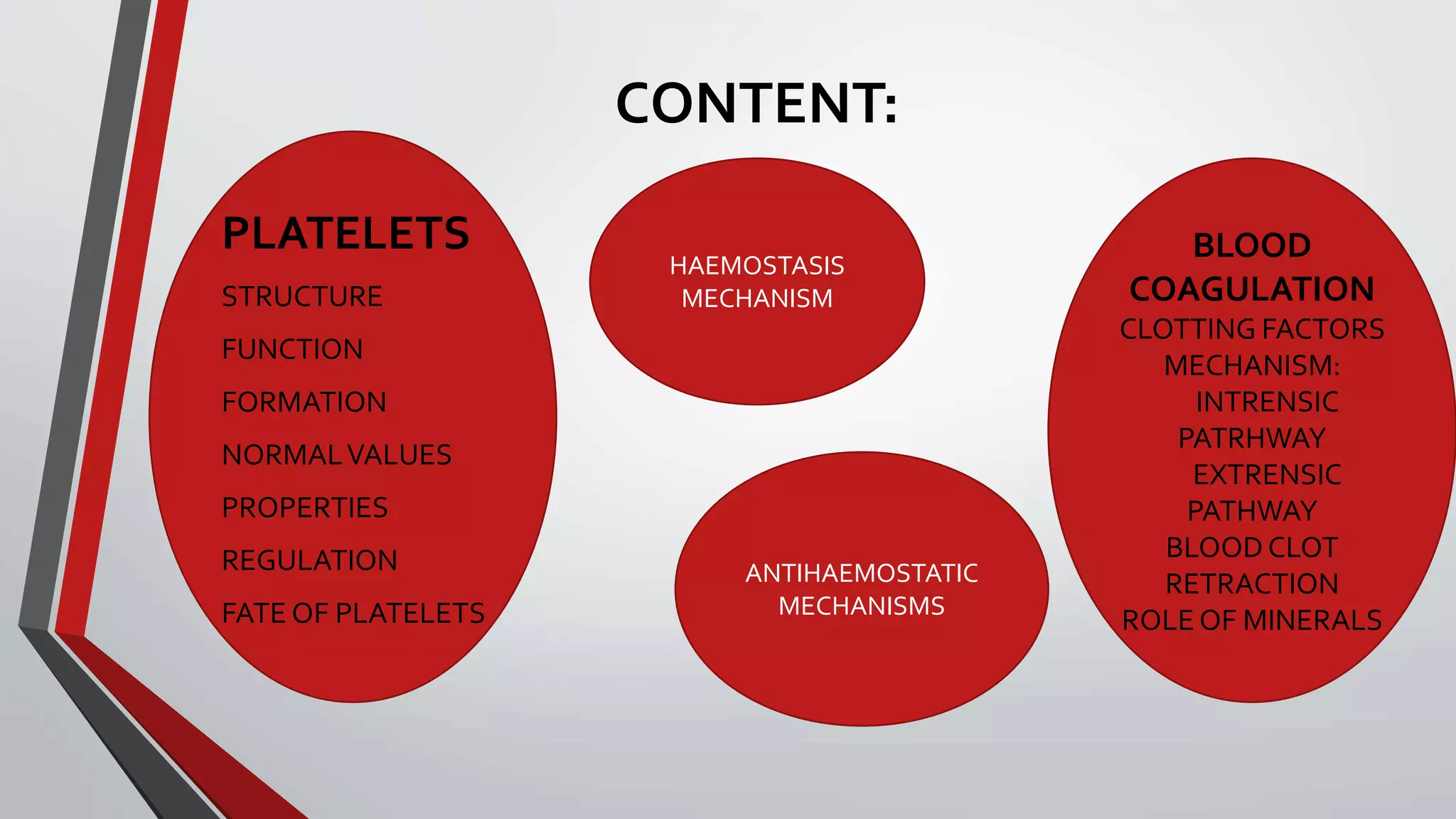
This document provides an overview of the physiology of blood coagulation. It discusses the structure and function of platelets, the clotting factors involved in coagulation, and the intrinsic and extrinsic pathways of coagulation. It also describes how fibrinogen is converted to fibrin to form a blood clot, as well as the roles of calcium, vitamins, and blood vessels. Mechanisms that prevent circulating blood from clotting and the anti-hemostatic factors involved are also summarized.
In Fibrinolytic system the clots are broken down regularly to maintain the blood flow. I case of certain disease this system is altered and produce coagulation abnormalities and diseases like MI , stroke etc.
the objectives from this ppt :-
1.Define haemostasis.
2.Describe the main mechanisms that prevent blood loss after an injury.
3.Describe role of platelets in haemostasis.
4.Outline the mechanism of platelet plug formation.
5.Describe the mechanisms of blood coagulation.
Platelets also called thrombocytes are tiny blood cells that help your body form clots to stop bleeding. If one of your blood vessels gets damaged, it sends out signals to the platelets. The platelets then rush to the site of damage. they form a plug (clot) to fix the damage.
Normal Blood count: 1.5‐4lakh/ μL of blood
Here's important & condensed ppt slides about hemostasis and its orchestrated steps and cogulation cascade, roles of endothelium,platelets and Coagulation protiens....!
Hemostasis and coagulation of blood by Pandian M, Tutor, Dept of Physiology, ...Pandian M
DEFINITION Hemostasis
STAGES OF HEMOSTASIS
VASOCONSTRICTION
PLATELET PLUG FORMATION
COAGULATION OF BLOOD DEFINITION
FACTORS INVOLVED IN BLOOD CLOTTING
SEQUENCE OF CLOTTING MECHANISM
BLOOD CLOT
ANTICLOTTING MECHANISM IN THE BODY
ANTICOAGULANTS
PHYSICAL METHODS TO PREVENT BLOOD CLOTTING
PROCOAGULANTS
TESTS FOR BLOOD CLOTTING
APPLIED PHYSIOLOGY
Hemostasis and coagulation of blood For M.Sc & Basic Medical Students by Pand...Pandian M
Blood coagulation
Mechanism of coagulation
STAGES OF HEMOSTASIS
Coagulation of blood
Factors involved in blood clotting
Enzyme cascade theory
Mechanisms for formation of prothrombin activator
Fibrinolysis
Anticlotting mechanism in the body
Applied physiology
Coagulation of blood right from haematopoiesis, platelets, endothelial injuries, development of clotting factors, coagulation cascade, applied aspect of coagulation related disorders and much more.
: Hemostasis is a complex process which causes the bleeding process to stop.
It refers to the process of keeping blood within a damaged blood vessel.
The endothelial cells of intact blood vessels prevent blood coagulation
Title: Sense of Taste
Presenter: Dr. Faiza, Assistant Professor of Physiology
Qualifications:
MBBS (Best Graduate, AIMC Lahore)
FCPS Physiology
ICMT, CHPE, DHPE (STMU)
MPH (GC University, Faisalabad)
MBA (Virtual University of Pakistan)
Learning Objectives:
Describe the structure and function of taste buds.
Describe the relationship between the taste threshold and taste index of common substances.
Explain the chemical basis and signal transduction of taste perception for each type of primary taste sensation.
Recognize different abnormalities of taste perception and their causes.
Key Topics:
Significance of Taste Sensation:
Differentiation between pleasant and harmful food
Influence on behavior
Selection of food based on metabolic needs
Receptors of Taste:
Taste buds on the tongue
Influence of sense of smell, texture of food, and pain stimulation (e.g., by pepper)
Primary and Secondary Taste Sensations:
Primary taste sensations: Sweet, Sour, Salty, Bitter, Umami
Chemical basis and signal transduction mechanisms for each taste
Taste Threshold and Index:
Taste threshold values for Sweet (sucrose), Salty (NaCl), Sour (HCl), and Bitter (Quinine)
Taste index relationship: Inversely proportional to taste threshold
Taste Blindness:
Inability to taste certain substances, particularly thiourea compounds
Example: Phenylthiocarbamide
Structure and Function of Taste Buds:
Composition: Epithelial cells, Sustentacular/Supporting cells, Taste cells, Basal cells
Features: Taste pores, Taste hairs/microvilli, and Taste nerve fibers
Location of Taste Buds:
Found in papillae of the tongue (Fungiform, Circumvallate, Foliate)
Also present on the palate, tonsillar pillars, epiglottis, and proximal esophagus
Mechanism of Taste Stimulation:
Interaction of taste substances with receptors on microvilli
Signal transduction pathways for Umami, Sweet, Bitter, Sour, and Salty tastes
Taste Sensitivity and Adaptation:
Decrease in sensitivity with age
Rapid adaptation of taste sensation
Role of Saliva in Taste:
Dissolution of tastants to reach receptors
Washing away the stimulus
Taste Preferences and Aversions:
Mechanisms behind taste preference and aversion
Influence of receptors and neural pathways
Impact of Sensory Nerve Damage:
Degeneration of taste buds if the sensory nerve fiber is cut
Abnormalities of Taste Detection:
Conditions: Ageusia, Hypogeusia, Dysgeusia (parageusia)
Causes: Nerve damage, neurological disorders, infections, poor oral hygiene, adverse drug effects, deficiencies, aging, tobacco use, altered neurotransmitter levels
Neurotransmitters and Taste Threshold:
Effects of serotonin (5-HT) and norepinephrine (NE) on taste sensitivity
Supertasters:
25% of the population with heightened sensitivity to taste, especially bitterness
Increased number of fungiform papillae
TEST BANK for Operations Management, 14th Edition by William J. Stevenson, Ve...kevinkariuki227
TEST BANK for Operations Management, 14th Edition by William J. Stevenson, Verified Chapters 1 - 19, Complete Newest Version.pdf
Ozempic: Preoperative Management of Patients on GLP-1 Receptor Agonists Saeid Safari
Preoperative Management of Patients on GLP-1 Receptor Agonists like Ozempic and Semiglutide
ASA GUIDELINE
NYSORA Guideline
2 Case Reports of Gastric Ultrasound
Title: Sense of Smell
Presenter: Dr. Faiza, Assistant Professor of Physiology
Qualifications:
MBBS (Best Graduate, AIMC Lahore)
FCPS Physiology
ICMT, CHPE, DHPE (STMU)
MPH (GC University, Faisalabad)
MBA (Virtual University of Pakistan)
Learning Objectives:
Describe the primary categories of smells and the concept of odor blindness.
Explain the structure and location of the olfactory membrane and mucosa, including the types and roles of cells involved in olfaction.
Describe the pathway and mechanisms of olfactory signal transmission from the olfactory receptors to the brain.
Illustrate the biochemical cascade triggered by odorant binding to olfactory receptors, including the role of G-proteins and second messengers in generating an action potential.
Identify different types of olfactory disorders such as anosmia, hyposmia, hyperosmia, and dysosmia, including their potential causes.
Key Topics:
Olfactory Genes:
3% of the human genome accounts for olfactory genes.
400 genes for odorant receptors.
Olfactory Membrane:
Located in the superior part of the nasal cavity.
Medially: Folds downward along the superior septum.
Laterally: Folds over the superior turbinate and upper surface of the middle turbinate.
Total surface area: 5-10 square centimeters.
Olfactory Mucosa:
Olfactory Cells: Bipolar nerve cells derived from the CNS (100 million), with 4-25 olfactory cilia per cell.
Sustentacular Cells: Produce mucus and maintain ionic and molecular environment.
Basal Cells: Replace worn-out olfactory cells with an average lifespan of 1-2 months.
Bowman’s Gland: Secretes mucus.
Stimulation of Olfactory Cells:
Odorant dissolves in mucus and attaches to receptors on olfactory cilia.
Involves a cascade effect through G-proteins and second messengers, leading to depolarization and action potential generation in the olfactory nerve.
Quality of a Good Odorant:
Small (3-20 Carbon atoms), volatile, water-soluble, and lipid-soluble.
Facilitated by odorant-binding proteins in mucus.
Membrane Potential and Action Potential:
Resting membrane potential: -55mV.
Action potential frequency in the olfactory nerve increases with odorant strength.
Adaptation Towards the Sense of Smell:
Rapid adaptation within the first second, with further slow adaptation.
Psychological adaptation greater than receptor adaptation, involving feedback inhibition from the central nervous system.
Primary Sensations of Smell:
Camphoraceous, Musky, Floral, Pepperminty, Ethereal, Pungent, Putrid.
Odor Detection Threshold:
Examples: Hydrogen sulfide (0.0005 ppm), Methyl-mercaptan (0.002 ppm).
Some toxic substances are odorless at lethal concentrations.
Characteristics of Smell:
Odor blindness for single substances due to lack of appropriate receptor protein.
Behavioral and emotional influences of smell.
Transmission of Olfactory Signals:
From olfactory cells to glomeruli in the olfactory bulb, involving lateral inhibition.
Primitive, less old, and new olfactory systems with different path
Lung Cancer: Artificial Intelligence, Synergetics, Complex System Analysis, S...Oleg Kshivets
RESULTS: Overall life span (LS) was 2252.1±1742.5 days and cumulative 5-year survival (5YS) reached 73.2%, 10 years – 64.8%, 20 years – 42.5%. 513 LCP lived more than 5 years (LS=3124.6±1525.6 days), 148 LCP – more than 10 years (LS=5054.4±1504.1 days).199 LCP died because of LC (LS=562.7±374.5 days). 5YS of LCP after bi/lobectomies was significantly superior in comparison with LCP after pneumonectomies (78.1% vs.63.7%, P=0.00001 by log-rank test). AT significantly improved 5YS (66.3% vs. 34.8%) (P=0.00000 by log-rank test) only for LCP with N1-2. Cox modeling displayed that 5YS of LCP significantly depended on: phase transition (PT) early-invasive LC in terms of synergetics, PT N0—N12, cell ratio factors (ratio between cancer cells- CC and blood cells subpopulations), G1-3, histology, glucose, AT, blood cell circuit, prothrombin index, heparin tolerance, recalcification time (P=0.000-0.038). Neural networks, genetic algorithm selection and bootstrap simulation revealed relationships between 5YS and PT early-invasive LC (rank=1), PT N0—N12 (rank=2), thrombocytes/CC (3), erythrocytes/CC (4), eosinophils/CC (5), healthy cells/CC (6), lymphocytes/CC (7), segmented neutrophils/CC (8), stick neutrophils/CC (9), monocytes/CC (10); leucocytes/CC (11). Correct prediction of 5YS was 100% by neural networks computing (area under ROC curve=1.0; error=0.0).
CONCLUSIONS: 5YS of LCP after radical procedures significantly depended on: 1) PT early-invasive cancer; 2) PT N0--N12; 3) cell ratio factors; 4) blood cell circuit; 5) biochemical factors; 6) hemostasis system; 7) AT; 8) LC characteristics; 9) LC cell dynamics; 10) surgery type: lobectomy/pneumonectomy; 11) anthropometric data. Optimal diagnosis and treatment strategies for LC are: 1) screening and early detection of LC; 2) availability of experienced thoracic surgeons because of complexity of radical procedures; 3) aggressive en block surgery and adequate lymph node dissection for completeness; 4) precise prediction; 5) adjuvant chemoimmunoradiotherapy for LCP with unfavorable prognosis.
These simplified slides by Dr. Sidra Arshad present an overview of the non-respiratory functions of the respiratory tract.
Learning objectives:
1. Enlist the non-respiratory functions of the respiratory tract
2. Briefly explain how these functions are carried out
3. Discuss the significance of dead space
4. Differentiate between minute ventilation and alveolar ventilation
5. Describe the cough and sneeze reflexes
Study Resources:
1. Chapter 39, Guyton and Hall Textbook of Medical Physiology, 14th edition
2. Chapter 34, Ganong’s Review of Medical Physiology, 26th edition
3. Chapter 17, Human Physiology by Lauralee Sherwood, 9th edition
4. Non-respiratory functions of the lungs https://academic.oup.com/bjaed/article/13/3/98/278874
263778731218 Abortion Clinic /Pills In Harare ,sisternakatoto
263778731218 Abortion Clinic /Pills In Harare ,ABORTION WOMEN’S CLINIC +27730423979 IN women clinic we believe that every woman should be able to make choices in her pregnancy. Our job is to provide compassionate care, safety,affordable and confidential services. That’s why we have won the trust from all generations of women all over the world. we use non surgical method(Abortion pills) to terminate…Dr.LISA +27730423979women Clinic is committed to providing the highest quality of obstetrical and gynecological care to women of all ages. Our dedicated staff aim to treat each patient and her health concerns with compassion and respect.Our dedicated group ABORTION WOMEN’S CLINIC +27730423979 IN women clinic we believe that every woman should be able to make choices in her pregnancy. Our job is to provide compassionate care, safety,affordable and confidential services. That’s why we have won the trust from all generations of women all over the world. we use non surgical method(Abortion pills) to terminate…Dr.LISA +27730423979women Clinic is committed to providing the highest quality of obstetrical and gynecological care to women of all ages. Our dedicated staff aim to treat each patient and her health concerns with compassion and respect.Our dedicated group of receptionists, nurses, and physicians have worked together as a teamof receptionists, nurses, and physicians have worked together as a team wwww.lisywomensclinic.co.za/
Report Back from SGO 2024: What’s the Latest in Cervical Cancer?bkling
Are you curious about what’s new in cervical cancer research or unsure what the findings mean? Join Dr. Emily Ko, a gynecologic oncologist at Penn Medicine, to learn about the latest updates from the Society of Gynecologic Oncology (SGO) 2024 Annual Meeting on Women’s Cancer. Dr. Ko will discuss what the research presented at the conference means for you and answer your questions about the new developments.
HOT NEW PRODUCT! BIG SALES FAST SHIPPING NOW FROM CHINA!! EU KU DB BK substit...GL Anaacs
Contact us if you are interested:
Email / Skype : kefaya1771@gmail.com
Threema: PXHY5PDH
New BATCH Ku !!! MUCH IN DEMAND FAST SALE EVERY BATCH HAPPY GOOD EFFECT BIG BATCH !
Contact me on Threema or skype to start big business!!
Hot-sale products:
NEW HOT EUTYLONE WHITE CRYSTAL!!
5cl-adba precursor (semi finished )
5cl-adba raw materials
ADBB precursor (semi finished )
ADBB raw materials
APVP powder
5fadb/4f-adb
Jwh018 / Jwh210
Eutylone crystal
Protonitazene (hydrochloride) CAS: 119276-01-6
Flubrotizolam CAS: 57801-95-3
Metonitazene CAS: 14680-51-4
Payment terms: Western Union,MoneyGram,Bitcoin or USDT.
Deliver Time: Usually 7-15days
Shipping method: FedEx, TNT, DHL,UPS etc.Our deliveries are 100% safe, fast, reliable and discreet.
Samples will be sent for your evaluation!If you are interested in, please contact me, let's talk details.
We specializes in exporting high quality Research chemical, medical intermediate, Pharmaceutical chemicals and so on. Products are exported to USA, Canada, France, Korea, Japan,Russia, Southeast Asia and other countries.
New Directions in Targeted Therapeutic Approaches for Older Adults With Mantl...i3 Health
i3 Health is pleased to make the speaker slides from this activity available for use as a non-accredited self-study or teaching resource.
This slide deck presented by Dr. Kami Maddocks, Professor-Clinical in the Division of Hematology and
Associate Division Director for Ambulatory Operations
The Ohio State University Comprehensive Cancer Center, will provide insight into new directions in targeted therapeutic approaches for older adults with mantle cell lymphoma.
STATEMENT OF NEED
Mantle cell lymphoma (MCL) is a rare, aggressive B-cell non-Hodgkin lymphoma (NHL) accounting for 5% to 7% of all lymphomas. Its prognosis ranges from indolent disease that does not require treatment for years to very aggressive disease, which is associated with poor survival (Silkenstedt et al, 2021). Typically, MCL is diagnosed at advanced stage and in older patients who cannot tolerate intensive therapy (NCCN, 2022). Although recent advances have slightly increased remission rates, recurrence and relapse remain very common, leading to a median overall survival between 3 and 6 years (LLS, 2021). Though there are several effective options, progress is still needed towards establishing an accepted frontline approach for MCL (Castellino et al, 2022). Treatment selection and management of MCL are complicated by the heterogeneity of prognosis, advanced age and comorbidities of patients, and lack of an established standard approach for treatment, making it vital that clinicians be familiar with the latest research and advances in this area. In this activity chaired by Michael Wang, MD, Professor in the Department of Lymphoma & Myeloma at MD Anderson Cancer Center, expert faculty will discuss prognostic factors informing treatment, the promising results of recent trials in new therapeutic approaches, and the implications of treatment resistance in therapeutic selection for MCL.
Target Audience
Hematology/oncology fellows, attending faculty, and other health care professionals involved in the treatment of patients with mantle cell lymphoma (MCL).
Learning Objectives
1.) Identify clinical and biological prognostic factors that can guide treatment decision making for older adults with MCL
2.) Evaluate emerging data on targeted therapeutic approaches for treatment-naive and relapsed/refractory MCL and their applicability to older adults
3.) Assess mechanisms of resistance to targeted therapies for MCL and their implications for treatment selection
3. WHAT IS COAGULATION?
• BLOOD REMAINS INTHE FLUID CONDITIONWITHINTHE BLOOD
VESSELSTHROUGHOUT LIFE,
• BUTWHENTHE BLOOD IS SHED FROMTHE BLOODVESSELS ARE
COLLECTED INTHE COINTAINER ,IT LOOSES ITS FLUIDITYWITHIN A FEW
MINUTES AND IS CONVERTEDTO JELLY-LIKE MASSWHICH IS CALLED
CLOT.
• THIS PHENOMENON IS CALLED COAGULATION OR CLOTTING OF
BLOOD.
12. CELL MEMBRANE
• IT CONSISTS OF LIPID,CARBOHYDRATES,PROTEINS,GLYCOPROTEINS
GLYCOPROTEINS:
FORMSTHE SURFACE COAT OFTHE PLATELET MEMBRANE
PREVENTS ADHERENCE OF PLATELETTO NORMAL ENDOTHELIUM
ACCELERATESTHE ADHERENCE OF PLATELETSTO COLLAGEN AND
DAMAGED ENDOTHELIUM IN INJURED BLOODVESSELS.
13. PHOSPOLIPIDS:
• PHOSPHOLIPIDS OFTHE PLATELET MEMBRANE CONTAIN PLATELET
FACTOR-3WHICH PLAYS AN IMPORTANT ROLE IN ACTIVATING ROLE IN
THE SEVERAL POINTS IN BLOOD CLOTTING PROCESS
14. CONTD…..
• INVAGINATION OFTHE SURFACE MEMBRANE FORMSTHE SO-CALLED
CANALICULAR SYSTEM ORTHE SURFACE CONNECTING SYSTEM
• RECEPTORS PRESENT ONTHE PLATELET MEMBRANE COMBAINS
WITH SPECIFIC SUBSTANCES LIKE COLLAGEN AND FIBRINOGEN.
15. PROPERTIES AND FUNCTION OF PLATELETS
• ADHESIVENESS
PLATELETS POSSESSTHE PROPERTY OF ADHESIVENESS
WHENTHEY COME IN CONTACT ANDWET SURFACE OR ROUGH
SURFACE ,THESE ARE ACTIVATED AND STICKTOTHE SURFACE
FACTORS RESPOSIBLE FOR ADHESIVENESS ARE : COLLAGEN,
THROMBIN, ADP ,THROMBOXANE A2 , Ca+ANDVON WILLEBRANT FACTOR
18. FUNCTIONS OF PLATELETS
ROLE IN HAEMOSTASIS
ROLE IN CLOT FORMATION
ROLE IN CLOT RETRACTION
ROLE IN REPAIR OF INJURED BLOODVESSELS
ROLE IN DEFENCE MECHANISAM
TRANSPORT AND STORAGE FUNCTIONS
20. PHGYSIOLOGICALVARIATIONS;
AGE LESS IN INFANTS(1 LAC – 2LAC/MM3)
ADULT LEVELS ARE REACHED BY 3RD MONTH OF AGE
SEX NORMALLY NO DIFFERENCE BETWEEN MALES AND FEMALES
DURING,MENSTRUATIONTHE COUNT IS REDUCED IN FEMALES
AFTER MEAL COUNT IS SLIGHTLY INCREASED
AFTER SEVERE
MUSCULAR
EXERCISE
MAY INCREASE
AT HIGH
ALTITUDE
COUNT IS INCREASED
21. FORMATION OF PLATELETS:
• FORMATION OR DEVELOPMENT OF PLATELETS IS CALLED
THROMBOPOIESIS
• THE PLATELETS ARE PRODUCED INTHE BONE MARROW
• THE PLURIPOTENT STEM CELLS FORMS PLATELETS IS CONVERTED
INTO COLONY FORMING UNITS CALLED “MEG-CFU”
• THROUGHVARIOUS STAGES IT DEVELOPS INTO PLATELET
23. MEGAKARYOBLAST:
• THE EARLIEST RECOGNIZABLE PRECURSOR OF PLATELETS INTHE
BONE MARROW IS MEGAKARYOBLAST
• IT ARISES FROM MEG-CFU BYTHE PROCESS OF DIFFERENTIATION
DIAMETER 20-30uM
CYTOPLASM SMALL,BLUE AND NON-GRANULAR
NUCLEUS LARGE ,OVAL OR KIDNEY SHAPED WITH
SEVERAL NUCLEOLI
24.
25. PROMEGAKARYOCYTES:
• PROMEGAKARYOCYTE IS FORMED FROMTHE MEGAKARYOBLAST
• THE LARGE CELL CONTAINING UPTO 32TIMESTHE NORMAL DIPLOID
CONTENT OF NUCLEAR DNA IS FORMED
• WHEN FURTHER NUCLEAR REPLICATION CEASES ,CYTOPLASM
BECOMES GRANULAR
• THE GRANULES ARE BASICALLY BASOPHILIC
26. ENDOREDUPLICATION:
• THE MEGAKARYOBLAST UNDERGOES ENDOREDUPLICATION OF
NUCLEAR CHROMATIN
• I.E)NUCLEAR CHROMATIN REPLICATES REPEATEDLY IN MULTIPLES OF
TWOWITHOUT DIVISION OF CELLS.
27. MEGAKARYOCYTES:
THE PROMEGAKARYOCYTE MATURES INTO MEGAKARYOCYTE
• PLATELETSARE FORMED FROMTHE PSEUDOPODIAOF MEGAKARYOCYTECYTOPLASM
WHICH GETS DETACHED INTOTHE BLOOD STREAM
• EACH MEGAKARYOCYTES FORMS -4000PLATELETS
• THE FORMATION OF PLATELETS FROMTHE STEMCELLSTAKES ABOUT 10 DAYS
DIAMETER LARGE CELL-30-90uM
NUCLEUS SINGLE MULTILOBED[4-16]
NUCLEUSWITH COARSLYCLUMPED
CHROMATIN
CYTOPLASM ABUDENT,LIGHT BLUE IN COLOUR
RED –PURPLE GRANULES
CELL MARGIN IRREGULAR –MANY PSEUDOPODIA
28.
29.
30. CONTROL OFTHROMBOPOIESIS:
• THROMBOPOIESIS SEEMSTO BE REGULATED BY FOLLOWING HUMORAL
FACTORS:
THROMBOPOIETIN
MEGAKARYOCYTE-COLONY STIMULATING ACTIVITY[MEG-CSA]
• THE FACTOR STIMULATING THE SYNTHESIS AND RELEASE OFTHESE AGENTS ARE
NOTYET KNOWN
31. LIFE SPAN AND FATE OF PLATELETS:
• LIFE SPAN OF PLATELETSVARIES FROM 8-12 DAYSWITH AN AVERAGE
OF 10 DAYS.
• PLATELETS ARE DESTROYED BYTHETISSUE MACROPHAGES SYSTEM
IN SPLEEN.
[NOTE: SPLENOMEGALY:REDUCTION INTHE PLATELET COUNT
SPLENECTOMY:INCREASE IN PLATELET COUNT]
43. BLOOD COAGULATION:
• COAGULATION FORMS AN INDISPENSABLE DEFENCE AGAINST
EXCESSIVE BLEEDING FROMTHEWOUND
• THE PROCESS OF BLOOD COAGULATION CONSISTS OF A CASCADE OF
REACTIONS.
• THE FINAL PRODUCT IS FORMATION OF FIBRIN NETWORK
44.
45. OTHERS;
• HMW-K HIGH MOLECULAR WEIGHT KININOGEN OR FITZGERALD
FACTOR
• PRE KA PREKALLIKREIN OR FLETCHER FACTOR
• KA KALLIKREIN
• PL PLATELET PGOSPHOLIPID
46. FACTOR I:
FIBRINOGEN:
• IT IS A SOLUBLE PLASMA PROTEIN
• MOLECULAR WGT: 3,40,000 DELTON
• SYNTHESIZED IN LIVER
• CONTAINS 6 POLY PEPTIDE CHAINS
• PLASMA CONC 0.3GM/100ML
• FIBRINOGEN ------------THROMBIN---------FIBRIN
47. FACTOR II:
PROTHROMBIN:
• IT IS A PLASMA PROTEIN[ALPHA 2 GLOBULIN]
• INACTIVE PRECURSOR OF ENZYMETHROMBIN
• MOLECULAR WGT: 69,000DELTON
• SYNTHESIZED IN LIVER WITHTHE PRESENCE OFVITAMIN-K
• PLASMA CONC 40MG/100ML
48. FACTOR III:
THROMBOPLASTIN:
• ALSO CALLEDTISSUE FACTOR ORTISSUETHROMBOPLASTIN
• RELEASED IN EXTRENSIC PATHWAY FORTHE FORMATION OF
PROTHROMBIN ACTIVATOR
49. FACTOR IV:
CALCIUM
• CALCIUM IONS ARE RESPONSIBLE FORTHE BLOOD COAGULATION
[NOTE:DISCUSSED IN MECHANISM IN DETAIL]
50. FACTORV:
LABILE FACTOR:
• ALSO CALLED PROACCELERIN
• UNSTABLE FACTOR OFTHE PLASMA
• PROTHROMBIN………………THROMBIN [EXTRENSIC AND INTRENSIC]
• CONSUMED DURING CLOTTING ,ABSENT IN SERUM
51. FACTORVII:
STABLE FACTOR:
• STABLE PROTEIN
• SYNTHESIZED IN LIVER IN PRESENCE OFVIT-K
• ACTIVATION OF FACTOR X IN EXTRENSIC PATHWAY
• NOT CONSUMED DURING CLOTTING, PRESENT IN SERUM AND PLASMA
52. FACTORVIII:
ANTI-HEMOPHILIC FACTOR:
• PROTEIN OF BETA 2 GLOBULINTYPE
• SYNTHESIZED IN LIVER
• ACTIVATION OF FACTOR X AND FORMATION OF PROTHROMBIN IN
INTRENSIC PATHWAY
• CONSUMED DURING CLOTTING
• ABSENT IN SERUM
53. FACTOR IX:
CHRISTMAS FACTOR:
• ALSO CALLED PLASMATHROMBOPLASTIN COMPONENT[PTC]/AUTO
PROTHROMBIN II
• SYNTHESIZED IN LIVER
• ACTIVATED BY XI a INTHE PRESENCE OF CA+
• FORMATION OF PROTHROMBIN ACTIVATOR INTHE INTRENSIC
PATHWAY
54. FACTOR X:
STAUART PROWER FACTOR:
• PROTEIN PRESENT IN PLASMA
• SYNTHESIZED IN LIVER
• ACTIVATED BY IX a INTHE PRESENCE OF FACTOR
VIII,CA+,PHOSPHOLIPIDS
• FORMS PROTHROMBIN ACTIVATOR COMPLEX (EXTRENSIC AND
INTRENSIC)
56. FACTOR XII:
HAGEMAN FACTOR:
• XII IS ACTIVATEDTO XII aWHEN IT COMES IN CONTACT WITH –VE
CHARGE OR FOREIGN BODIES OR ROUGH SURFACES
• FEEDBACK ACTIVATION
58. MECHANISM OF COAGULATION:
• CLOT FORMATION IS INITIATED UNDER FOLLOWING SUTIATIONS:
TRAUMATOTHEVASCULARWALL AND ADJACENTTISSUE
TRAUMATO BLOOD
CONTACT OF BLOODTOTHE DAMAGED ENDOTHELIUM OR
COLLAGEN OR OTHERTISSUE ELEMENTS OUTSIDETHEVESSELS
59. THE PROCESS OF COAGULATION:
• FORMATION OF PROTHROMBIN ACTIVATOR
• CONVERSION OF PROTHROMBIN------------- THROMBIN
• CONVERSION OF FIBRINOGEN …………..….FIBRIN
61. EXTRINSIC PATHWAY:
• RELEASE OFTISSUETHROMBOPLASTINS
• ACTIVATION OF FACTOR X--------X a
• EFFECTS OF X a ----------------------PROTHROMBIN
62.
63. INTRINSIC PATHWAY:
• ACTIVATION FACTOR XII
• ACTIVATION FACTOR XI
• ACTIVATION FACTOR IX
• ACTIVATION FACTOR X
• FPRMATION OF PROTHROMBIN ACTIVATOR
70. WHY CIRCULATING BLOOD DOES NOT
CLOT?
VELOCITY OF CIRCULATION
SURFACE EFFECTS OF ENDOTHELIUM
CIRCULATING ANTI-COAGULANTS
FIBRINOLYTIC MECHANISM
REMOVAL OF ACTIVATED CLOTTING
FACTOR
74. CIRCULATING ANTI-COAGULANTS:
• THE NATURAL ANTI-COAGULANTS CIRCULATING INTHE BLOOD
CONSTITUETHE ANTO-COAGULANT MECHANISM OFTHE BODY
HEPARIN
ANTITHROMBIN II
PROTEIN C
75. HEPARIN
• IT ISTHE POWERFULL NATURAL ACTING ANTI-COAGULANT’
• FIRST ISOLATED FROM LIVER SO,CALLED HEPARIN[HEPAR=LIVER]
• POLYSACCHARIDE CONTAINING SULPHATE GROUP
• MOLECULAR WEIGHT=15,000-18,000
77. MECHANISM OF ACTION:
• IT IS PRESENT ONTHE LUMINAL SURFACE OFVASCULAR ENDOTHELIUM
PREVENTS ACTIVATION OF PROTHROMBINTOTHROMBIN
INHIBITSTHE ACTION OFTHROMBIN ON FIBRINOGEN
FACILITATESTHE ACTION OF ANTITHROMBIN III
[INHIBITSTHE ACTIVE FORMS OF CLOTTING FACTOR IX,X,XI,XII]
79. FIBRINOLYTIC MECHANISM:
• FIBRINOLYSIS REFERSTOTHE PROCESSTHAT BRINGS ABOUTTHE
DISSOLUTION OF FIBRIN.
• THE IMPORTANT COMPONENT OFTHE FIBRINOLYTIC SYSTEM IS
PLASMIN OR FIBRINOLYSIN
• PRESENT INTHE BLOOD IN AN INACTIVE FORM CALLED PLASMINOGEN
• ALSO CALLED PLASMIN SYSTEM
80. PGYSIOLOGICAL ROLE OF FIBRINOLYSIS
SYSTEM:
• CLEANINGTHE MINUTE CLOTS OFTINYVESSELS
• PROMOTE NORMAL HEALING PROCESS
• LIQUEFACTION OF MENSTRUAL CLOT
• LIQUEFACTION OF SPERMS INTHE EPIDIDYMIS