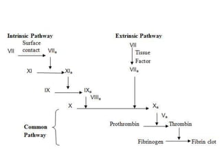
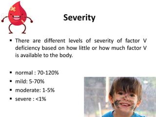
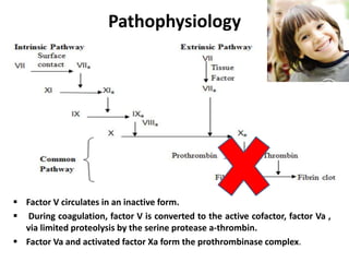
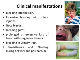
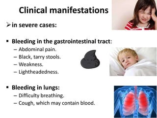
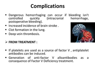
Factor V Deficiency
(Owren’s disease)
Dr. Leen Doya
Department of pediatric
Tishreen university
Introduction
• Hemostasis is the process of forming clots in the
walls damaged blood vessels and preventing blood
loss while maintaining blood in a fluid state within
the vascular system .
Background
 Factor V, or proaccelerin, is an essential component
in the blood coagulation cascade.
 Is a protein made in liver that helps convert
prothrombin into thrombin.
 No enough factor V or it doesn’t work properly,
blood may not clot effectively enough to stop from
bleeding.
 Not to be confused with Factor V Leiden, which is a
type of Thrombophilia.
.
History
 first described in a Norwegian patient in
1943.
 Identified by Dr. Paul Owren as ( severe
bleeding tendency due to the deficiency
of a previously unknown coagulation
factor), so it is also called as Owren’s
disease, parahemophilia .
Epidemiology
 Fewer than 200 cases of congenital factor V
deficiency have been reported worldwide since
1943.
 rare, occurring in approximately 1 per million
population.
 M=F.
Epidemiology
 affects persons of all ages(age at presentation
indirectly varies with the severity of disease).
 No apparent racial predilection.
 This condition is more common in countries such as
Iran and southern India, where it occurs up to ten
times more frequently than in western countries.
subtypes
I. Genetic (Inherited ):
 the most common type , is caused by mutation(s)
in the F5 gene
 inheritance of factor V deficiency is autosomal
recessive (observed in families with factor V
deficiency).
 Heterozygotes have lowered levels of factor V but
probably never bleed abnormally .
subtypes
II. Acquired :
 rare clinical condition in which the development of
antibodies to factor V (factor V inhibitors) leads to
hemorrhagic complications of varying severity.
 addition of normal plasma cannot correct the
prolonged PT and (aPTT).
 can occur after surgery, childbirth, use of bovine
thrombin or medications , and in patients with
autoimmune diseases (SLE) , certain neoplasms,
Factor V Deficiency
classified
I. Type 1 ( quantitative Factor V Deficiency):
the levels of factor V are low.
II. Type 2 (qualitative Factor V Deficiency):
the levels are normal or near-normal, but
the activity of factor V is impaired.
Severity
 There are different levels of severity of factor V
deficiency based on how little or how much factor V
is available to the body.
 normal : 70-120%
 mild: 5-70%
 moderate: 1-5%
 severe : <1%
Pathophysiology
 Factor V circulates in an inactive form.
 During coagulation, factor V is converted to the active cofactor, factor Va ,
via limited proteolysis by the serine protease a-thrombin.
 Factor Va and activated factor Xa form the prothrombinase complex.
Clinical manifestations
 Bleeding into the skin.
 Excessive bruising with minor
injuries.
 Nose bleeds.
 Bleeding gums.
 prolonged or excessive loss of
blood with surgery or trauma.
 Bleeding in urinary tract.
 Hemarthrosis and Bleeding
during delivery and postpartum .
Clinical manifestations
in severe cases:
 Bleeding in the gastrointestinal tract:
– Abdominal pain.
– Black, tarry stools.
– Weakness.
– Lightheadedness.
 Bleeding in lungs:
– Difficulty breathing.
– Cough, which may contain blood.
Bleeding in the joints :
– Swelling
– Stiffness and pain
– Tingling sensation
– The joint may feel warm to touch
• Bleeding into the brain:
– Headaches
– Seizures
– Numbness
– Nausea and vomiting
– Vision changes
– Confusion
Clinical manifestations
Acquired Factor V Deficiency, the signs and symptoms
may include those of the pre-existing condition that
caused Factor V Deficiency.
Physical
 The most common:
 Ecchymoses.
 bleeding from mucosal surfaces.
 pallor secondary to blood loss.
 Petechiae are uncommon because platelet numbers
and function are not affected.
Laboratory Studies
 Factor V assay (decreased activity) .
 Bleeding time (prolonged in severe case).
 aPTT (Prolonged).
 PT (Prolonged).
 Thrombin time (Normal).
 Stypven time (Russell viper venom time [RVVT]):
(Prolonged).
 Correction of PT or partial thromboplastin time
(PTT) with the mixing of equal amounts of normal
and patient plasma.
Imaging Studies
 Early and aggressive imaging studies (for
hemorrhage, after coagulation therapy is initiated) .
 Head and Body CT scan with or without intravenous
contrast (Assess spontaneous or traumatic
hemorrhage).
 Head and spinal column MRI .
 Radiograph for joint assessment.
 angiography and nucleotide bleeding scan as
clinically indicated.
Treatment
 usually only needed for severe bleeds or before surgery.
 there is no concentrate containing only factor V.
 fresh plasma or (FFP) infusions are used to correct the
deficiency temporarily and should be given daily during
a bleeding episode.
 loading dose of FFP is 15-20 mL/kg and then 3-6 mL/kg
daily (depend on monitoring the factor V level ).
 The half-life ranges from 24-36 hours
 aim being a factor V level of 25%.
 Complication : Fluid overload and viral transmission.
Treatment
 platelet transfusions are emerging as an alternative
to FFP.
 Factor V stored within platelet alpha granules has
greater procoagulant potential and is released locally
at sites of vascular injury.
Preoperative and postoperative
 The safe level of factor V for adequate surgical
hemostasis is 25% of the activity of factor V in
normal control plasma.
 Postoperatively, FFP should be administered for 3-10
days, with careful observation of wound bleeding.
 Tooth extraction in a patient with factor V hereditary
deficiency is safely performed with both
supplementation of FFP and application of local
hemostasis.
Treatment
 Corticosteroids have been used successfully in
acquired factor V deficiency.
 Further Outpatient Care:
 should monitor individuals with severe factor V
deficiency.
Prevention
 no prevention exists (inherited disorder).
 Immunize patients who may require plasma-derived
coagulation factor concentrates with hepatitis B vaccines.
 no use (NSAIDs) as this greatly increases the risk of
bleeding.
 Caution is needed for injections should be given SC rather
than IM ( reduce the risk of hematoma developing).
 Some activities may need to be avoided, such as contact
sports that carry a high risk of head injury .
 It is always advisable to wear a medical alert identity
necklace or bracelet .
Complications
 Dangerous hemorrhaging can occur if bleeding isn't
controlled quickly (intracranial hemorrhage,
postoperative bleeding).
 Increased incidence of brain stroke .
 Clot formation in the lung.
 Deep vein thrombosis.
 FROM TREATMENT :
 If platelets are used as a source of factor V , antiplatelet
antibodies can be induced.
 Generation of anti-factor V alloantibodies as a
consequence of Factor V Deficiency treatment.
Combined deficiency of
factors V and VIII
 Factor V deficiency may also occur at the same time as
factor VIII deficiency, producing more severe bleeding
problems.
 Rare bleeding disorder described in 1954 by Oeri et al.
 Several families ( about 100 have been reported).
Background
Prevalence
 estimated between 1/100,000 and 1/1,000,000.
 more prevalent in the Mediterranean area and in
areas where consanguineous marriages are common.
Etiology
 Inheritance is autosomal recessive.
 Associated with mutations of LMAN-1 (ERGIC-53)
approximately 70% of cases or MCFD2 approximately
30% of cases , both of which regulate intracellular
trafficking of V and VIII.
Clinical description
 can manifest at any age.
 usually mild symptoms
 Epistaxis, easy bruising, post-surgical
or post partum bleeding and
menorrhagia are the most common
symptoms.
 Hemarthrosis and muscular
hematomas may occur.
Diagnostic methods
 Prolonged PT and aPTT .
 Levels of factor V and factor VIII range from as low as
1% to as high as 46%.
 generally fall between 5% and 30%.
Management
and Prognosis
 Management:
 aims at controlling the bleeding
 includes treatments with FFP and desmopressin
administration.
 Prognosis:
 The prognosis is favorable for moderate forms of the
disease.
 Management of patients with more severe forms
should be carried out at a specialized center.
THE END

Factor v deficiency

  • 1.
    Factor V Deficiency (Owren’sdisease) Dr. Leen Doya Department of pediatric Tishreen university
  • 2.
    Introduction • Hemostasis isthe process of forming clots in the walls damaged blood vessels and preventing blood loss while maintaining blood in a fluid state within the vascular system .
  • 6.
    Background  Factor V,or proaccelerin, is an essential component in the blood coagulation cascade.  Is a protein made in liver that helps convert prothrombin into thrombin.  No enough factor V or it doesn’t work properly, blood may not clot effectively enough to stop from bleeding.  Not to be confused with Factor V Leiden, which is a type of Thrombophilia. .
  • 7.
    History  first describedin a Norwegian patient in 1943.  Identified by Dr. Paul Owren as ( severe bleeding tendency due to the deficiency of a previously unknown coagulation factor), so it is also called as Owren’s disease, parahemophilia .
  • 8.
    Epidemiology  Fewer than200 cases of congenital factor V deficiency have been reported worldwide since 1943.  rare, occurring in approximately 1 per million population.  M=F.
  • 9.
    Epidemiology  affects personsof all ages(age at presentation indirectly varies with the severity of disease).  No apparent racial predilection.  This condition is more common in countries such as Iran and southern India, where it occurs up to ten times more frequently than in western countries.
  • 10.
    subtypes I. Genetic (Inherited):  the most common type , is caused by mutation(s) in the F5 gene  inheritance of factor V deficiency is autosomal recessive (observed in families with factor V deficiency).  Heterozygotes have lowered levels of factor V but probably never bleed abnormally .
  • 11.
    subtypes II. Acquired : rare clinical condition in which the development of antibodies to factor V (factor V inhibitors) leads to hemorrhagic complications of varying severity.  addition of normal plasma cannot correct the prolonged PT and (aPTT).  can occur after surgery, childbirth, use of bovine thrombin or medications , and in patients with autoimmune diseases (SLE) , certain neoplasms,
  • 12.
    Factor V Deficiency classified I.Type 1 ( quantitative Factor V Deficiency): the levels of factor V are low. II. Type 2 (qualitative Factor V Deficiency): the levels are normal or near-normal, but the activity of factor V is impaired.
  • 13.
    Severity  There aredifferent levels of severity of factor V deficiency based on how little or how much factor V is available to the body.  normal : 70-120%  mild: 5-70%  moderate: 1-5%  severe : <1%
  • 14.
    Pathophysiology  Factor Vcirculates in an inactive form.  During coagulation, factor V is converted to the active cofactor, factor Va , via limited proteolysis by the serine protease a-thrombin.  Factor Va and activated factor Xa form the prothrombinase complex.
  • 15.
    Clinical manifestations  Bleedinginto the skin.  Excessive bruising with minor injuries.  Nose bleeds.  Bleeding gums.  prolonged or excessive loss of blood with surgery or trauma.  Bleeding in urinary tract.  Hemarthrosis and Bleeding during delivery and postpartum .
  • 16.
    Clinical manifestations in severecases:  Bleeding in the gastrointestinal tract: – Abdominal pain. – Black, tarry stools. – Weakness. – Lightheadedness.  Bleeding in lungs: – Difficulty breathing. – Cough, which may contain blood.
  • 17.
    Bleeding in thejoints : – Swelling – Stiffness and pain – Tingling sensation – The joint may feel warm to touch • Bleeding into the brain: – Headaches – Seizures – Numbness – Nausea and vomiting – Vision changes – Confusion Clinical manifestations
  • 18.
    Acquired Factor VDeficiency, the signs and symptoms may include those of the pre-existing condition that caused Factor V Deficiency.
  • 19.
    Physical  The mostcommon:  Ecchymoses.  bleeding from mucosal surfaces.  pallor secondary to blood loss.  Petechiae are uncommon because platelet numbers and function are not affected.
  • 20.
    Laboratory Studies  FactorV assay (decreased activity) .  Bleeding time (prolonged in severe case).  aPTT (Prolonged).  PT (Prolonged).  Thrombin time (Normal).  Stypven time (Russell viper venom time [RVVT]): (Prolonged).  Correction of PT or partial thromboplastin time (PTT) with the mixing of equal amounts of normal and patient plasma.
  • 21.
    Imaging Studies  Earlyand aggressive imaging studies (for hemorrhage, after coagulation therapy is initiated) .  Head and Body CT scan with or without intravenous contrast (Assess spontaneous or traumatic hemorrhage).  Head and spinal column MRI .  Radiograph for joint assessment.  angiography and nucleotide bleeding scan as clinically indicated.
  • 22.
    Treatment  usually onlyneeded for severe bleeds or before surgery.  there is no concentrate containing only factor V.  fresh plasma or (FFP) infusions are used to correct the deficiency temporarily and should be given daily during a bleeding episode.  loading dose of FFP is 15-20 mL/kg and then 3-6 mL/kg daily (depend on monitoring the factor V level ).  The half-life ranges from 24-36 hours  aim being a factor V level of 25%.  Complication : Fluid overload and viral transmission.
  • 23.
    Treatment  platelet transfusionsare emerging as an alternative to FFP.  Factor V stored within platelet alpha granules has greater procoagulant potential and is released locally at sites of vascular injury.
  • 24.
    Preoperative and postoperative The safe level of factor V for adequate surgical hemostasis is 25% of the activity of factor V in normal control plasma.  Postoperatively, FFP should be administered for 3-10 days, with careful observation of wound bleeding.  Tooth extraction in a patient with factor V hereditary deficiency is safely performed with both supplementation of FFP and application of local hemostasis.
  • 25.
    Treatment  Corticosteroids havebeen used successfully in acquired factor V deficiency.  Further Outpatient Care:  should monitor individuals with severe factor V deficiency.
  • 26.
    Prevention  no preventionexists (inherited disorder).  Immunize patients who may require plasma-derived coagulation factor concentrates with hepatitis B vaccines.  no use (NSAIDs) as this greatly increases the risk of bleeding.  Caution is needed for injections should be given SC rather than IM ( reduce the risk of hematoma developing).  Some activities may need to be avoided, such as contact sports that carry a high risk of head injury .  It is always advisable to wear a medical alert identity necklace or bracelet .
  • 27.
    Complications  Dangerous hemorrhagingcan occur if bleeding isn't controlled quickly (intracranial hemorrhage, postoperative bleeding).  Increased incidence of brain stroke .  Clot formation in the lung.  Deep vein thrombosis.  FROM TREATMENT :  If platelets are used as a source of factor V , antiplatelet antibodies can be induced.  Generation of anti-factor V alloantibodies as a consequence of Factor V Deficiency treatment.
  • 28.
  • 29.
     Factor Vdeficiency may also occur at the same time as factor VIII deficiency, producing more severe bleeding problems.  Rare bleeding disorder described in 1954 by Oeri et al.  Several families ( about 100 have been reported). Background
  • 30.
    Prevalence  estimated between1/100,000 and 1/1,000,000.  more prevalent in the Mediterranean area and in areas where consanguineous marriages are common.
  • 31.
    Etiology  Inheritance isautosomal recessive.  Associated with mutations of LMAN-1 (ERGIC-53) approximately 70% of cases or MCFD2 approximately 30% of cases , both of which regulate intracellular trafficking of V and VIII.
  • 32.
    Clinical description  canmanifest at any age.  usually mild symptoms  Epistaxis, easy bruising, post-surgical or post partum bleeding and menorrhagia are the most common symptoms.  Hemarthrosis and muscular hematomas may occur.
  • 33.
    Diagnostic methods  ProlongedPT and aPTT .  Levels of factor V and factor VIII range from as low as 1% to as high as 46%.  generally fall between 5% and 30%.
  • 34.
    Management and Prognosis  Management: aims at controlling the bleeding  includes treatments with FFP and desmopressin administration.  Prognosis:  The prognosis is favorable for moderate forms of the disease.  Management of patients with more severe forms should be carried out at a specialized center.
  • 35.