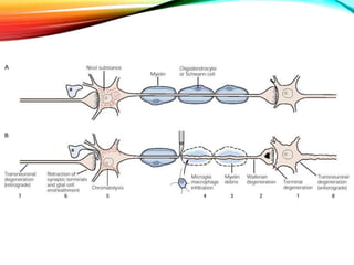
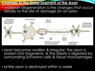
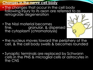
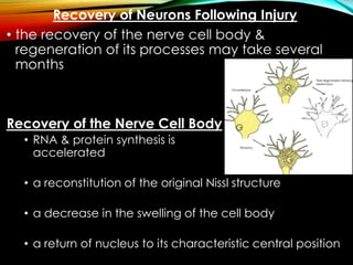
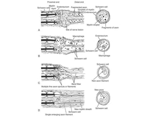
Neurons can be injured through cutting, crushing, pulling or pressure. There are different classifications of nerve injury depending on the extent of damage. Wallerian degeneration occurs after the axon is damaged, causing the distal segment to degenerate within a week. The proximal segment and cell body also exhibit retrograde degeneration changes. Recovery is possible if the nerve sheath remains intact to guide regeneration. In the peripheral nervous system, Schwann cells and macrophages clear debris to allow new axon growth. However, regeneration is more limited in the central nervous system due to inhibitory factors and scar tissue formation by astrocytes.
1. Response of neurons & nerve fibers to injury
Types of injury
Wallerian degeneration, regeneration &
Factors for regeneration
RK Goit, Lecturer
Department of Physiology
2. • Neuron is the basic function unit of the nervous
system.
• In the mature human, if it is destroyed, it is not
replaced.
• It may be injured due to various reasons(cutting,
crushing, pull & pressure).
• These injuries may damage a nerve variously &
the injury is classified according to the extent of
the damage.
3.
4.
5. Seddon (1944) described 3 clinical types of nerve injury:
• Neurapraxia (Class I)
• the term applied to a transient block
• paralysis is incomplete, recovery is rapid & complete,
& there is no nerve degeneration
• pressure is the most common cause
• Axonotmesis (Class II)
• the term applied to a nerve lesion in which the axons
are damaged but the surrounding connective tissue
sheaths remain intact
• Wallerian degeneration occurs peripherally
• functional recovery is more rapid & more complete
than after complete section of the nerve trunk
• crush injuries, traction, & compression are the most
common causes
6. • Neurotmesis (Class III)
• the term applied to complete section of the
nerve trunk
• occur on severe contusion, stretch, laceration
Sunderland (1951) expanded Seddon’s classification
to 5 degrees:
• First-degree (Class I)
• Seddon’s neurapraxia & first-degree are the same
• Second-degree (Class II)
• Seddon’s axonotmesis & second-degree are the
same
7. • Third-degree (Class II)
• nerve fiber interruption
• there is a lesion of the endoneurium, but the
epineurium & perineurium remain intact
• recovery from a third-degree injury is possible, but
surgical intervention may be required
• Fourth-degree (Class II)
• only the epineurium remain intact
• surgical repair is required
• Fifth-degree (Class III)
• lesion of complete transection of the nerve
• Recovery is not possible without an appropriate
surgical treatment
8.
9. Injury of the Nerve Cell Body
• Severe damage of the nerve cell body may
result in degeneration of the entire neuron
• In the CNS, the tissue macrophages (microglial
cells) remove the debris, & the neighboring
astrocytes replace the neuron with scar tissue
• In the PNS, the tissue macrophages remove the
debris, & the local fibroblasts replace the neuron
with scar tissue
10. Injury of the Nerve Cell Process
• if the axon of the nerve cell is
divided, degenerative changes will take place
in
1. distal segment of the axon
2. a portion of the axon proximal to the injury
3. the cell body from which the axon arises
11. Changes in the Distal Segment of the Axon
• wallerian degeneration is the changes that occur
distally to the site of damage on an axon
• axon becomes swollen & irregular; the axon is
broken into fragments, & the debris is digested by
surrounding Schwann cells & tissue macrophages
• entire axon is destroyed within a week
12. • myelin sheath is converted into lipid droplets
• the droplets are extruded from the Schwann cell
& subsequently are phagocytosed by tissue
macrophages
• Schwann cells now begin to proliferate rapidly &
axonal sprouts grow from the proximal
stump, enter the distal stump, & grow toward the
nerve's end-organs
• if regeneration does not occur, the axon & the
Schwann cells are replaced by fibrous tissue
produced by local fibroblasts
13. Changes in the Proximal Segment of the Axon
• The changes in the proximal segment of the
axon are similar to those that take place in the
distal segment but extend only proximally above
the lesion as far as the first node of Ranvier
14. Changes in the nerve cell body
• The changes that occur in the cell body
following injury to its axon are referred to as
retrograde degeneration
• The Nissl material becomes
fine, granular, & dispersed throughout
the cytoplasm (chromatolysis)
• the nucleus moves toward the periphery of the
cell, & the cell body swells & becomes rounded
• Synaptic terminals are replaced by Schwann
cells in the PNS & microglial cells or astrocytes in
the CNS
15. Recovery of Neurons Following Injury
• the recovery of the nerve cell body &
regeneration of its processes may take several
months
Recovery of the Nerve Cell Body
• RNA & protein synthesis is
accelerated
• a reconstitution of the original Nissl structure
• a decrease in the swelling of the cell body
• a return of nucleus to its characteristic central position
16. Regeneration of Axons in Peripheral Nerves
• depend on endoneurial tubes & possessed by
Schwann cells
• The following mechanisms are involved:
1. the axons are attracted by chemotropic factors
secreted by the Schwann cells in the distal stump,
2. growth-stimulating factors exist within the distal
stump, &
3. inhibitory factors are present in the perineurium to
inhibit the axons from leaving the nerve
17. Regeneration of Axons in the CNS
• Central axons may not be as good at
regeneration as peripheral axons
• The regeneration process is aborted by
• failure of oligodendrocytes to serve in the same
manner as Schwann cells,
• laying down of scar tissue by the active astrocytes
• absence of nerve growth factors in the CNS
• neuroglial cells may produce nerve growth-inhibiting
factors
18.
19. References
• Clinical Neuroanatomy, 7/E Snell
• Essentials of Medical Physiology, 3/E Mahapatra
• Principles of Neural Science, 5/E Kandel ER,
Schwartz JH, Jessell TM (editors)
Thank You