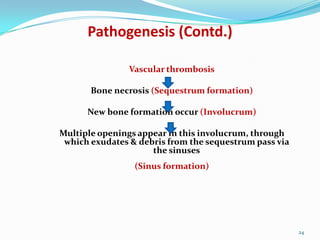
Osteomyelitis

Dr.punita adajania

1
 NELATON (1834) : coined osteomyelitis

 The root words osteon (bone) and myelo

(marrow) are combined with itis (inflammation)
to define the clinical state in which bone is
infected with microorganisms.

2
Defination of Osteomyelitis

Osteomyelitis is defined as an
acute or chronic inflammatory
process of bone, bone marrow
and its structure secondary to
infection with micro organisms.

3
CLASSIFICATION
 Duration , Mechanism & Host response.
 Duration

- Acute / Subacute / Chronic
 Mechanism - Heamatogenous(Blood)
- Exogenous (open fractures)
 Host response - Pyogenic / Granulomatous

4


Duration of infection

Acute ( <2 WEEKS)
2. Subacute (2-3 WEEKS)
3. Chronic ( >3 WEEKS)
1.

5
ACUTE OSTEOMYELITIS
 Age : Infancy and childhood.
 Sex : Males predominate 4:1

 Location : Metaphysis of long bone.
 Cause: Poor nutrition, unhygienic surroundings.

6
Etiological Agents
 Infants < 1 year – Group B streptococci

Staph aureus
E.coli
 1- 16 years – S. aureus , S. pyogens , H. Influenza
 > 16 years – S.aureus , S.epidermidis , Gram –ve bacteria

7
Pathogenesis
Introduction of bacteria from :
 Outside through a wound or continuity from a

neighboring soft tissue infection
 Hematogenous spread from a pre existing focus (most

common route of infection)

8
PATHOLOGY OF ACUTE
OSTEOMYELITIS
 Staph. aureus is the commonest causative organism.
 Others: strepto and pneumococci

9
MICRO ORGANISMS

REACH BONE VIA BLOOD
CIRCULATION

BACTERIA GET LODGED IN THE
METAPHYSIS

10
HOST BONE INITIATES AN
INFLAMMATORY REACTION

LEADS TO BONE DESTRUCTION
AND PRODUCTION OF
INFLAMMATORY EXUDATE
(PUS)

AFTER SUFFICIENT PUS
FORMATION,IT SPREADS INTO
DIFFERENT
DIRECTIONS(MEDULLARY
CAVITY, CORTEX)

11
CLINICAL FEATURES

 Fever (High Grade)

 Child refuses to use limb (pseudoparalysis)
 Local redness , swelling , warmth , oedema
 Newborn – failure to thrive , drowsy , irritable.

12
Laboratory Tests
 Elevations in the peripheral white blood cell count (WBC)
 Erythrocyte sedimentation rate (ESR) is elevated.
 The C-reactive protein level usually is elevated.
 Blood culture is positive in most of the cases.

13
 X-RAYS: Earliest sign to appear is periosteal new bone

deposition at metaphysis.(7-10 days)
 BONE SCAN
 Aspiration of bone using thick needle: for pus removal

14
15
Differential Diagnosis
 Acute septic arthritis(tenderness and swelling at joint rather

than at metaphysis)
 Acute rheumatic arthritis( features same as septic arthritis but

blood levels helps in diagnosis)
 Scurvy(mimics O.M, but absence of pain, tenderness and fever

points towards scurvy)
 Acute poliomyelitis(presence of fever and muscle tenderness but

bones are not tender)

16
Management of acute osteomyelitis.
 Bone abscess
 Septic Arthritis
 Septicemia
 Fracture

 Growth arrest
 Overlying soft-tissue cellulitis
 Chronic infection

18
Chronic Osteomyelitis
 Definition:

“ A severe, persistent and incapacitating infection of
bone and bone marrow ”

Term used for chronic pyogenic osteomyelitis.

19
3 types:
 Chronic osteomyelitis secondary to acute osteomyelitis
 Garre’s osteomyelitis

 Brodie’s abscess

20
Aetiological Agents
Usual organisms (with time there is always a mixed
infection)
 Staph.aureus(commonest)
 Staph.pyogenes
 E.coli
 Pseudomonas
 Staph.epidermidis
(commonest in surgical implant)
21
Clinical Features
Pain
Pyrexia
Redness
Tenderness
Discharging sinus
(seropurulent discharge
to thick pus)
Lower end of
femur(commonest site)

a)
b)
c)
d)
e)

22
Pathogenesis

Inadequate treatment of acute OM /Foreign
implant /
Open fracture
Inflammatory process continues with time
together with persistent infection by
Staphylococcus aureus
Persistent infection in the bone leads to
increase in intramedullary pressure due
to inflammatory exudates (pus)
stripping the periosteum
23
Pathogenesis (Contd.)
Vascular thrombosis
Bone necrosis (Sequestrum formation)
New bone formation occur (Involucrum)
Multiple openings appear in this involucrum, through
which exudates & debris from the sequestrum pass via
the sinuses
(Sinus formation)

24
Radiographic Findings
1)




X-ray examination
Thickening and irregularity of the cortex
Patchy sclerosis (honey combed appearance)
Sequestrum seen. Appears denser than the
surrounding normal bone.

2) CT scan & MRI
- Show the extent of bone destruction, reactive
oedema, hidden abscess and sequestra

25
 BLOOD: ESR may be slightly elevated.

Total blood counts are increased.
 Pus culture

26
Treatment - Antibiotics
-

Chronic infection is seldom
eradicated by antibiotics alone.

-

Antibiotic (IV route) is given for 10
days prior to surgery.

-

Bactericidal drugs are important
to:
a) Stop the spread of infection to
healthy bone
b) Control acute flares

-

After the major debridement
surgery, antibiotic is
continued for another 6 weeks
(min) but usually >3months.
[treat until inflammatory
parameters (ESR) are normal]

-

Antibiotics used in treating
chronic osteomyelitis
(Fusidic acid, Clindamycin,
Cefazolin)

27
Surgical Treatmentsequestrum.
 SEQUESTRECTOMY: Removal of
A window is made in the overlying involucrum
and the sequestrum is removed.
 SAUCERIZATION: Bone cavity is converted

into a “saucer” by removing its wall.
Allows free drainage of the infected material.

28
 CURETTAGE: The wall of the cavity, lined by

infected granulation tissue is curetted until the
underlying normal-looking bone is seen.

 EXCISION OF AN INFECTED BONE: Excision

of the infected bone segment without
compromising the functions of the limb, and
building up the gap by transporting a segment of
the bone from adjacent part.

29
 AMPUTATION: Very rarely done. Preferred in

case of long standing discharging sinus (
especially when the sinus undergoes a
malignant change).
 In many cases, combination of these procedures

are also performed.
 After surgery, wound is closed over a

“continuous irrigation system”.

30
Complications
1) Pathological Fracture
- This occurs in the bone weakened by chronic
osteomyelitis
2) Deformity
–
In children the focus of osteomyelitis destroys part of
the epiphysis growth plate.
3) Shortening/ lengthening
Destruction of growth plate arrest growth.
Stimulation of growth plate due to hyperemia.

31
 The type of rehabilitation for osteomyelitis depends on

PHYSIOTHERAPY of the infected bone and the underlying cause
MANAGEMENT
the location
of infection.
 Splinting or cast immobilization: This may be

necessary to immobilize the affected bone and nearby
joints in order to avoid further trauma and to help the
area heal adequately.
 Splinting and cast immobilization are frequently done in

children, although motion of joints after initial control is
important to prevent stiffness and atrophy.
32
 Rehabilitation is aimed at restoring normal range
of motion, flexibility, strength, and endurance.
 The goal of rehabilitation for progressive

osteomyelitis is to maintain function and
enhance mobility.
 Active range of motion initially helps maintain
flexibility and strength and relieves the
musculoskeletal pain associated with muscular
weakness, paralysis, and immobility.

33
 As the therapy progresses, passive range of motion

exercises are preferable to avoid overexertion or possible
damage to the muscles.
 In the event of muscle weakness to the legs, balance

exercises may be utilized.
 As strength continues to progress, endurance becomes a

focus in the individual's rehabilitation program for
osteomyelitis.

34
 Aerobic exercises that increase cardiovascular

fitness are recommended.

 The American Heart Association recommends 30 to

60 minutes of aerobic activity 3 or 4 times a week.

35
36

Osteomyelitis

  • 1.
  • 2.
     NELATON (1834): coined osteomyelitis  The root words osteon (bone) and myelo (marrow) are combined with itis (inflammation) to define the clinical state in which bone is infected with microorganisms. 2
  • 3.
    Defination of Osteomyelitis Osteomyelitisis defined as an acute or chronic inflammatory process of bone, bone marrow and its structure secondary to infection with micro organisms. 3
  • 4.
    CLASSIFICATION  Duration ,Mechanism & Host response.  Duration - Acute / Subacute / Chronic  Mechanism - Heamatogenous(Blood) - Exogenous (open fractures)  Host response - Pyogenic / Granulomatous 4
  • 5.
     Duration of infection Acute( <2 WEEKS) 2. Subacute (2-3 WEEKS) 3. Chronic ( >3 WEEKS) 1. 5
  • 6.
    ACUTE OSTEOMYELITIS  Age: Infancy and childhood.  Sex : Males predominate 4:1  Location : Metaphysis of long bone.  Cause: Poor nutrition, unhygienic surroundings. 6
  • 7.
    Etiological Agents  Infants< 1 year – Group B streptococci Staph aureus E.coli  1- 16 years – S. aureus , S. pyogens , H. Influenza  > 16 years – S.aureus , S.epidermidis , Gram –ve bacteria 7
  • 8.
    Pathogenesis Introduction of bacteriafrom :  Outside through a wound or continuity from a neighboring soft tissue infection  Hematogenous spread from a pre existing focus (most common route of infection) 8
  • 9.
    PATHOLOGY OF ACUTE OSTEOMYELITIS Staph. aureus is the commonest causative organism.  Others: strepto and pneumococci 9
  • 10.
    MICRO ORGANISMS REACH BONEVIA BLOOD CIRCULATION BACTERIA GET LODGED IN THE METAPHYSIS 10
  • 11.
    HOST BONE INITIATESAN INFLAMMATORY REACTION LEADS TO BONE DESTRUCTION AND PRODUCTION OF INFLAMMATORY EXUDATE (PUS) AFTER SUFFICIENT PUS FORMATION,IT SPREADS INTO DIFFERENT DIRECTIONS(MEDULLARY CAVITY, CORTEX) 11
  • 12.
    CLINICAL FEATURES  Fever(High Grade)  Child refuses to use limb (pseudoparalysis)  Local redness , swelling , warmth , oedema  Newborn – failure to thrive , drowsy , irritable. 12
  • 13.
    Laboratory Tests  Elevationsin the peripheral white blood cell count (WBC)  Erythrocyte sedimentation rate (ESR) is elevated.  The C-reactive protein level usually is elevated.  Blood culture is positive in most of the cases. 13
  • 14.
     X-RAYS: Earliestsign to appear is periosteal new bone deposition at metaphysis.(7-10 days)  BONE SCAN  Aspiration of bone using thick needle: for pus removal 14
  • 15.
  • 16.
    Differential Diagnosis  Acuteseptic arthritis(tenderness and swelling at joint rather than at metaphysis)  Acute rheumatic arthritis( features same as septic arthritis but blood levels helps in diagnosis)  Scurvy(mimics O.M, but absence of pain, tenderness and fever points towards scurvy)  Acute poliomyelitis(presence of fever and muscle tenderness but bones are not tender) 16
  • 17.
    Management of acuteosteomyelitis.
  • 18.
     Bone abscess Septic Arthritis  Septicemia  Fracture  Growth arrest  Overlying soft-tissue cellulitis  Chronic infection 18
  • 19.
    Chronic Osteomyelitis  Definition: “A severe, persistent and incapacitating infection of bone and bone marrow ” Term used for chronic pyogenic osteomyelitis. 19
  • 20.
    3 types:  Chronicosteomyelitis secondary to acute osteomyelitis  Garre’s osteomyelitis  Brodie’s abscess 20
  • 21.
    Aetiological Agents Usual organisms(with time there is always a mixed infection)  Staph.aureus(commonest)  Staph.pyogenes  E.coli  Pseudomonas  Staph.epidermidis (commonest in surgical implant) 21
  • 22.
    Clinical Features Pain Pyrexia Redness Tenderness Discharging sinus (seropurulentdischarge to thick pus) Lower end of femur(commonest site) a) b) c) d) e) 22
  • 23.
    Pathogenesis Inadequate treatment ofacute OM /Foreign implant / Open fracture Inflammatory process continues with time together with persistent infection by Staphylococcus aureus Persistent infection in the bone leads to increase in intramedullary pressure due to inflammatory exudates (pus) stripping the periosteum 23
  • 24.
    Pathogenesis (Contd.) Vascular thrombosis Bonenecrosis (Sequestrum formation) New bone formation occur (Involucrum) Multiple openings appear in this involucrum, through which exudates & debris from the sequestrum pass via the sinuses (Sinus formation) 24
  • 25.
    Radiographic Findings 1)    X-ray examination Thickeningand irregularity of the cortex Patchy sclerosis (honey combed appearance) Sequestrum seen. Appears denser than the surrounding normal bone. 2) CT scan & MRI - Show the extent of bone destruction, reactive oedema, hidden abscess and sequestra 25
  • 26.
     BLOOD: ESRmay be slightly elevated. Total blood counts are increased.  Pus culture 26
  • 27.
    Treatment - Antibiotics - Chronicinfection is seldom eradicated by antibiotics alone. - Antibiotic (IV route) is given for 10 days prior to surgery. - Bactericidal drugs are important to: a) Stop the spread of infection to healthy bone b) Control acute flares - After the major debridement surgery, antibiotic is continued for another 6 weeks (min) but usually >3months. [treat until inflammatory parameters (ESR) are normal] - Antibiotics used in treating chronic osteomyelitis (Fusidic acid, Clindamycin, Cefazolin) 27
  • 28.
    Surgical Treatmentsequestrum.  SEQUESTRECTOMY:Removal of A window is made in the overlying involucrum and the sequestrum is removed.  SAUCERIZATION: Bone cavity is converted into a “saucer” by removing its wall. Allows free drainage of the infected material. 28
  • 29.
     CURETTAGE: Thewall of the cavity, lined by infected granulation tissue is curetted until the underlying normal-looking bone is seen.  EXCISION OF AN INFECTED BONE: Excision of the infected bone segment without compromising the functions of the limb, and building up the gap by transporting a segment of the bone from adjacent part. 29
  • 30.
     AMPUTATION: Veryrarely done. Preferred in case of long standing discharging sinus ( especially when the sinus undergoes a malignant change).  In many cases, combination of these procedures are also performed.  After surgery, wound is closed over a “continuous irrigation system”. 30
  • 31.
    Complications 1) Pathological Fracture -This occurs in the bone weakened by chronic osteomyelitis 2) Deformity – In children the focus of osteomyelitis destroys part of the epiphysis growth plate. 3) Shortening/ lengthening Destruction of growth plate arrest growth. Stimulation of growth plate due to hyperemia. 31
  • 32.
     The typeof rehabilitation for osteomyelitis depends on PHYSIOTHERAPY of the infected bone and the underlying cause MANAGEMENT the location of infection.  Splinting or cast immobilization: This may be necessary to immobilize the affected bone and nearby joints in order to avoid further trauma and to help the area heal adequately.  Splinting and cast immobilization are frequently done in children, although motion of joints after initial control is important to prevent stiffness and atrophy. 32
  • 33.
     Rehabilitation isaimed at restoring normal range of motion, flexibility, strength, and endurance.  The goal of rehabilitation for progressive osteomyelitis is to maintain function and enhance mobility.  Active range of motion initially helps maintain flexibility and strength and relieves the musculoskeletal pain associated with muscular weakness, paralysis, and immobility. 33
  • 34.
     As thetherapy progresses, passive range of motion exercises are preferable to avoid overexertion or possible damage to the muscles.  In the event of muscle weakness to the legs, balance exercises may be utilized.  As strength continues to progress, endurance becomes a focus in the individual's rehabilitation program for osteomyelitis. 34
  • 35.
     Aerobic exercisesthat increase cardiovascular fitness are recommended.  The American Heart Association recommends 30 to 60 minutes of aerobic activity 3 or 4 times a week. 35
  • 36.