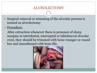
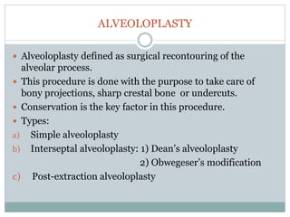
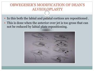
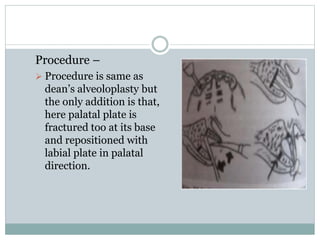
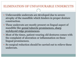
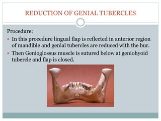
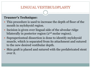
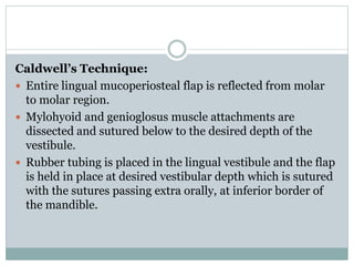
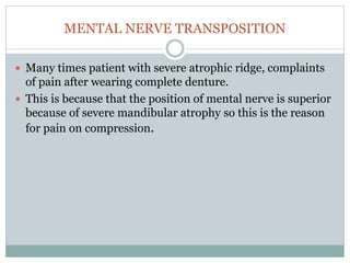
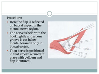
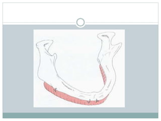
2. CONTENTS:
INTRODUCTION
DEFINITION
OBJECTIVES
AIMS OF PRE-PROSTHETIC SURGERY
CHARACTERISTICS OF IDEAL DENTURE BASE
PATIENT EVALUATION AND TREATMENT PLANNING
PRE-PROSTHETIC SURGICAL PROCEDURES
CONCLUSION
REFERENCES
3. INTRODUCTION
Prosthetics is the replacement of missing teeth (lost or
congenitally absent) and contiguous oral and maxillofacial
tissues, with artificial substitute.
Now, there remains significant number of patients, who can
never be made to use dentures effectively, because of -
- Bone atrophy
- Soft tissue hypertrophy
- or localized soft and hard tissue problems.
In these patients pre-prosthetic surgery offers significant
contribution by removing hindrance for prosthesis stability
and retention.
4. It is always hoped that the results of the preprosthetic
surgery are acceptable both surgically & prosthodontically.
In this vein, the services of an oral and maxillofacial
surgeon may be required, especially as the surgical
preparation becomes more complicated. In these
instances, a team approach is needed with the surgeon
and the prosthodontist serving as equal members of the
team.
5. DEFINITION
Pre-prosthetic surgery is carried out to reform/redesign
soft/hard tissues, by eliminating biological hindrances to
receive comfortable and stable prosthesis.
Preprosthetic surgery is defined as surgical procedures
designed to facilitate fabrication of a prosthesis or to
improve the prognosis of prosthodontic care.
(GPT8)
6. AIMS OF PRE-PROSTHETIC SURGERY
Provide adequate bony tissue support for the placement
of complete dentures.
Provide adequate soft tissue support, optimal vestibular
depth.
Elimination of pre-existing bony deformities e.g. tori,
prominent mylohyoid ridge, genial tubercle.
Correction of maxillary and mandibular ridge
relationship.
Elimination of pre-existing soft tissue deformities, e.g.
epulis, flabby ridges, hyperplastic tissues.
Maintain function
Esthetics
7. CHARACTERRISTICS OF IDEAL DENTURE BASE
Adequate bony support
Soft tissue coverage
No undercuts or protuberances
No sharp ridges
Adequate sulci
Absence of peripheral scar bands
No muscle fibres to mobilize prosthesis
No soft tissue folds/hypertrophies
No neoplastic lesions
Proper maxillomandibular arch relationships
Adequate palatal vault
8. PATIENT EVALUATION AND TREATMENT
PLANNING
Preprosthetic surgical treatment must begin with a proper
case history and physical examination
Special attention should be given to systemic diseases that
may be responsible for the severe degree of bone
resorption.
Esthetic and functional goals of the patient must be
assessed carefully.
Long term maintenance of the underlying tissues as well as
prosthetic appliances should be kept in mind.
9. PRE-PROSTHETIC SURGICAL PROCEDURES
CAN BE CLASSIFIED AS:
a) Basic procedures: can be carried out under local
anaesthesia on a day care basis.
b) Advanced surgery procedures: require hospitalization and
general anaesthesia.
Procedures are carried out for the following:
1. Alveolar ridge correction
2. Alveolar ridge extension
3. Alveolar ridge augmentation.
10. I. ALVEOLAR RIDGE CORRECTION
Bony surgeries –
i. Alveolectomy
ii. Alveoloplasty
iii. Elimination of unfavourable undercuts
- Reduction of genial tubercles
- Reduction of mylohyoid ridge
iv. Excision of tori
v. Maxillary tuberosity reduction and exostosis removal.
Soft tissue surgeries –
i. Removal of redundant crestal soft tissues
ii. Frenectomy
iii. Excision of epulis fissurata
iv. Excision palatal papillary hyperplasia.
12. ALVEOLECTOMY
Surgical removal or trimming of the alveolar process is
termed as alveolectomy
Procedure:
After extraction whenever there is presence of sharp
margins at interdental, interseptal or labiobuccal alveolar
crest, they should be trimmed with bone rounger or round
bur and smoothened with bone file.
13. ALVEOLOPLASTY
Alveoloplasty defined as surgical recontouring of the
alveolar process.
This procedure is done with the purpose to take care of
bony projections, sharp crestal bone or undercuts.
Conservation is the key factor in this procedure.
Types:
a) Simple alveoloplasty
b) Interseptal alveoloplasty: 1) Dean’s alveoloplasty
2) Obwegeser’s modification
c) Post-extraction alveoloplasty
14. SIMPLE ALVEOLOPLASTY
Procedure:
Bony areas requiring recontouring should be exposed using an
envelop type of flap.
A mucoperiosteal incision along the crest of the ridge with
adequate A-P extension is given
Adequate visualization and access to the alveolar ridge
obtained
Vertical incisions given if necessary
Excessive flap reflection may result in devitalized areas of bone
which may resorb rapidly after surgery
Recontouring can be accomplished with
Rongeur
Bone file
Bone bur in hand piece
15. Copious saline irrigation should be done throughout the
recontouring procedure to avoid overheating and bone
necrosis
After this the edges of the flap are trimmed and then sutured with
continuous or noncontinuous sutures.
16. DEAN’S INTERSEPTAL ALVEOLOPLASTY
Only done in maxillary anterior region to reduce gross maxillary over
jet.
Mostly done immediately after extraction of anterior teeth.
This technique is best used in an area where the ridge is of relative
regular contour and adequate height but presents an undercut to the
depth of the labial vestibule.
Advantages:
Labial prominence is reduced without reducing the height of the
ridge
The periosteal attachment to the bone can be maintained hereby
reducing bone resorption
Muscle attachments are left undisturbed
Disadvantage:
× Decrease in ridge thickness
17. Procedure:
After anterior teeth extraction,
Interseptal bone is cut with the
bur from canine to canine
region.
With the same bur vertical cuts
are made only in the labial cortex
at distal end of the canine
extraction socket bilaterally
without perforation of labial
mucosa.
Now labial cortex is fractured
with periosteal elevator and
compressed into palatal
direction in approximation with
palatal plate.
After removing any sharp
margin, suturing is done.
18. OBWEGESER’S MODIFICATION OF DEAN’S
ALVEOLOPLASTY
In this both the labial and palatal cortices are repositioned .
This is done when the anterior over jet is too gross that can
not be reduced by labial plate repositioning.
19. Procedure –
Procedure is same as
dean’s alveoloplasty but
the only addition is that,
here palatal plate is
fractured too at its base
and repositioned with
labial plate in palatal
direction.
20. ELIMINATION OF UNFAVOURABLE UNDERCUTS
Unfavourable undercuts are developed due to severe
atrophy of the mandible which hinders in proper denture
construction.
These undercuts are mostly present on lingual aspect of
mandible like genial tubercle prominences, sharp
mylohyoid ridge prominences.
Most of the times, patient wearing old dentures comes with
the complaint of ulceration or inflammation on these
lingual prominences.
So surgical reduction should be carried out to relieve these
undercuts.
21. REDUCTION OF GENIAL TUBERCLES
Procedure:
In this procedure lingual flap is reflected in anterior region
of mandible and genial tubercles are reduced with the bur.
Then Genioglossus muscle is sutured below at geniohyoid
tubercle and flap is closed.
22. REDUCTION OF MYLOHYOID RIDGE
Procedure:
Linear incision is made over the crest of the ridge in the
posterior aspect of the mandible
Full thickness mucoperiosteal flap is elevated to expose the
muscles
Bone file is used to remove the sharp prominance of the
mylohyoid ridge
Mylohyoid muscle is sutured below and flap is closed.
23. EXCISION OF TORI – PALATAL TORUS
Palatal tori are usually present on the midline of the hard
palate. Most palatal tori are less than 2 cm in diameter, but their
size can change throughout life.
Indication:
An extremely large torus filling the
palatal vault.
A torus that extend beyond the posterior
dam area.
Traumatized mucosa over the torus.
Deep bony undercuts interfering with denture insertion and stability.
Interference with function (speech, deglutition).
Small tori can be relieved during denture construction but large
tori should be surgically removed.
24. Procedure:
In this, midline incision is
given in palate and flap is
reflected with Y-shaped
releasing incisions.
Torus is removed by
making multiple cuts of it
and flap is sutured.
A palatal splint is given to
prevent hematoma
formation.
25. EXCISION OF TORI – MANDIBULAR TORUS
Torus mandibular is an exostosis found on the lingual
surface of the mandible opposite the canine and premolars
region.
They too interfere with denture retention because of the
loss of marginal seal in premolar region.
Indication:
Tori causing lingual undercuts and
interfering with lingual flange extension
of the planned prosthesis.
When the mucosal covering is ulcerated.
Large tori interfering with speech and
deglutition
26. Procedure:
• Bilateral lingual and inferior alveolar anesthesia is given
• Incision extending from 1 to 1.5cms beyond each tori is
given
• Always leave behind a band of tissue attached to the
midline between the anterior extent of the 2 incisions.
• When the torus has a small pedunculated base, a mallet and
an osteotome is used to cleave the tori from the medial
aspect of the mandible
• The direction of the initial bur is parallel to the medial
aspect of the mandible to prevent fracture of the lingual or
inferior cortex
27. • A bone file is then used to smoothen the lingual cortex
• Palpation is done to check for proper contour and presence
of any undercuts
• Continuous suturing is done and gauze packs are placed
and retained for the next 12 hrs
28. MAXILLARY TUBEROSITY REDUCTION AND EXOSTOSIS
REMOVAL
The main reason for tubercle overgrowth is extraction of
opposing mandibular 3rd molars and subsequent
supraeruption of maxillary 3rd molar, where remains as
bony overgrowth after maxillary 3rd molar extraction.
Maxillary tubercle interfere with denture construction
because it decreases inter-arch space.
29. Procedure:
Crestal incision from tuberosity to pre-molar area should
be given and hyperplastic soft tissue or bony overgrowth
should be removed with the help of chisel, mallet or burs.
After desired contour is achieved , the excess soft tissue is
trimmed and flap is sutured followed by splint over it.
31. REDUNDANT CRESTAL SOFT TISSUE REMOVAL
The presence of the fibrous, hyperplasic tissue gives rise to
flabby ridge form.
These flabby ridges results in unstable base for dentures.
In maxilla – enlarged tuberosity
In mandible – enlarged retro molar pad
Surgical removal is done by particular requirements of the
tissue growth.
32. FRENECTOMY
Many times there is high frenum attached near to the crest
of the ridge which may be too broad which interfere in
getting proper peripheral seal in denture.
Lingual frenum may be too short and attached till the tip of
the tongue which interfere with normal tongue movements
and causes speech problem to the patient , so surgical
correction is advocated in these cases.
33. LABIAL FRENECTOMY (MAXILLARY)
Procedure-
Cross-diamond excision:
i . This is done when there is lot of tissue is
available. Here base of the frenum at the alveolar crest
is grasped with haemostat and incision is taken above
and below the haemostat.
ii . The surgical defect is created by excision of
fibrous band. The closure is done by interrupted
sutures and small defect at alveolar crest is left to
granulate.
34. Z- plasty:
i. This procedure is used when the frenum is broad
and the vestibule is short.
V-Y incisions:
i. These incisions are used for lengthening localized
area.
Semi lunar incisions:
i. These incisions are used for broad premolar and
molar region freni.
35.
36. LINGUAL FRENECTOMY
Procedure:
Here lingual frenum is reduced by giving cross-diamond
incision.
After incision sub mucosal dissection is done on either side
and vertical suturing is given.
37. EXCISION OF EPULIS FISSURATUM
These are the benign, pedunculated lesions present as
excessive or redundant tissue of the vestibule, frequently
associated with over extended denture border.
These lesions are removed by Sharpe excision, electro
cauterization, cryosurgery or laser excision.
38. EXCISION OF PALATAL PAPILLARY HYPERPLASIA
This happens because of chronic denture irritation, because
of ill-fitting dentures.
There can be superimposed Candida infection.
Denture should be relieved in this region and antifungal
agent should be applied.
Supraperiosteal excision with a electrocautary can be done.
39. II. ALVEOLAR RIDGE EXTENSION
Whenever there is an inadequate vestibular depth present,
(due to mandibular atrophy and high muscle or soft tissue
attachment) so to increase retention and stability of
denture, deepening of vestibule is considered.
Sufficient amount of bone should be present (min 15mm
bone height) for alveolar ridge extension/ vestibuloplasty
procedure.
This procedure can be done in both jaws.
40. VESTIBULOPLASTY
Labial vestibuloplasty :
For mandibular ridge:
i. Kazanjian technique (1924).
ii. Godwin's modification (1947)
iii. Clark’s technique
iv. Obwegeser’s modification (1959)
For maxillary ridge:
i. Maxillary pocket inlay vestibuloplasty
Lingual vestibuloplasty:
i. Trauner’s technique
ii. Caldwell’s technique
* Mental nerve transposition
41. LABIAL VESTIBULOPLASTY (MANDIBULAR RIDGE)
Kazanjian technique:
Mucosal flap from inner aspect of the lower lip is used to
increase the vestibular depth in anterior mandibular labial
vestibule (premolar to premolar region).
Raw area is left on the lip side to be healed by secondary
intentions.
Periosteum of bone is left intact.
Drawback – scaring of mucosa with subsequent decreased
flexibility of lower lip.
42. Godwin’s modification:
In this procedure, flap is reflected from the inner aspect of
the lip till the alveolar crest and periosteum is reflected
from crest of the ridge till the desired depth of the vestibule.
This periosteum is now sutured to the lip mucosal margin
and then lip flap is sutured at the required vestibular depth.
Stent or splint is used for adaptation .
Advantage: less scaring of the lip mucosa.
43. Clark’s technique:
Here flap is reflected from alveolar crest till vermilian
border of the lip.
Supraperiosteal dissection is done till desired vestibular
depth and edge of the mobilized flap is pushed into
vestibular depth.
This flap is held in position with sutures passed through
the chin area extraorally and tied around the rubber
catheter.
Here alveolar bone is covered by periosteum which heals
quickly by granulation.
44. Obwegeser’s modification:
Here everything is same but the only modification is that
the alveolar bone with periosteal attachment is covered
with the split thickness skin graft or mucosal graft.
45. LABIAL VESTIBULOPLASTY (MAXILLARY RIDGE)
Maxillary pocket-inlay vestibuloplasty:
Here incision is made in the vestibule from molar to molar
region and supraperiosteal dissection is carried out.
Now a split thickness graft is placed on the extended
flanges of prefabricated denture and this denture is
positioned in extended vestibular depth which is fixed with
circumzygomatic wiring.
Wound margins are sutured to the graft.
New denture will be constructed after 6 weeks.
Advantages:
• Better retention of the dentures.
• Helps to restore deficiency in the region of nasolabial fold
with improved contour.
46. LINGUAL VESTIBULOPLASTY
Trauner’s Technique:
This procedure is used to increase the depth of floor of the
mouth in mylohyoid region.
Incision is given over lingual side of the alveolar ridge
bilaterally in posterior region (2nd molar region).
Supraperiosteal dissection is done to identify mylohyoid
muscle, which is separated from its attachment and sutured
to the new desired vestibular depth.
Skin graft is placed and sutured with the prefabricated stent
over it.
47. Caldwell’s Technique:
Entire lingual mucoperiosteal flap is reflected from molar
to molar region.
Mylohyoid and genioglosus muscle attachments are
dissected and sutured below to the desired depth of the
vestibule.
Rubber tubing is placed in the lingual vestibule and the flap
is held in place at desired vestibular depth which is sutured
with the sutures passing extra orally, at inferior border of
the mandible.
48. MENTAL NERVE TRANSPOSITION
Many times patient with severe atrophic ridge, complaints
of pain after wearing complete denture.
This is because that the position of mental nerve is superior
because of severe mandibular atrophy so this is the reason
for pain on compression.
49. Procedure:
Here the flap is reflected
on buccal aspect in the
mental nerve region.
The nerve is held with the
hook lightly and a bony
groove is cut below
mental foramen only in
buccal cortex.
Then nerve is positioned
in that groove secured in
place with gelfoam and
flap is sutured.
50. III. ALVEOLAR RIDGE AUGMENTATION
This procedure is done when alveolar bone has been
completely disappeared to the point where in maxilla a flat
surface is present between vestibule and palate and in
mandible mental nerve is positioned almost at the crest.
Here alveolar bone height is less that 15 mm.
So vestibuloplasty is out of consideration in this case until
the replacement of necessary supportive bone is done.
So we have two options available with us :
a) Augmentation of alveolar bone.
b) Place the implant.
51. Aims:
Restoration of optimum ridge height, width, ridge form,
vestibular depth and optimum denture bearing area.
Protection of neurovascular bundle.
Establishment of proper interarch relationship
Improvement of retention and stability of denture.
Improve the patient comfort for wearing the denture.
52. Materials used for ridge augmentation:
1. Autogeneous bone graft: iliac crest, rib grafts.
2. Allogenic bone grafts: freeze dried cadaver bone.
3. Alloplastic material: hydroxyapatite.
4. Metal mesh with autogenous cancellous bone.
5. Metal mesh with hydroxyapatite.
53. RIDGE AUGMENTATION PROCEDURE
1. Superior border augmentation
2. Inferior border augmentation
3. Interpositional or sandwich bone grafts
4. Onlay grafting
5. Visor osteotomy
6. Modified visor osteotomy
54. MANDIBULAR AUGMENTATION
Superior border grafting:
First described by Davis in 1970.
Remaining bone < 10 mm
Here two autogenous bone grafts
of 15 cm each are used.
One rib is scored to the cortex followed by giving shape
of the mandible and attached at the superior border of
the mandible by circummandibular wiring.
The other rib graft is made into corticocancellous
particles and moulded around the first rib graft.
Surgical flap is then closed.
56. Disadvantages –
× Donor site morbidity.
× Second surgical site is necessary.
× Continued resorption at the graft sites
57. Inferior border grafting:
This procedure is indicated when alveolar ridge height is less
than 5 to 8mm and is at risk of pathological fracture.
In this procedure, a cadever mandible used for grafting which
is filled with cancellous graft material for revascularization.
This mandible is then fixed to the inferior border with vicryl
sutures, by circummandibular fixation and neck flap is
closed.
Osseointegrated implants can be placed after 4-6 months.
58.
59. Advantages
Does not obliterate the vestibule.
Interim denture can be worn.
No change in vertical dimension.
Graft is not subjected to direct masticatory force.
60. Disadvantages:
× It will not correct the abnormalities of denture bearing
area.
× It will not protect a highly placed mental nerve.
× Donor site morbidity.
× Resorption of the graft.
× Presence of scar.
61. INTERPOSITIONAL BONE GRAFT (SANDWICH
GRAFTING)
In this procedure, a horizontal osteotomy is performed by
splitting of the maxilla or mandible and bone is grafted in
the gap.
In mandible, this procedure is mainly used in anterior
mandible.
Prosthetic appliance is given after 3-5 months.
Advantages:
Less resorption than onlay grafting.
More predictable long term results.
Decreased incidence of nerve paresthesia than the visor
osteotomy.
Can be used in conjuction with implants.
62.
63. ONLAY GRAFTING
This procedure helps in increasing width of the ridge.
Here graft material is placed on the buccal cortex either
in putty form by mixing with saline/blood or in the
form of blocks or split thickness rib/illiac crest graft.
Advantages:
Improves width and to some extent height too.
Can be used in anterior and posterior region.
64. VISOR OSTEOTOMY
Was originated by Harle and modified by Peterson and
Slade.
It is used where insufficient vertical mandibular bone
height is present for the horizontal osteotomy technique
but adequate bone width (approximately 10mm) is
present.
65. The mandible is split vertically and the lingual section
is elevated to increase the mandibular height.
Cancellous bone or particulate bone and marrow is
placed to correct the contours and fill in the gaps on
the facial side of the elevated segment.
Transosteal wires hold the segments in place for a
period of 3-4 months before vestibuloplasties are
performed.
The disadvantage is unavoidable nerve trauma and the
resultant parasthesia.
66. MODIFIED VISOR OSTEOTOMY
The procedures of choice for mandibular ridge
augmentation include the combination of osteotomy
techniques (horizontal or vertical) with interpositional
bone grafting.
These procedures involve the movement of a pedicle of
bone (not technically a graft) along with its blood supply.
Theoretically, the viability of the bone will be greater and
the resorption decreased because the blood supply to the
bone is maintained.
67. Procedure:
Vertical osteotomy cut is made in the posterior region to
divide the segments buccolingually.
A horizontal osteotomy is performed in the anterior
mandible to divide the anterior segment superiorly and
inferiorly, and bone grafting was done into the
osteotomised gap.
Two osteotomised segments are fixed with wires.
68.
69. Advantages:
Increased bone height which is relatively stable.
Shortened post-operative period (3 months).
Rate of resorption is less when compared to onlay grafts.
Disadvantages:
× Nerve trauma
× Parasthesia
× Mandibular fracture
× Flap dehiscence.
70. PROBLEMS ENCOUNTERED WITH
AUGMENTION PROCEDURE
1. Inadequate soft tissue cover.
2. Rejection of the auto grafts (failure of union with the host
bone).
3. Dehiscence of overlying mucosa.
4. Migration of the graft material.
5. Resorption of the graft.
71. CONCLUSION
Accurate diagnosis of the problem areas during denture
construction and determination of the necessity of surgery
is accomplished by careful evaluation of the information
systematically obtained from the patient.
As conservation is the philosophy of surgical patient
management. Therefore every attempt should be made to
preserve as much as oral structures as possible.
Proper knowledge of the available surgical procedures
helps in achieving the best results.
72. REFERENCES
BOUCHER`S –prosthodontic treatment for edentulous
patients 11th edition .
CHARLES HEARTWELL & ARTHUR O RAHN –Syllabus
of complete dentures 4th edition.
JOHN J SHARRY- Complete denture prosthodontics 2nd
edition.
SHELDON WINKLER- Essentials of complete dentures 2nd
edition
ZARB, BOLENDER – Prosthodontic treatment for
edentulous patients 12th edition.
Principles of oral & maxillofacial surgery-Peterson